

A cell-based model exhibiting branching and anastomosis during tumor-induced angiogenesis
- PMID: 17277180
- PMCID: PMC1852370
- DOI: 10.1529/biophysj.106.101501
A cell-based model exhibiting branching and anastomosis during tumor-induced angiogenesis
Abstract
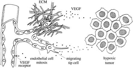
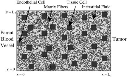
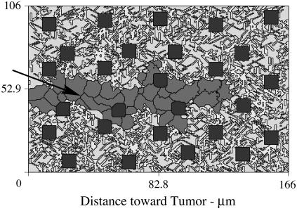
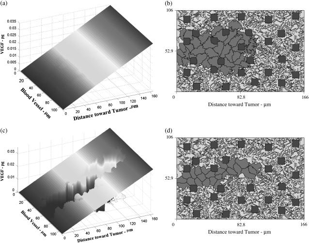
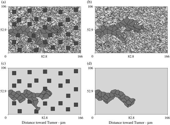
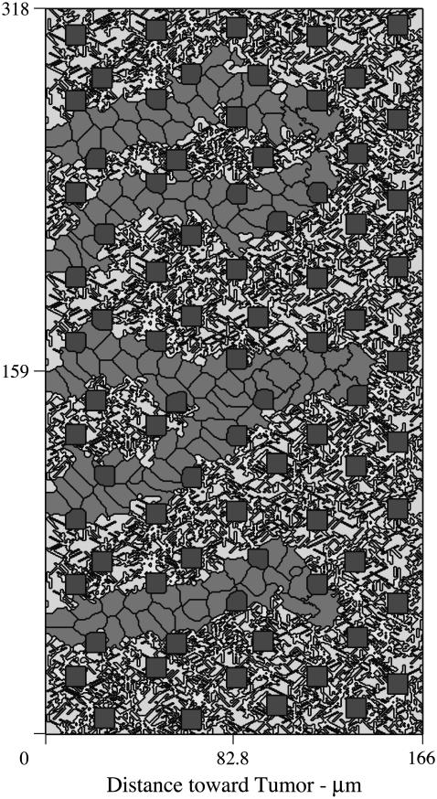
This work describes the first cell-based model of tumor-induced angiogenesis. At the extracellular level, the model describes diffusion, uptake, and decay of tumor-secreted pro-angiogenic factor. At the cellular level, the model uses the cellular Potts model based on system-energy reduction to describe endothelial cell migration, growth, division, cellular adhesion, and the evolving structure of the stroma. Numerical simulations show: 1), different tumor-secreted pro-angiogenic factor gradient profiles dramatically affect capillary sprout morphology; 2), average sprout extension speeds depend on the proximity of the proliferating region to the sprout tip, and the coordination of cellular functions; and 3), inhomogeneities in the extravascular tissue lead to sprout branching and anastomosis, phenomena that emerge without any prescribed rules. This model provides a quantitative framework to test hypotheses on the biochemical and biomechanical mechanisms that control tumor-induced angiogenesis.
Figures

Similar articles
-
Continuous and discrete mathematical models of tumor-induced angiogenesis.Bull Math Biol. 1998 Sep;60(5):857-99. doi: 10.1006/bulm.1998.0042. Bull Math Biol. 1998. PMID: 9739618
-
Mathematical modeling of tumor-induced angiogenesis.Annu Rev Biomed Eng. 2006;8:233-57. doi: 10.1146/annurev.bioeng.8.061505.095807. Annu Rev Biomed Eng. 2006. PMID: 16834556 Review.
-
Evolution of uncontrolled proliferation and the angiogenic switch in cancer.Math Biosci Eng. 2012 Oct;9(4):843-76. doi: 10.3934/mbe.2012.9.843. Math Biosci Eng. 2012. PMID: 23311425
-
Mathematical modelling, simulation and prediction of tumour-induced angiogenesis.Invasion Metastasis. 1996;16(4-5):222-34. Invasion Metastasis. 1996. PMID: 9311387 Review.
-
Mathematical modelling of angiogenesis.J Neurooncol. 2000 Oct-Nov;50(1-2):37-51. doi: 10.1023/a:1006446020377. J Neurooncol. 2000. PMID: 11245280 Review.
Cited by
-
A computational model of in vitro angiogenesis based on extracellular matrix fibre orientation.Comput Methods Biomech Biomed Engin. 2013;16(7):790-801. doi: 10.1080/10255842.2012.662678. Epub 2012 Apr 19. Comput Methods Biomech Biomed Engin. 2013. PMID: 22515707 Free PMC article.
-
Towards integration of time-resolved confocal microscopy of a 3D in vitro microfluidic platform with a hybrid multiscale model of tumor angiogenesis.PLoS Comput Biol. 2023 Jan 18;19(1):e1009499. doi: 10.1371/journal.pcbi.1009499. eCollection 2023 Jan. PLoS Comput Biol. 2023. PMID: 36652468 Free PMC article.
-
Theoretical models for coronary vascular biomechanics: progress & challenges.Prog Biophys Mol Biol. 2011 Jan;104(1-3):49-76. doi: 10.1016/j.pbiomolbio.2010.10.001. Epub 2010 Oct 30. Prog Biophys Mol Biol. 2011. PMID: 21040741 Free PMC article. Review.
-
From single cells to tissue architecture-a bottom-up approach to modelling the spatio-temporal organisation of complex multi-cellular systems.J Math Biol. 2009 Jan;58(1-2):261-83. doi: 10.1007/s00285-008-0172-4. Epub 2008 Apr 2. J Math Biol. 2009. PMID: 18386011
-
A Review of Cell-Based Computational Modeling in Cancer Biology.JCO Clin Cancer Inform. 2019 Feb;3:1-13. doi: 10.1200/CCI.18.00069. JCO Clin Cancer Inform. 2019. PMID: 30715927 Free PMC article. Review.
References
-
- Straume, O., H. B. Salvesen, and L. A. Akslen. 1999. Angiogenesis is prognostically important in vertical growth phase melanomas. Int. J. Oncol. 15:595–599. - PubMed
-
- Asahara, T., C. Kalka, and J. M. Isner. 2000. Stem cell therapy and gene transfer for regeneration. Gene Ther. 7:451–457. - PubMed
-
- Carmeliet, P., and R. K. Jain. 2000. Angiogenesis in cancer and other diseases. Nature. 407:249–257. - PubMed
-
- Leith, J. T., and S. Michelson. 1995. Secretion rates and levels of vascular endothelial growth factor in clone-a or Hct-8 human colon tumor cells as a function of oxygen concentration. Cell Prolif. 28:415–430. - PubMed
-
- Carmeliet, P., V. Ferreira, G. Breier, S. Pollefeyt, L. Kieckens, M. Gertsenstein, M. Fahrig, A. Vandenhoeck, K. Harpal, C. Eberhardt, C. Declercq, J. Pawling, L. Moons, D. Collen, W. Risau, and A. Nagy. 1996. Abnormal blood vessel development and lethality in embryos lacking a single VEGF allele. Nature. 380:435–439. - PubMed
Publication types
MeSH terms
Substances
LinkOut - more resources
Full Text Sources
