

Neurexin-neuroligin signaling in synapse development
- PMID: 17275284
- PMCID: PMC2820508
- DOI: 10.1016/j.conb.2007.01.011
Neurexin-neuroligin signaling in synapse development
Abstract
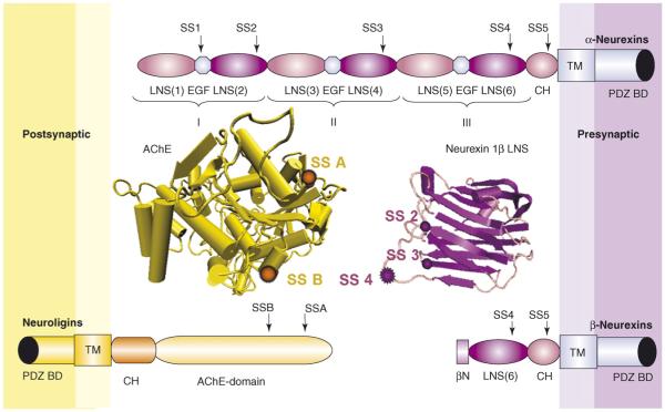
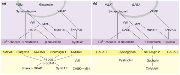
Neurexins and neuroligins are emerging as central organizing molecules for excitatory glutamatergic and inhibitory GABAergic synapses in mammalian brain. They function as cell adhesion molecules, bridging the synaptic cleft. Remarkably, each partner can trigger formation of a hemisynapse: neuroligins trigger presynaptic differentiation and neurexins trigger postsynaptic differentiation. Recent protein interaction assays and cell culture studies indicate a selectivity of function conferred by alternative splicing in both partners. An insert at site 4 of beta-neurexins selectively promotes GABAergic synaptic function, whereas an insert at site B of neuroligin 1 selectively promotes glutamatergic synaptic function. Initial knockdown and knockout studies indicate that neurexins and neuroligins have an essential role in synaptic transmission, particularly at GABAergic synapses, but further studies are needed to assess the in vivo functions of these complex protein families.
Figures
Similar articles
-
Induction of GABAergic postsynaptic differentiation by alpha-neurexins.J Biol Chem. 2008 Jan 25;283(4):2323-34. doi: 10.1074/jbc.M703957200. Epub 2007 Nov 15. J Biol Chem. 2008. PMID: 18006501 Free PMC article.
-
Alternative splicing controls selective trans-synaptic interactions of the neuroligin-neurexin complex.Neuron. 2006 Jul 20;51(2):171-8. doi: 10.1016/j.neuron.2006.06.005. Neuron. 2006. PMID: 16846852
-
A splice code for trans-synaptic cell adhesion mediated by binding of neuroligin 1 to alpha- and beta-neurexins.Neuron. 2005 Oct 20;48(2):229-36. doi: 10.1016/j.neuron.2005.08.026. Neuron. 2005. PMID: 16242404
-
GABA and neuroligin signaling: linking synaptic activity and adhesion in inhibitory synapse development.Curr Opin Neurobiol. 2008 Feb;18(1):77-83. doi: 10.1016/j.conb.2008.05.008. Epub 2008 May 29. Curr Opin Neurobiol. 2008. PMID: 18513949 Free PMC article. Review.
-
A matter of balance: role of neurexin and neuroligin at the synapse.Neurochem Res. 2013 Jun;38(6):1174-89. doi: 10.1007/s11064-013-1029-9. Epub 2013 Apr 5. Neurochem Res. 2013. PMID: 23559421 Review.
Cited by
-
Identification of visual cortex cell types and species differences using single-cell RNA sequencing.Nat Commun. 2022 Nov 12;13(1):6902. doi: 10.1038/s41467-022-34590-1. Nat Commun. 2022. PMID: 36371428 Free PMC article.
-
Neuroligin-3-Mediated Synapse Formation Strengthens Interactions between Hippocampus and Barrel Cortex in Associative Memory.Int J Mol Sci. 2024 Jan 5;25(2):711. doi: 10.3390/ijms25020711. Int J Mol Sci. 2024. PMID: 38255783 Free PMC article.
-
Dystroglycan Mediates Clustering of Essential GABAergic Components in Cerebellar Purkinje Cells.Front Mol Neurosci. 2020 Aug 28;13:164. doi: 10.3389/fnmol.2020.00164. eCollection 2020. Front Mol Neurosci. 2020. PMID: 32982691 Free PMC article.
-
Synaptic Organizers in Alzheimer's Disease: A Classification Based on Amyloid-β Sensitivity.Front Cell Neurosci. 2020 Sep 2;14:281. doi: 10.3389/fncel.2020.00281. eCollection 2020. Front Cell Neurosci. 2020. PMID: 32982693 Free PMC article. Review.
-
The cell adhesion molecule neuroplastin-65 is a novel interaction partner of γ-aminobutyric acid type A receptors.J Biol Chem. 2012 Apr 20;287(17):14201-14. doi: 10.1074/jbc.M111.293175. Epub 2012 Mar 2. J Biol Chem. 2012. PMID: 22389504 Free PMC article.
References
-
- Ushkaryov YA, Petrenko AG, Geppert M, Sudhof TC. Neurexins: synaptic cell surface proteins related to the alpha-latrotoxin receptor and laminin. Science. 1992;257:50–56. - PubMed
-
- Scheiffele P, Fan J, Choih J, Fetter R, Serafini T. Neuroligin expressed in nonneuronal cells triggers presynaptic development in contacting axons. Cell. 2000;101:657–669. - PubMed
-
-
Graf ER, Zhang X, Jin SX, Linhoff MW, Craig AM. Neurexins induce differentiation of GABA and glutamate postsynaptic specializations via neuroligins. Cell. 2004;119:1013–1026.Using co-cultures of neurons with COS cells expressing neurexins, this study showed that signaling occurs from neurexins to the postsynaptic site to mediate clustering of receptors and scaffolding proteins through neuroligins. In contrast to prior work focusing on glutamate synapses, these authors showed that neurexin signaling occurs at GABA synapses selectively through neuroligin 2.
-
-
-
Prange O, Wong TP, Gerrow K, Wang YT, El-Husseini A. A balance between excitatory and inhibitory synapses is controlled by PSD-95 and neuroligin. Proc Natl Acad Sci USA. 2004;101:13915–13920.These authors altered the levels of neuroligin 1 and PSD-95 in cultured neurons and showed that the ratio of these proteins affects not only glutamate synapses but also GABA synapses. For example, increased expression of PSD-95 not only enhanced glutamate-mediated transmission but also reduced GABA-mediated input. Thus, the level of neuroligins and key interacting partners can control the balance of glutamate versus GABA inputs on a neuron.
-
-
-
Chih B, Engelman H, Scheiffele P. Control of excitatory and inhibitory synapse formation by neuroligins. Science. 2005;307:1324–1328.This study followed on key earlier work [2] showing that presentation of neuroligins to glutamatergic axons is sufficient to induce presynaptic differentiation. Here, overexpression and knockdown of neuroligins in cultured neurons was reported to alter glutamatergic and GABAergic inputs. The major functional effect of knockdown of neuroligins 1, 2 and 3 together was a reduction in mIPSC-like events.
-
Publication types
MeSH terms
Substances
Grants and funding
LinkOut - more resources
Full Text Sources
Other Literature Sources
