

Release factors 2 from Escherichia coli and Thermus thermophilus: structural, spectroscopic and microcalorimetric studies
- PMID: 17272297
- PMCID: PMC1849895
- DOI: 10.1093/nar/gkl696
Release factors 2 from Escherichia coli and Thermus thermophilus: structural, spectroscopic and microcalorimetric studies
Abstract
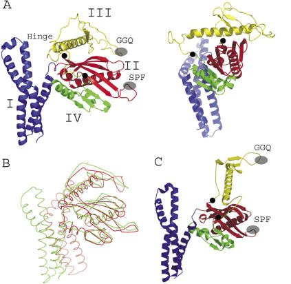
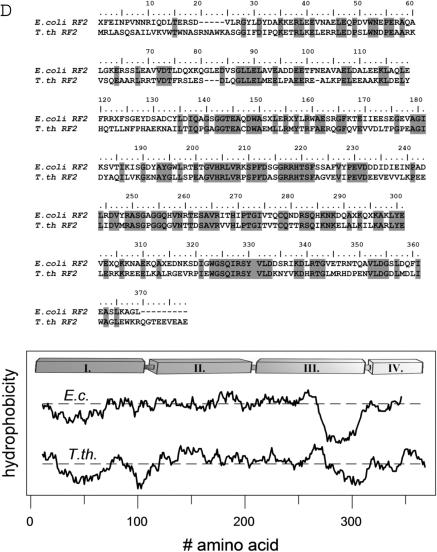
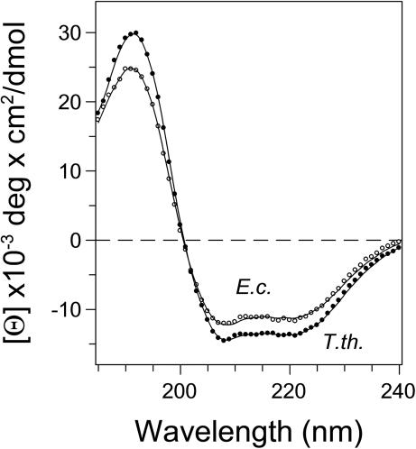
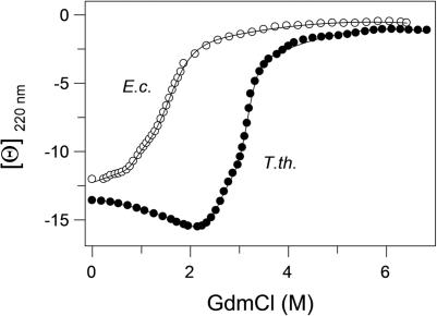
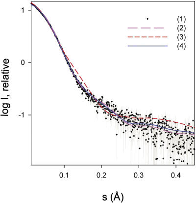
Prokaryotic class I release factors (RFs) respond to mRNA stop codons and terminate protein synthesis. They interact with the ribosomal decoding site and the peptidyl-transferase centre bridging these 75 A distant ribosomal centres. For this an elongated RF conformation, with partially unfolded core domains II.III.IV is required, which contrasts the known compact RF crystal structures. The crystal structure of Thermus thermophilus RF2 was determined and compared with solution structure of T. thermophilus and Escherichia coli RF2 by microcalorimetry, circular dichroism spectroscopy and small angle X-ray scattering. The structure of T. thermophilus RF2 in solution at 20 degrees C is predominantly compact like the crystal structure. Thermodynamic analysis point to an initial melting of domain I, which is independent from the melting of the core. The core domains II.III.IV melt cooperatively at the respective physiological temperatures for T. thermophilus and E. coli. Thermodynamic analyses and the X-ray scattering results for T. thermophilus RF2 in solution suggest that the compact conformation of RF2 resembles a physiological state in absence of the ribosome.
Figures

Similar articles
-
Structure of the Escherichia coli ribosomal termination complex with release factor 2.Nature. 2003 Jan 2;421(6918):90-4. doi: 10.1038/nature01225. Nature. 2003. PMID: 12511961
-
The SAXS solution structure of RF1 differs from its crystal structure and is similar to its ribosome bound cryo-EM structure.Mol Cell. 2005 Dec 22;20(6):929-38. doi: 10.1016/j.molcel.2005.11.022. Mol Cell. 2005. PMID: 16364917
-
Molecular basis for bacterial class I release factor methylation by PrmC.Mol Cell. 2005 Dec 22;20(6):917-27. doi: 10.1016/j.molcel.2005.10.025. Mol Cell. 2005. PMID: 16364916
-
Multidomain initiation factor 2 from Thermus thermophilus consists of the individual autonomous domains.Biochemistry. 2008 Apr 29;47(17):4992-5005. doi: 10.1021/bi702295g. Epub 2008 Apr 5. Biochemistry. 2008. PMID: 18393450
-
A cryo-electron microscopic study of ribosome-bound termination factor RF2.Nature. 2003 Jan 2;421(6918):87-90. doi: 10.1038/nature01224. Nature. 2003. PMID: 12511960
Cited by
-
Diversity and Similarity of Termination and Ribosome Rescue in Bacterial, Mitochondrial, and Cytoplasmic Translation.Biochemistry (Mosc). 2021 Sep;86(9):1107-1121. doi: 10.1134/S0006297921090066. Biochemistry (Mosc). 2021. PMID: 34565314 Free PMC article. Review.
-
Inhibition of translation termination by small molecules targeting ribosomal release factors.Sci Rep. 2019 Oct 28;9(1):15424. doi: 10.1038/s41598-019-51977-1. Sci Rep. 2019. PMID: 31659219 Free PMC article.
-
Consensus among flexible fitting approaches improves the interpretation of cryo-EM data.J Struct Biol. 2012 Feb;177(2):561-70. doi: 10.1016/j.jsb.2011.10.002. Epub 2011 Oct 13. J Struct Biol. 2012. PMID: 22019767 Free PMC article.
-
The codon specificity of eubacterial release factors is determined by the sequence and size of the recognition loop.RNA. 2010 Aug;16(8):1623-33. doi: 10.1261/rna.2117010. Epub 2010 Jun 28. RNA. 2010. PMID: 20584893 Free PMC article.
-
Mechanism of ribosome rescue by ArfA and RF2.Elife. 2017 Mar 16;6:e23687. doi: 10.7554/eLife.23687. Elife. 2017. PMID: 28300532 Free PMC article.
References
-
- Ogle J.M., Ramakrishnan V. Structural insights into translational fidelity. Annu. Rev. Biochem. 2005;74:129–177. - PubMed
-
- Schuwirth B.S., Borovinskaya M.A., Hau C.W., Zhang W., Vila-Sanjurjo A., Holton J.M., Cate J.H. Structures of the bacterial ribosome at 3.5 A resolution. Science. 2005;310:827–834. - PubMed
-
- Gao H., Sengupta J., Valle M., Korostelev A., Eswar N., Stagg S.M., Van Roey P., Agrawal R.K., Harvey S.C., Sali A., et al. Study of the structural dynamics of the Escherichia coli 70S ribosome using real-space refinement. Cell. 2003;113:789–801. - PubMed
-
- Frank J., Sengupta J., Gao H., Li W., Valle M., Zavialov A., Ehrenberg M. The role of tRNA as a molecular spring in decoding, accommodation, and peptidyl transfer. FEBS Lett. 2005;579:959–962. - PubMed
-
- Schmeing T.M., Huang K.S., Strobel S.A., Steitz T.A. An induced-fit mechanism to promote peptide bond formation and exclude hydrolysis of peptidyl-tRNA. Nature. 2005;438:520–524. - PubMed
Publication types
MeSH terms
Substances
Associated data
- Actions
LinkOut - more resources
Full Text Sources
Molecular Biology Databases
