Comparative Study
. 2007 Feb 21;26(4):1055-67.
doi: 10.1038/sj.emboj.7601551. Epub 2007 Feb 1.

c-Myc primed mitochondria determine cellular sensitivity to TRAIL-induced apoptosis

Affiliations
Comparative Study

c-Myc primed mitochondria determine cellular sensitivity to TRAIL-induced apoptosis

Anni I Nieminen et al. EMBO J. .

Abstract

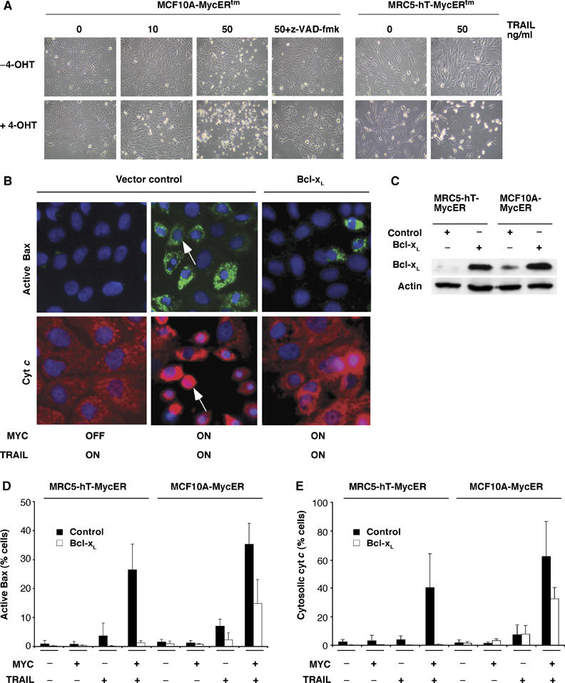
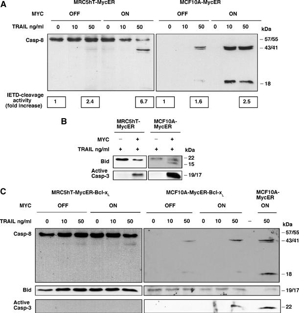
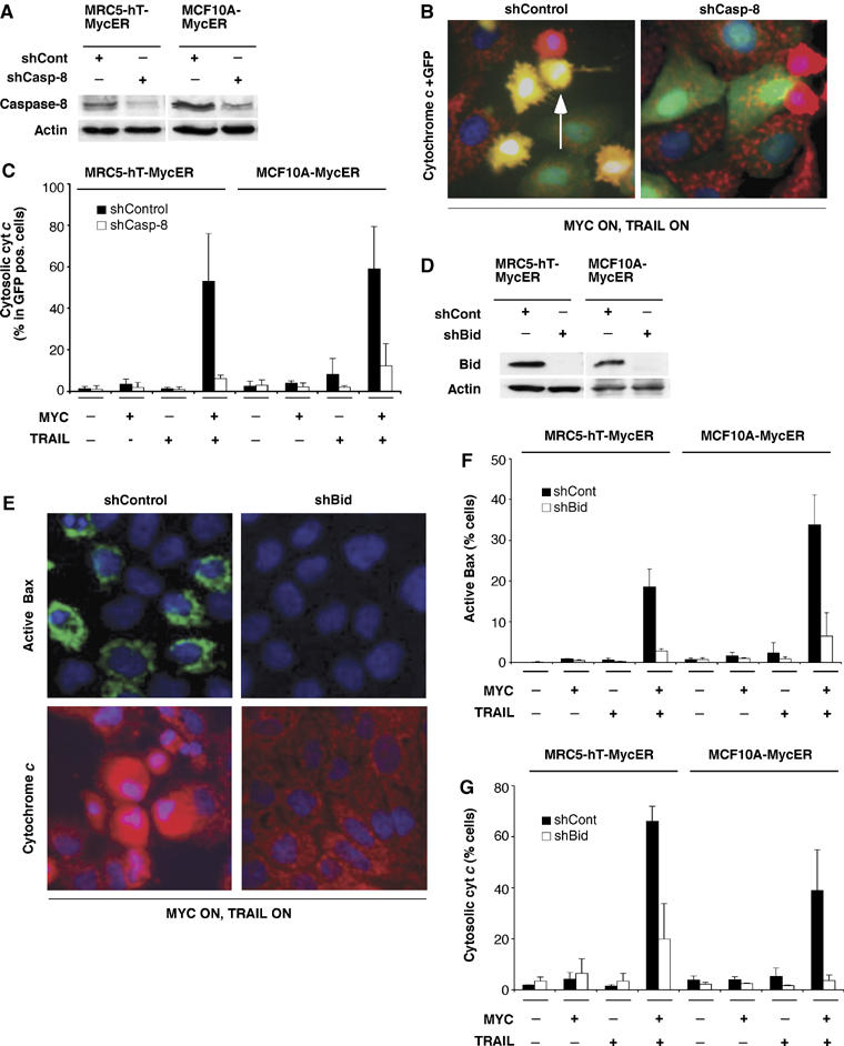
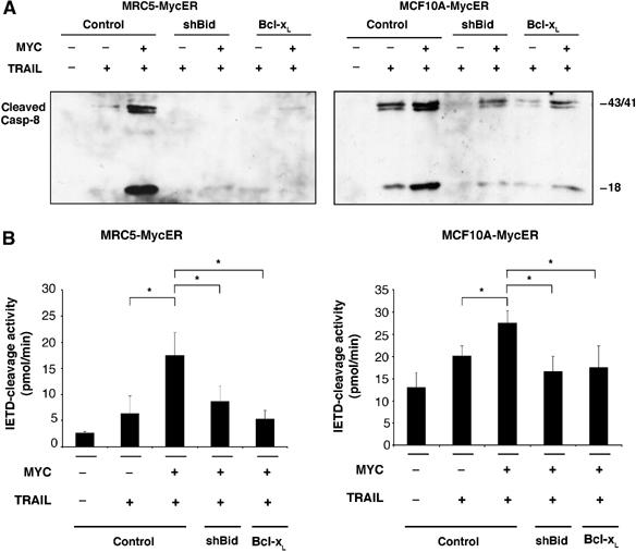
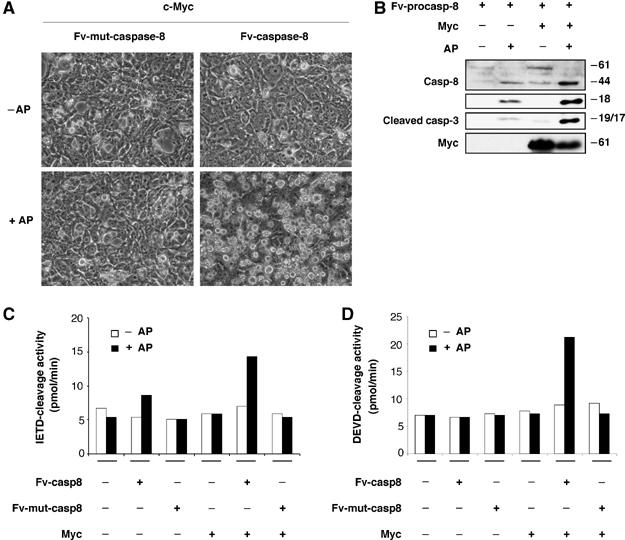
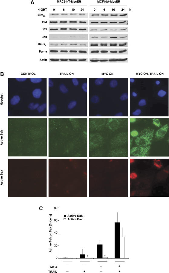
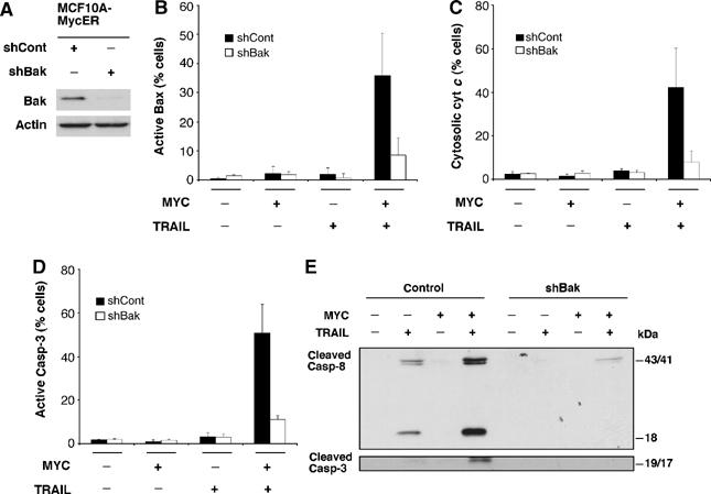
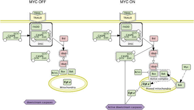
Oncogenic c-Myc renders cells sensitive to TRAIL-induced apoptosis, and existing data suggest that c-Myc sensitizes cells to apoptosis by promoting activation of the mitochondrial apoptosis pathway. However, the molecular mechanisms linking the mitochondrial effects of c-Myc to the c-Myc-dependent sensitization to TRAIL have remained unresolved. Here, we show that TRAIL induces a weak activation of procaspase-8 but fails to activate mitochondrial proapoptotic effectors Bax and Bak, cytochrome c release or downstream effector caspase-3 in non-transformed human fibroblasts or mammary epithelial cells. Our data is consistent with the model that activation of oncogenic c-Myc primes mitochondria through a mechanism involving activation of Bak and this priming enables weak TRAIL-induced caspase-8 signals to activate Bax. This results in cytochrome c release, activation of downstream caspases and postmitochondrial death-inducing signaling complex -independent augmentation of caspase-8-Bid activity. In conclusion, c-Myc-dependent priming of the mitochondrial pathway is critical for the capacity of TRAIL-induced caspase-8 signals to activate effector caspases and for the establishment of lethal caspase feedback amplification loop in human cells.


Figures

Figure 1
Figure 1
TRAIL-induced activation of Bax and release of cytochrome c (cyt c) requires active c-Myc and is blocked by Bcl-xL. (A) c-Myc sensitizes MCF10A mammary epithelial cells and MRC5 fibroblasts to TRAIL-induced apoptosis. To activate Myc, MCF10A-MycERtm and MRC5-hT-MycERtm cells were treated for 24 h with 10 nM 4-OHT. Control cells were treated with ethanol carrier. Subsequently, the cells were treated with 0, 10 or 50 ng/ml of TRAIL for 2 h. Representative photomicrographs of the cell cultures are shown in the picture. zVAD-fmk (50 μM) was used to block caspases. (B) c-Myc was activated in the control and Bcl-xL-overexpressing MCF10A-MycERtm cells and the cells were treated with 50 ng/ml of TRAIL as described in (A). Fixed cells were immunostained either with anti-active Bax or anti-cyt c antibody and counterstained with Hoechst 33258. Shown are immunofluorescence images of the cells treated with ethanol carrier (MYC OFF) or 4-OHT (MYC ON) and TRAIL. Note the punctate, mitochondrial staining pattern of active Bax in the cells with active c-Myc and TRAIL pathways (white arrow). Cyt c normally exhibits punctate mitochondrial staining pattern, which becomes diffuse upon release of cyt c to the cytosol (white arrow). (C) Western immunoblot analysis showing ectopic expression level of Bcl-xL. (D, E) Quantitation of cells with active Bax or cytosolic cyt c. Cells were treated (MYC +/−, 50 ng/ml TRAIL +/−) and stained as in B. Images were collected and analyzed as described in Materials and methods. Between 300–700 cells were analyzed at each treatment. The graph values represent mean±s.d. of minimum of three independent experiments.
Figure 2
Figure 2
TRAIL induces activation of caspase-8, which is augmented by active c-Myc via Bcl-xL regulated pathway. (A) The cells were treated as in Figure 1A and the cell lysates were analyzed for procaspase-8 cleavage and for proteolytic activity towards Ac-IETD-pNa. The immunoblot analysis shows that active c-Myc enhances TRAIL-induced cleavage of procaspase-8 into the active subunits (p43/p41 and p18). The caspase-8 antibody used recognized full-length procaspase-8 in MRC5 cells, but for some reason only weakly in MCF10A cells. However, the cleavage products that were diagnostic for caspase-8 activation were reliably detected in both cell types. The boxed values represent relative caspase-8 activities extracted from the datasets in Figure 4B. (B) Bid and caspase-3 activation. The immunoblot analyses of Bid cleavage using an antibody recognizing both full length and truncated forms of Bid and caspase-3 activation with an antibody recognizing active processed form. (C) The cells overexpressing Bcl-xL were treated as in A and analyzed for procaspase-8 processing. The immunoblots demonstrate that c-Myc fails to augment TRAIL-induced processing of procaspase-8 in the cells overexpressing Bcl-xL.
Figure 3
Figure 3
Caspase-8 and Bid are required for induction of cyt c release and Bax activation by c-Myc and TRAIL. (A) Western immunoblot demonstrates lentiviral shRNA silencing of endogenous procaspase-8. The efficiency of lentiviral infection was about 80%. (B) Immunofluorescence detection of cyt c release specifically in EGFP-positive transduced cells. Control and caspase-8 shRNA-transduced MCF10A-MycERtm cells were immunostained with anti-cyt c antibody (red) and the nuclei were counterstained with Hoechst (blue). Transduced EGFP expressing cells are green in the images. In merged images, diffuse yellow staining pattern (white arrow) indicates cytosolic cyt c in EGFP-positive cells. (C) Quantitation of transduced cells with cytosolic cyt c. The cells were treated as in Figure 1 and scored from merged images. The graph values show percentage of EGFP-positive cells with cytosolic cyt c. (D) Western immunoblot demonstrating lentiviral shRNA silencing of endogenous Bid. (E) Control and Bid shRNA-transduced cells were treated and analyzed as described in Figure 1. Representative immunofluorescence images are shown. (F, G) Quantitation of Bid-deficient cells with active Bax or cytosolic cyt c. Cells were scored as in Figure 1. All graph values show percentages and represent mean±s.d. of at least three independent experiments.
Figure 4
Figure 4
c-Myc fails to augment TRAIL-induced activation of procaspase-8 in the cells with impaired mitochondrial Bid pathway. (A) c-Myc and TRAIL pathways were activated in the cell strains lacking Bid or overexpressing Bcl-xL as described in Figure 1 and the cell lysates were analyzed for procaspase-8 cleavage. (B) The cell lysates were analyzed for proteolytic activity towards Ac-IETD-pNa substrate. Lysates (40 g) from the above experiments were analyzed. The graph values represent mean±s.d. of three independent experiments (*P-value <0.05).
Figure 5
Figure 5
c-Myc augments cleavage of procaspase-8 outside the DISC. The chemically dimerizable Fv-caspase-8 expression constructs were cotransfected with mock or c-Myc expression plasmid into 293FT cells. After 48 h of transfection, the Fv constructs were dimerized by incubation of the cells with 100 nM AP20187 for four additional hours. (A) Phase contrast photomicrographs of the cells. (B) The immunoblots demonstrate that a chemically induced, non-DISC mediated processing of Fv-caspase-8 into the active subunits is enhanced in the cells overexpressing c-Myc (compare +AP, −Myc and +AP, +Myc). The blots also show that active Fv-caspase-8 cooperates with c-Myc to induce activation of caspase-3. (C) caspase-8-like activity (Ac-IETD-pNa) and (D) caspase-3-like activity (Ac-DEVD-pNa). The caspase activity assays were performed as described in Figure 4.
Figure 6
Figure 6
Chemical inhibition of antiapoptotic Bcl-2 proteins (Bcl-xL, Bcl-2 and Bcl-w) sensitizes cells to TRAIL. (A) Photomicrographs of controls and MCF10A-MycERtm cells pretreated with 1 μM ABT-737 compound for 24 h followed by further 2 h treatment with 50 ng/ml TRAIL. (B) The immunoblots demonstrate that ABT-737 augments TRAIL-induced processing of procaspase-8. (C) The cell lysates were analyzed for proteolytic activity towards Ac-IETD-pNa substrate as described in Figure 4. The graph values represent mean±s.d. of three independent experiments (*P-value <0.05).
Figure 7
Figure 7
c-Myc transiently upregulates Bak protein expression and induces Bak activation (A) Western immunoblot showing effects of c-Myc activation on the protein expression levels of six Bcl-2 proteins. Cells were treated with 4-OHT for 024 h before lysis. (B) MCF10A-MycERtm cells were treated with ethanol carrier or 4-OHT for 24 h and subsequently, 50 ng/ml of TRAIL was added for 2 h as indicated. The cells were fixed and costained with anti-active Bax and anti-active Bak antibodies. Note the weak Bak antibody signal in the cells with active c-Myc and an increased strength of the signal in cells exposed to Myc and TRAIL. (C) Quantitation of cells exhibiting immunoreactivity towards active Bax or active Bak. Cells treated as in (B) were fixed and immunostained and images were collected from randomly chosen fields using fixed exposure time on each channel. Positive cells were scored from non-merged images (examples of images shown in B). The graph values represent mean±s.d. of three independent experiments.
Figure 8
Figure 8
Bak is required for apoptotic events induced by c-Myc and TRAIL. (A) Western immunoblot demonstrating lentiviral shRNA silencing of endogenous Bak in MCF10A-MycERtm cells. Lysates were made after hygromycin selection. (B–D) Bax activation, cyt c release and capase-3 activation were quantitated as described in Figure 1. The graph values in (B, C) represent mean±s.d. of three independent experiments and of two experiments in (D). (E) The immunoblot shows that c-Myc fails to augment TRAIL-induced processing of procaspase-8 in Bak-deficient cells. The analyses were performed as in Figure 4.
Figure 9
Figure 9
Mitochondria-priming model. Binding of TRAIL to its cognate receptors induces sublethal level of caspase-8 activity in human epithelial and fibroblast cells. In healthy cells, this weak caspase-8 activity is insufficient to trigger Bid-mediated Bax or Bak activation. However, oncogenic c-Myc or specific drugs can preactivate Bak and in these conditions even a weak activation of caspase-8 and Bid fully activates formation of Bak/Bax complexes, which triggers release of cyt c and subsequently recruits downstream effector caspases. Downstream effector caspases cleave vital cellular substrates and generate substantially more caspase-8 activity through interchain cleavage, which results in strong Bid activation. We propose that the onset of such caspase feedback loop represents a phase transition, where apoptosis becomes a TRAIL-independent cell autonomous process. These mechanisms may have evolved to ensure that the progression of late stage apoptosis is not dependent on the extracellular availability of death ligands. The model is illustrated in the figure as a process diagram with graphical notation system (Kitano et al, 2005). The symbols are: closed arrow, state transition; open arrow, translocation; closed arrow and dotted line, unknown transition; circle-headed line, promotion of transition; solid line surrounding protein complexes, known protein complexes; dotted line surrounding protein complexes, hypothetical protein complexes; dotted line surrounding single protein, active protein.

Similar articles

Cited by

References

    1. Algeciras-Schimnich A, Barnhart BC, Peter ME (2002) Apoptosis-independent functions of killer caspases. Curr Opin Cell Biol 14: 721–726 - PubMed
    1. Annis MG, Soucie EL, Dlugosz PJ, Cruz-Aguado JA, Penn LZ, Leber B, Andrews DW (2005) Bax forms multispanning monomers that oligomerize to permeabilize membranes during apoptosis. EMBO J 24: 2096–2103 - PMC - PubMed
    1. Aza-Blanc P, Cooper CL, Wagner K, Batalov S, Deveraux QL, Cooke MP (2003) Identification of modulators of TRAIL-induced apoptosis via RNAi-based phenotypic screening. Mol Cell 12: 627–637 - PubMed
    1. Boatright KM, Renatus M, Scott FL, Sperandio S, Shin H, Pedersen IM, Ricci JE, Edris WA, Sutherlin DP, Green DR, Salvesen GS (2003) A unified model for apical caspase activation. Mol Cell 11: 529–541 - PubMed
    1. Certo M, Moore Vdel G, Nishino M, Wei G, Korsmeyer S, Armstrong SA, Letai A (2006) Mitochondria primed by death signals determine cellular addiction to antiapoptotic BCL-2 family members. Cancer Cell 9: 351–365 - PubMed

Publication types

MeSH terms