

Human bone marrow mesenchymal stromal cells express the neural ganglioside GD2: a novel surface marker for the identification of MSCs
- PMID: 17264296
- PMCID: PMC1885494
- DOI: 10.1182/blood-2006-08-039347
Human bone marrow mesenchymal stromal cells express the neural ganglioside GD2: a novel surface marker for the identification of MSCs
Erratum in
- Blood. 2007 Aug 1;110(3):826
Abstract
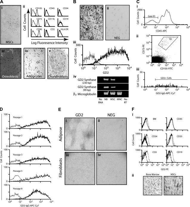
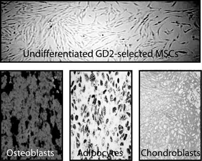
Mesenchymal stromal cells (MSCs) have enormous potential for the regeneration of bone, cartilage, and other tissues derived from primitive mesoderm. Despite extensive research, there is still no single marker that reliably identifies MSCs within the bone marrow. Using immunocytochemistry and flow cytometry, we demonstrate here that the neural ganglioside GD2 is expressed by MSCs either newly isolated from bone marrow or expanded in tissue culture; this finding was supported by reverse transcriptase-polymerase chain reaction (RT-PCR) analysis showing expression of the mRNA for GD2 synthase, an essential enzyme for GD2 biosynthesis. GD2 was also expressed on MSCs isolated from adipose tissue, but not on foreskin fibroblasts. Importantly, MSCs were the only cells within normal marrow that expressed this marker. Thus, GD2 appears to be the first reported single surface marker that uniquely distinguishes MSCs from other marrow elements. GD2 may prove valuable to study MSC biology and for the preparation of MSCs for clinical applications.
Figures
Similar articles
-
Neural ganglioside GD2(+) cells define a subpopulation of mesenchymal stem cells in adult murine bone marrow.Cell Physiol Biochem. 2013;32(4):889-98. doi: 10.1159/000354492. Epub 2013 Sep 27. Cell Physiol Biochem. 2013. PMID: 24107641
-
Neural ganglioside GD2 identifies a subpopulation of mesenchymal stem cells in umbilical cord.Cell Physiol Biochem. 2009;23(4-6):415-24. doi: 10.1159/000218188. Epub 2009 May 6. Cell Physiol Biochem. 2009. PMID: 19471109
-
GD2 expression is closely associated with neuronal differentiation of human umbilical cord blood-derived mesenchymal stem cells.Cell Mol Life Sci. 2010 Jun;67(11):1845-58. doi: 10.1007/s00018-010-0292-z. Epub 2010 Feb 18. Cell Mol Life Sci. 2010. PMID: 20165901 Free PMC article.
-
ALCAM (CD166) as a gene expression marker for human mesenchymal stromal cell characterisation.Gene. 2020 Dec;763S:100031. doi: 10.1016/j.gene.2020.100031. Epub 2020 Mar 14. Gene. 2020. PMID: 34493362
-
Molecular targeting regulation of proliferation and differentiation of the bone marrow-derived mesenchymal stem cells or mesenchymal stromal cells.Curr Drug Targets. 2012 Apr;13(4):561-71. doi: 10.2174/138945012799499749. Curr Drug Targets. 2012. PMID: 22443584 Review.
Cited by
-
Mesenchymal Stromal/Stem Cells in Regenerative Medicine and Tissue Engineering.Stem Cells Int. 2018 Aug 19;2018:8031718. doi: 10.1155/2018/8031718. eCollection 2018. Stem Cells Int. 2018. PMID: 30210552 Free PMC article. Review.
-
Dental pulp stem cells in regenerative medicine.Br Dent J. 2018 May 4. doi: 10.1038/sj.bdj.2018.348. Online ahead of print. Br Dent J. 2018. PMID: 29725075
-
An MRI-visible non-viral vector bearing GD2 single chain antibody for targeted gene delivery to human bone marrow mesenchymal stem cells.PLoS One. 2013 Oct 7;8(10):e76612. doi: 10.1371/journal.pone.0076612. eCollection 2013. PLoS One. 2013. PMID: 24116127 Free PMC article.
-
Comparison of molecular profiles of human mesenchymal stem cells derived from bone marrow, umbilical cord blood, placenta and adipose tissue.Int J Mol Med. 2016 Jan;37(1):115-25. doi: 10.3892/ijmm.2015.2413. Epub 2015 Nov 19. Int J Mol Med. 2016. PMID: 26719857 Free PMC article.
-
Human mesenchymal stem cells induced to differentiate as chondrocytes follow a biphasic pattern of extracellular matrix production.J Orthop Res. 2018 Jun;36(6):1757-1766. doi: 10.1002/jor.23820. Epub 2017 Dec 22. J Orthop Res. 2018. PMID: 29194731 Free PMC article.
References
-
- Horwitz E, Le BK, Dominici M, et al. Clarification of the nomenclature for MSC: the International Society for Cellular Therapy position statement. Cytotherapy. 2005;7:393–395. - PubMed
-
- Dominici M, Le BK, Mueller I, et al. Minimal criteria for defining multipotent mesenchymal stromal cells: the International Society for Cellular Therapy position statement. Cytotherapy. 2006;8:315–317. - PubMed
-
- Le Blanc K, Rasmusson I, Sundberg B, et al. Treatment of severe acute graft-versus-host disease with third party haploidentical mesenchymal stem cells. Lancet. 2004;363:1439–1441. - PubMed
-
- Studeny M, Marini FC, Dembinski JL, et al. Mesenchymal stem cells: potential precursors for tumor stroma and targeted-delivery vehicles for anticancer agents. J Natl Cancer Inst. 2004;96:1593–1603. - PubMed
Publication types
MeSH terms
Substances
Grants and funding
LinkOut - more resources
Full Text Sources
Other Literature Sources
