. 2007 Jan 29;176(3):283-94.
doi: 10.1083/jcb.200604141.

Pericentric heterochromatin reprogramming by new histone variants during mouse spermiogenesis

Affiliations

Pericentric heterochromatin reprogramming by new histone variants during mouse spermiogenesis

Jérôme Govin et al. J Cell Biol. .

Abstract

During male germ cell postmeiotic maturation, dramatic chromatin reorganization occurs, which is driven by completely unknown mechanisms. For the first time, we describe a specific reprogramming of mouse pericentric heterochromatin. Initiated when histones undergo global acetylation in early elongating spermatids, this process leads to the establishment of new DNA packaging structures organizing the pericentric regions in condensing spermatids. Five new histone variants were discovered, which are expressed in late spermiogenic cells. Two of them, which we named H2AL1 and H2AL2, specifically mark the pericentric regions in condensing spermatids and participate in the formation of new nucleoprotein structures. Moreover, our investigations also suggest that TH2B, an already identified testis-specific H2B variant of unknown function, could provide a platform for the structural transitions accompanying the incorporation of these new histone variants.


Figures
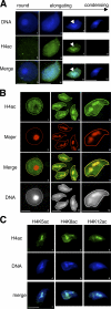
Figure 1.
Figure 1.
Pericentric chromatin becomes acetylated in elongating spermatids. (A) H4 acetylation pattern in germ cells was analyzed by IF on spermatids at the indicated stages of maturation, using an anti–tetra-acetylated H4 antibody (H4ac). DNA was counterstained with DAPI. Arrows indicate the redistribution of the acetylated signal in an intensely DAPI-stained region in elongating spermatids. (B) Immuno-FISH assays, detecting H4 acetylation (H4ac) by IF and major satellites by FISH, were performed on round (1–4) and elongating (5–12) spermatids. Acquisition of the H4 acetylation staining has been enhanced to increase the signal intensity, compared with A, to show the hypoacetylation state of pericentric heterochromatin in round spermatids. (C) The acetylation of lysines 5, 8, or 12 of histone H4 (H4K5ac, H4K8ac, and H4K12ac, respectively) were analyzed in elongating spermatids by IF as in A using the corresponding antibodies. Bars, 5 μm.
Figure 2.
Figure 2.
A restricted domain of pericentric chromatin harbors both euchromatic and heterochromatic marks in elongating spermatids. (A) HP1β (red) and tetra-acetylated H4 (H4ac; green) were analyzed by IF in round (1–4), elongating (5–8), and condensing (9–12) spermatids. Merge corresponds to HP1β and H4ac codetection. Bars, 5 μm. (B) Acetylation of histones (Ac; red) and trimethylation of H3K9 (H3K9me3; green) were analyzed by IF in round (1–4), elongating (5–8), and condensing (9–12) spermatids. Merge corresponds to H4ac and H3K9me3 codetection. Bars, 5 μm. (C and D) Intensity of histone Ac (red; Alexa 546) and H3K9me3 (green; Alexa 488) fluorescence signals were quantified on step 9 spermatids using an analysis software (MetaMorph). For each detection, a region containing values >50% of maximal fluorescence was delimited (C, 4–8; red and green borders for Ac and H3K9me3 signals, respectively), and quantification performed along the gray axis (D, 1–3) was reported along a diagram. A representative picture of the analysis of five different cells shows that the acetylated domain and the H3K9me3 do not perfectly overlap. Bar, 2.5 μm.
Figure 3.
Figure 3.
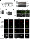
Two MNase-resistant structures are present in condensing spermatids and mainly correspond to pericentric heterochromatin. (A) Schematic representation of germ cells at different stages of spermatogenesis (Sc, spermatocytes; R, round spermatids; E, elongating spematids; C, condensing and condensed spermatids [steps 12–16]) used in MNase-digestion assays. (B) Chromatin of purified step 12–16 spermatids (top) or somatic cells (bottom) were submitted to MNase digestion during the indicated times. The DNA fragments present in MNase-resistant nucleoprotein structures were purified and analyzed by electrophoresis on a 2% agarose gel. Lane 1 corresponds to the size markers. (C) MNase-resistant DNA fragments obtained as in B, after 15 min of MNase digestion, were analyzed on a 4% agarose gel, with small size markers. (D) The small DNA fragment (Sm) and the nucleosomal DNA fragment (Nuc) obtained after 8 and 30 min, respectively, of MNase digestion of condensing spermatids chromatin were purified to be used as probes for FISH. The control probe (Ctl) corresponds to the genome of whole testis cells entirely digested into mononucleosomes by a prolonged action of MNase. (E and F) FISH using probes described in C (green) were performed on mouse metaphase chromosomes (E) and on male germ cells in codetection with a major satellite probe (F, red). DNA was counterstained with DAPI. Arrows in E indicate centric and pericentric chromatin domains. Bars, 5 μm.
Figure 4.
Figure 4.
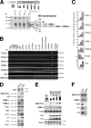
Identification of three new testis-specific histone variants specifically expressed during late spermiogenesis. (A) Acidosoluble extracts were prepared from male germ cells purified according to their maturation stages (Sc, spermatocytes; R, or round; E, elongating and condensing; C, condensing and condensed spermatids) and separated on 15% SDS-PAGE stained with Coomassie blue. Proteins present in step 12–16 spermatid extracts (lane 4) have been excised from the gel and identified by MS. (B) PCR using specific primers of the newly identified H2AL1, H2AL2, H2AL3, H2BL1, and H2BL2, as well as H3t and GAPDH as controls, were performed on reverse-transcription products (+) obtained from total RNA of the indicated mouse tissues, in comparison to negative controls without (−) the reverse transcriptase (RT). (C) Reverse transcription was performed on total RNA extracted from either whole testis or fractions enriched in germ cells at different maturation stages. Enrichment of the indicated cDNA in each fraction compared with the spermatocytes was evaluated by qPCR using the appropriate primers. The mean value of at least two independent assays is presented. Error bars indicate mean ± SD. (D, E, and F) Western blots using the indicated antibodies were performed on various testis or germ cell extracts. Coomassie staining of the extracts is also shown (Stain panels). (D) Whole testis extract (WTE) and step 12–16 spermatid nuclei. CS, condensing/condensed spermatids. Note that after a longer exposure, the anti-H2AL1/L2, -H2BL1, -Prm2, -TP1, and -TP2 blots all showed a weak signal in the whole testis extract (not depicted). (E) Germ cells fractionated according to their maturation stages by sedimentation on BSA gradient. (F) Step 12–16 spermatid nuclei (CS) and mature epididymal sperm heads (Spz).
Figure 5.
Figure 5.
H2AL1/L2 are localized on pericentric heterochromatin in early condensing spermatids. (A) H2AL1/L2 were analyzed by IF on round (1–3), elongating (4–6), early condensing (7–12), and condensing and condensed (13–18) spermatids in codetection with acetylated histones (Ac; 1–9) or protamine (Prm; 10–18). The DNA panels correspond to the DAPI counterstaining. Arrows indicate the distribution of H2AL1/L2 on densely DAPI-stained regions. Bars, 5 μm. (B) H2AL1/L2 were analyzed by IH on testis sections. The tubule sections were staged according to the association of the corresponding germinal cells (Russell et al., 1990). The stage of each section is indicated with Roman numbers. The antibodies stained the spermatids found in stages IX, X, and XI tubules, respectively (corresponding to step 9, 10, and 11 spermatids). Bars, 20 μm. (C) TP1 or TP2 were detected by IF on early condensing spermatids. DNA panels correspond to the DAPI counterstaining. Bars, 5 μm.
Figure 6.
Figure 6.
In condensing spermatids, H2AL1/L2 are associated with the new nucleoprotein structure identified by MNase digestion. Nucleoprotein structures released from step 12–16 spermatids after MNase digestion were fractionated by ultracentrifugation on a sucrose gradient. DNA fragments and proteins of each collected fraction were analyzed, respectively, by electrophoresis on an agarose gel (DNA) and by Western blot using the appropriate antibodies. A and B correspond to two independent experiments. In B, fractions 3–5 (containing the small fragment [Sm]) and fractions 8–10 (containing the nucleosomal fragment [Nuc]) of the gradient were pooled for Western blots (bottom, lanes 2 and 3) and Coomassie staining (top, lanes 1 and 2) analyses, respectively. To confirm the absence of H3 and H4, the proteins from the small fragment fraction were also loaded in tenfold excess (Western blot [WB], lane 4).
Figure 7.
Figure 7.
H2AL2–TH2B dimers can be assembled into nucleosomes when ectopically expressed in somatic cells. (A) Cos7 cells were cotransfected with vectors expressing GFP-fused H2A, H2AL1, H2AL2, or GFP alone as a control, and HA-tagged H2B or TH2B, or an empty vector (−). After immunoprecipitation (IP) performed on whole cell extracts using an anti-HA antibody, the immunoprecipitated proteins were denatured in Laemmli loading buffer and analyzed by Western blot (WB). A Western blot probed with an anti-HA antibody showed the efficiency of the immunoprecipitations (top). The coimmunoprecipitation of histones H2A, H2AL1, or H2AL2 was detected by Western blot with an anti-GFP antibody and compared with the input. (B) Oligonucleosomes were prepared by MNase digestion from Cos7 cells expressing HA-H2AL2 and Flag-TH2B and fractionated by ultracentrifugation on a sucrose gradient. DNA fragments and proteins of each collected fraction were analyzed, respectively, by electrophoresis on an agarose gel (DNA) and by Western blots using the indicated antibodies. (C) Oligonucleosomes prepared from Cos7 cells expressing HA-H2A/Flag-H2B or HA-H2AL2/Flag-TH2B were captured by hydroxyapatite. After elution by increasing NaCl concentrations, endogenous somatic histones were detected on SDS-PAGE by Coomassie staining (top) and the ectopically expressed H2A/H2B or H2AL2/TH2B by Western blots.

Similar articles

Cited by

References

    1. Aul, R.B., and R.J. Oko. 2001. The major subacrosomal occupant of bull spermatozoa is a novel histone H2B variant associated with the forming acrosome during spermiogenesis. Dev. Biol. 239:376–387. - PubMed
    1. Caron, C., C. Pivot-Pajot, L.A. Van Grunsven, E. Col, C. Lestrat, S. Rousseaux, and S. Khochbin. 2003. Cdyl: a new transcriptional co-repressor. EMBO Rep. 4:877–882. - PMC - PubMed
    1. Caron, C., J. Govin, S. Rousseaux, and S. Khochbin. 2005. How to pack the genome for a safe trip. Prog. Mol. Subcell. Biol. 38:65–89. - PubMed
    1. Churikov, D., J. Siino, M. Svetlova, K. Zhang, A. Gineitis, E. Morton Bradbury, and A. Zalensky. 2004. Novel human testis-specific histone H2B encoded by the interrupted gene on the X chromosome. Genomics. 84:745–756. - PubMed
    1. Faure, A.K., C. Pivot-Pajot, A. Kerjean, M. Hazzouri, R. Pelletier, M. Peoc'h, B. Sele, S. Khochbin, and S. Rousseaux. 2003. Misregulation of histone acetylation in Sertoli cell-only syndrome and testicular cancer. Mol. Hum. Reprod. 9:757–763. - PubMed

Publication types