

Increased apoptosis, p53 up-regulation, and cerebellar neuronal degeneration in repair-deficient Cockayne syndrome mice
- PMID: 17229834
- PMCID: PMC1783131
- DOI: 10.1073/pnas.0610619104
Increased apoptosis, p53 up-regulation, and cerebellar neuronal degeneration in repair-deficient Cockayne syndrome mice
Abstract
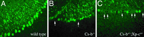
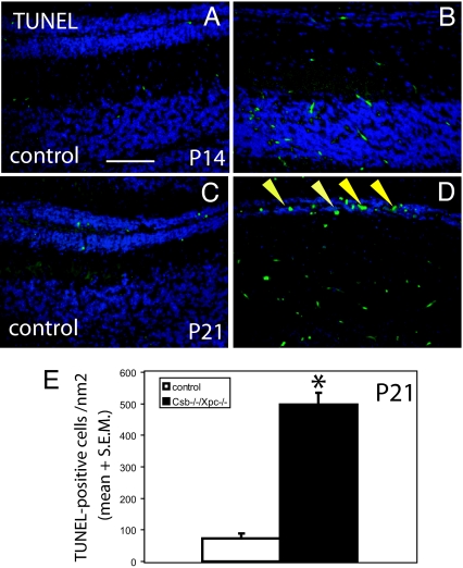
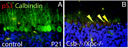
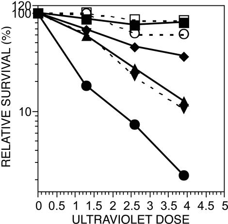
Cockayne syndrome (CS) is a rare recessive childhood-onset neurodegenerative disease, characterized by a deficiency in the DNA repair pathway of transcription-coupled nucleotide excision repair. Mice with a targeted deletion of the CSB gene (Csb-/-) exhibit a much milder ataxic phenotype than human patients. Csb-/- mice that are also deficient in global genomic repair [Csb-/-/xeroderma pigmentosum C (Xpc)-/-] are more profoundly affected, exhibiting whole-body wasting, ataxia, and neural loss by postnatal day 21. Cerebellar granule cells demonstrated high TUNEL staining indicative of apoptosis. Purkinje cells, identified by the marker calbindin, were severely depleted and, although not TUNEL-positive, displayed strong immunoreactivity for p53, indicating cellular stress. A subset of animals heterozygous for Csb and Xpc deficiencies was more mildly affected, demonstrating ataxia and Purkinje cell loss at 3 months of age. Mouse, Csb-/-, and Xpc-/- embryonic fibroblasts each exhibited increased sensitivity to UV light, which generates bulky DNA damage that is a substrate for excision repair. Whereas Csb-/-/Xpc-/- fibroblasts were more UV-sensitive than either single knockout, double-heterozygote fibroblasts had normal UV sensitivity. Csb-/- mice crossed with a strain defective in base excision repair (Ogg1) demonstrated no enhanced neurodegenerative phenotype. Complete deficiency in nucleotide excision repair therefore renders the brain profoundly sensitive to neurodegeneration in specific cell types of the cerebellum, possibly because of unrepaired endogenous DNA damage that is a substrate for nucleotide but not base excision repair.
Conflict of interest statement
The authors declare no conflict of interest.
Figures


Similar articles
-
Disruption of the Cockayne syndrome B gene impairs spontaneous tumorigenesis in cancer-predisposed Ink4a/ARF knockout mice.Mol Cell Biol. 2001 Mar;21(5):1810-8. doi: 10.1128/MCB.21.5.1810-1818.2001. Mol Cell Biol. 2001. PMID: 11238917 Free PMC article.
-
Cockayne syndrome exhibits dysregulation of p21 and other gene products that may be independent of transcription-coupled repair.Neuroscience. 2007 Apr 14;145(4):1300-8. doi: 10.1016/j.neuroscience.2006.08.074. Epub 2006 Oct 19. Neuroscience. 2007. PMID: 17055654 Free PMC article.
-
Mutations in Cockayne Syndrome-Associated Genes (Csa and Csb) Predispose to Cisplatin-Induced Hearing Loss in Mice.J Neurosci. 2016 Apr 27;36(17):4758-70. doi: 10.1523/JNEUROSCI.3890-15.2016. J Neurosci. 2016. PMID: 27122034 Free PMC article.
-
The role of Cockayne Syndrome group B (CSB) protein in base excision repair and aging.Mech Ageing Dev. 2008 Jul-Aug;129(7-8):441-8. doi: 10.1016/j.mad.2008.04.009. Epub 2008 Apr 30. Mech Ageing Dev. 2008. PMID: 18541289 Free PMC article. Review.
-
Cockayne Syndrome Group B (CSB): The Regulatory Framework Governing the Multifunctional Protein and Its Plausible Role in Cancer.Cells. 2021 Apr 10;10(4):866. doi: 10.3390/cells10040866. Cells. 2021. PMID: 33920220 Free PMC article. Review.
Cited by
-
Xeroderma pigmentosum and other diseases of human premature aging and DNA repair: molecules to patients.Mech Ageing Dev. 2011 Jun-Jul;132(6-7):340-7. doi: 10.1016/j.mad.2011.06.004. Epub 2011 Jun 25. Mech Ageing Dev. 2011. PMID: 21708183 Free PMC article. Review.
-
An abundant evolutionarily conserved CSB-PiggyBac fusion protein expressed in Cockayne syndrome.PLoS Genet. 2008 Mar 21;4(3):e1000031. doi: 10.1371/journal.pgen.1000031. PLoS Genet. 2008. PMID: 18369450 Free PMC article.
-
How does suppression of IGF-1 signaling by DNA damage affect aging and longevity?Mech Ageing Dev. 2008 May;129(5):243-53. doi: 10.1016/j.mad.2008.02.005. Epub 2008 Feb 17. Mech Ageing Dev. 2008. PMID: 18374391 Free PMC article. Review.
-
Blinded by the UV light: how the focus on transcription-coupled NER has distracted from understanding the mechanisms of Cockayne syndrome neurologic disease.DNA Repair (Amst). 2013 Aug;12(8):656-71. doi: 10.1016/j.dnarep.2013.04.018. Epub 2013 May 16. DNA Repair (Amst). 2013. PMID: 23683874 Free PMC article.
-
Disorders of nucleotide excision repair: the genetic and molecular basis of heterogeneity.Nat Rev Genet. 2009 Nov;10(11):756-68. doi: 10.1038/nrg2663. Epub 2009 Oct 7. Nat Rev Genet. 2009. PMID: 19809470 Review.
References
-
- Nance MA, Berry SA. Am J Med Genet. 1992;42:68–84. - PubMed
-
- Leech RW, Brumback RA, Miller RH, Otsuka F, Tarone RE, Robbins JH. J Neuropathol Exp Neurol. 1985;44:507–519. - PubMed
-
- Wood RD, Mitchell M, Sgouros J, Lindahl T. Science. 2001;291:1284–1289. - PubMed
-
- Bohr VA, Sander M, Kraemer KH. DNA Repair (Amsterdam) 2004;4:293–302. - PubMed
Publication types
MeSH terms
Substances
Grants and funding
LinkOut - more resources
Full Text Sources
Research Materials
Miscellaneous
