. 2007 Apr;81(7):3058-67.
doi: 10.1128/JVI.02082-06. Epub 2007 Jan 17.

Influenza A virus NS1 protein activates the PI3K/Akt pathway to mediate antiapoptotic signaling responses

Affiliations

Influenza A virus NS1 protein activates the PI3K/Akt pathway to mediate antiapoptotic signaling responses

Christina Ehrhardt et al. J Virol. 2007 Apr.

Abstract

Recently we have shown that influenza A virus infection leads to activation of the phosphatidylinositol 3-kinase (PI3K)/Akt pathway and that this cellular reaction is dependent on the expression of the viral nonstructural protein 1 (NS1). These data also suggested that PI3K activation confers a virus-supporting activity at intermediate stages of the infection cycle. So far it is not known which process is regulated by the kinase that supports virus replication. It is well established that upon infection with influenza A virus, the expression of the viral NS1 keeps the induction of beta interferon and the apoptotic response within a tolerable limit. On a molecular basis, this activity of NS1 has been suggested to preclude the activation of cellular double-stranded RNA receptors as well as impaired modulation of mRNA processing. Here we present a novel mode of action of the NS1 protein to suppress apoptosis induction. NS1 binds to and activates PI3K, which results in the activation of the PI3K effector Akt. This leads to a subsequent inhibition of caspase 9 and glycogen synthase-kinase 3beta and limitation of the virus-induced cell death program. Thus, NS1 not only blocks but also activates signaling pathways to ensure efficient virus replication.


Figures
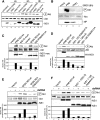
FIG. 1.
FIG. 1.
Influenza A virus NS1 induces activation of the PI3K/Akt signaling pathway. (A) A549 cells were left untreated (lane 1), were mock infected (lane 11), or were infected with influenza A virus strain Victoria/3/75 (H3N2) (lanes 2, 5, and 8), Thailand/KAN-1/2004 (H5N1) (lanes 3, 6, and 9), or PR8 (H1N1) (lane 4, 7, and 10) at a MOI of 5 for the indicated times. (B) A549 cells were left untreated (lane 4), mock infected (lane 1), or infected with influenza A virus strain PR8 (lane 2) or delNS1 (ΔNS1) (lane 3) at a MOI of 5 for 8 h. (C) A549 cells were transfected with empty vectors (lanes 1 and 4) or expression constructs for wild-type PR8 NS1 (PR8 NS1wt) (lane 2), NS-IAmut1 expressing a NS1 protein of wild-type length with five amino acid replacements at positions 181 to 185 (LIGGL to KQRRS) (lane 3), wild-type WSN NS1 (WSN NS1wt) (lane 5), and the corresponding NS1 with a R38AK41A mutation in the dsRNA binding site (lane 6). (D) A549 cells were transfected with constructs expressing NS1wt (lane 2) or truncated NS1 consisting of the N-terminal amino acids 1 to 125 (lane 3) or 1 to 126(+3) (lane 5), as well as their corresponding dsRNA binding site mutants bearing an R38AK41A mutation (lanes 4 and 6). (E) A549 cells were transfected with an empty vector or expression constructs for WSN NS1wt (lanes 3 and 4) and the corresponding NS1 with a R38AK41A mutation (lanes 5 and 6). Cells were left untreated (lanes 1, 3, and 5) or were transfected with the dsRNA analog poly(IC) (5 μg/ml) in the presence of DOTAP for 45 min and subsequently harvested (lanes 2, 4, and 6). (F) A549 cells were transfected with constructs expressing NS1wt (lanes 3 and 4) or truncated NS1 consisting of the N-terminal amino acids 1 to 126(+3) (lanes 5 and 6) or the corresponding dsRNA binding site mutant bearing an R38AK41A mutation (lanes 7 and 8). Note that cells in panels C to F were cotransfected with a plasmid expressing wild-type Akt. In all assays phosphorylated Akt (Ser473) was detected by Western blotting. Equal protein loading of the kinase was verified in Akt Western blots. Ongoing viral replication was demonstrated by accumulation of the viral NP in panel B. Equal NS1 production from plasmids was monitored in Western blots detecting NS1 (panel A, lanes 2 to 10; panel C, lanes 2, 3, 5, and 6; and panel E, lanes 3 to 6) or HA-tagged NS1 (panel D, lanes 2 to 6, and panel F, lanes 3 to 8). Relative Akt phosphorylation was normalized to Akt and NS1 content, upon quantification with the Lumi-Analyst program (Boehringer Mannheim). Akt phosphorylation levels of vector-transfected cells (panel C, lane 4, and panel E, lane 1) were arbitrarily set at 1, while phosphorylation of Akt in untreated WSN NSwt-expressing cells (panel D, lane 3) or untreated PR8 NS1wt-expressing cells (panel F, lane 1) was arbitrarily set at 100%.
FIG. 2.
FIG. 2.
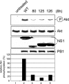
Reduced Akt phosphorylation upon infection with virus mutants bearing C-terminal deletions in the NS1 protein. A549 cells were infected with a recombinant PR8 strain (wild type [WT]) (lane 2) or corresponding virus mutants expressing truncated NS1 proteins of amino acids 1 to 80 (80, lane 3), 1 to 125 (125, lane 4), or 1 to 126 (126, lane 5) at a MOI of 5 for 8 h. Phosphorylated Akt (Ser473) was detected by Western blotting. Equal protein loading of the kinase was verified in Akt Western blots. Equal viral replication status was demonstrated by accumulation of the viral polymerase protein PB1. Expression levels of NS1 and mutants were monitored in NS1 Western blots. Relative Akt phosphorylation was normalized to Akt and NS1 content, upon quantification with the Lumi-Analyst program (Boehringer Mannheim), Akt phosphorylation levels in cells infected with the recombinant PR8 strain (lane 2) were arbitrarily set at 100%.
FIG. 3.
FIG. 3.
NS1 coimmunopreciptates with p85α and -β, two isoforms of the regulatory subunit of PI3K. (A and B) HEK 293 cells were transfected with a plasmid expressing a YFP-tagged version of p85α (p85α-YFP). At 24 h posttransfection cells were left untreated or infected with the influenza A virus strain PR8 (A) or the reassortant WSN-HK (B) (MOI = 5) for 4 h and subsequently harvested. Cells were subjected to immunoprecipitation (IP) with an anti-GFP antibody directed against the YFP tag (lanes 1 and 2) or a serum control (lanes 3). Coimmunoprecipitated NS1 was detected by Western blotting (WB). Equal protein loads of p85α-YFP in the immunoprecipitates were verified using the anti-GFP antibody detecting the YFP tag. The viral NS1 protein input of crude cell lysates served as control (lower panels). (C to F) A549 cells (C and D) or HEK 293 cells (E and F) were left untreated or infected with the influenza A virus strain PR8 (MOI = 5) for 4 h and subsequently harvested. Cells were subjected to immunoprecipitation of endogenous p85α with an anti-p85α antibody (panel C, lanes 2 and 3, and panel E, lanes 1 and 2), an anti-NS1 antiserum (panel D, lanes 2 and 3), an anti-p85β (panel F, lanes 1 and 2), or serum as a control (panels C and D, lanes 4, and panels E and F, lanes 3). Coimmunoprecipitated NS1 (C, E, and F) or coimmunoprecipitated p85β (D) was detected by Western blotting. Equal protein loads in the immunoprecipitates were verified in p85α (C and E), p85β (F), or NS1 (D) Western blots. The viral NS1 protein and endogenous p85α or -β input of crude cell lysate served as a control.
FIG. 4.
FIG. 4.
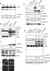
An active PI3K/Akt pathway suppresses viral apoptosis induction via inactivation of the Akt effectors GSK-3β and caspase 9. (A) MDCK cells were infected with a recombinant PR8 strain (wild type [WT]) (lane 4) or corresponding virus mutants expressing truncated NS1 proteins of amino acids 1 to 80 (80) (lane 2) and 1 to 126 (126) (lane 3) at a MOI of 1 for 8 h. (B, C, and D) MDCK cells were treated with the specific PI3K inhibitor LY294002 (50 μM) or equal volumes of the solvent (DMSO) right after infection with influenza A virus strain PR8 at a MOI of 20 (C) or with FPV at a MOI of 0.01 (B) or 1 (D) for 6 h (C and D) or 18 h (B). Thereafter cells were lysed and phosphorylated Akt (Ser473) was detected by Western blotting. Induction of apoptosis-regulating markers, such as cleavage of PARP and caspase 9 or phosphorylation of GSK-3β, was detected with specific antibodies against PARP (cleaved [cl.] or uncleaved), phosphorylated GSK-3β, and caspase 9. Note that in panels B and D the caspase 9 antibody detected only the full-length form of the caspase. Equal protein loads were verified with Akt or ERK2 blots. Ongoing viral replication was demonstrated by accumulation of the viral NS1 or PB1 protein in Western blots. Additionally, ongoing viral replication was verified via NP staining of MDCK cells which were infected with FPV and/or treated with LY294002 (50 μM) under the same conditions as described for the experiments shown in panel B. (E and F) Vero cells were treated with the specific PI3K inhibitor LY294002 (50 μM) or equal volumes of the solvent (DMSO) right after infection with the influenza A virus strain FPV (MOI = 1) (E) or PR8 (MOI = 7) for 6 h. Phosphorylated Akt (Ser473) and induction of PARP cleavage were detected as described above. Equal protein loads were verified with ERK2 blots. Ongoing viral replication was demonstrated by accumulation of the viral NS1 protein.
FIG. 4.
FIG. 4.
An active PI3K/Akt pathway suppresses viral apoptosis induction via inactivation of the Akt effectors GSK-3β and caspase 9. (A) MDCK cells were infected with a recombinant PR8 strain (wild type [WT]) (lane 4) or corresponding virus mutants expressing truncated NS1 proteins of amino acids 1 to 80 (80) (lane 2) and 1 to 126 (126) (lane 3) at a MOI of 1 for 8 h. (B, C, and D) MDCK cells were treated with the specific PI3K inhibitor LY294002 (50 μM) or equal volumes of the solvent (DMSO) right after infection with influenza A virus strain PR8 at a MOI of 20 (C) or with FPV at a MOI of 0.01 (B) or 1 (D) for 6 h (C and D) or 18 h (B). Thereafter cells were lysed and phosphorylated Akt (Ser473) was detected by Western blotting. Induction of apoptosis-regulating markers, such as cleavage of PARP and caspase 9 or phosphorylation of GSK-3β, was detected with specific antibodies against PARP (cleaved [cl.] or uncleaved), phosphorylated GSK-3β, and caspase 9. Note that in panels B and D the caspase 9 antibody detected only the full-length form of the caspase. Equal protein loads were verified with Akt or ERK2 blots. Ongoing viral replication was demonstrated by accumulation of the viral NS1 or PB1 protein in Western blots. Additionally, ongoing viral replication was verified via NP staining of MDCK cells which were infected with FPV and/or treated with LY294002 (50 μM) under the same conditions as described for the experiments shown in panel B. (E and F) Vero cells were treated with the specific PI3K inhibitor LY294002 (50 μM) or equal volumes of the solvent (DMSO) right after infection with the influenza A virus strain FPV (MOI = 1) (E) or PR8 (MOI = 7) for 6 h. Phosphorylated Akt (Ser473) and induction of PARP cleavage were detected as described above. Equal protein loads were verified with ERK2 blots. Ongoing viral replication was demonstrated by accumulation of the viral NS1 protein.
FIG. 5.
FIG. 5.
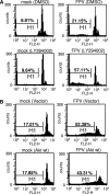
Inhibition of PI3K enhances virus-induced apoptosis, while overexpression of Akt results in reduced cell death. (A) MDCK cells were treated with LY294002 (50 μM) or equivalent volumes of the solvent (DMSO) right after infection with FPV (MOI = 0.005). (B) MDCK cells were transfected with plasmids expressing empty vector or wild-type Akt. At 48 h posttransfection cells were infected with the influenza A virus strain FPV (MOI = 0.005). (A and B) Apoptotic hypodiploid nuclei were measured in Nicoletti assays at 16 h p.i. as described in Materials and Methods. Note that basal apoptotic rates in transfected cells in panel B were higher than those in panel A due to the transfection procedure. (C) A549 cells were treated with the specific PI3K inhibitor LY294002 (50 μM) or equal volumes of the solvent (DMSO) right after infection with PR8 (MOI = 20). At 6 h postinfection, apoptotic leakage of the nuclear membrane was determined via nuclear PI inclusion. Cell bodies were visualized via actin staining.
FIG. 5.
FIG. 5.
Inhibition of PI3K enhances virus-induced apoptosis, while overexpression of Akt results in reduced cell death. (A) MDCK cells were treated with LY294002 (50 μM) or equivalent volumes of the solvent (DMSO) right after infection with FPV (MOI = 0.005). (B) MDCK cells were transfected with plasmids expressing empty vector or wild-type Akt. At 48 h posttransfection cells were infected with the influenza A virus strain FPV (MOI = 0.005). (A and B) Apoptotic hypodiploid nuclei were measured in Nicoletti assays at 16 h p.i. as described in Materials and Methods. Note that basal apoptotic rates in transfected cells in panel B were higher than those in panel A due to the transfection procedure. (C) A549 cells were treated with the specific PI3K inhibitor LY294002 (50 μM) or equal volumes of the solvent (DMSO) right after infection with PR8 (MOI = 20). At 6 h postinfection, apoptotic leakage of the nuclear membrane was determined via nuclear PI inclusion. Cell bodies were visualized via actin staining.
FIG. 6.
FIG. 6.
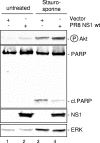
NS1 expression results in reduced PARP cleavage induced by the nonviral apoptosis stimulus staurosporine. MDCK cells were transfected with plasmids expressing empty vectors or the PR8 NS1 wild-type protein (PR8 NS1 wt). At 24 h posttransfection cells were left untreated or treated with 1 μM staurosporine for 5 h. Phosphorylated Akt (Ser473) was detected by Western blotting. Induction of PARP cleavage as a hallmark of apoptosis was detected with specific antibodies against PARP (cleaved [cl.] or uncleaved). Protein loads were controlled with an ERK2 blot. Equal viral protein expression was verified in NS1 Western blots.

Similar articles

Cited by

References

    1. Alessi, D. R., M. Andjelkovic, B. Caudwell, P. Cron, N. Morrice, P. Cohen, and B. A. Hemmings. 1996. Mechanism of activation of protein kinase B by insulin and IGF-1. EMBO J. 15:6541-6551. - PMC - PubMed
    1. Basler, C. F., X. Wang, E. Muhlberger, V. Volchkov, J. Paragas, H. D. Klenk, A. Garcia-Sastre, and P. Palese. 2000. The Ebola virus VP35 protein functions as a type I IFN antagonist. Proc. Natl. Acad. Sci. USA 97:12289-12294. - PMC - PubMed
    1. Brazil, D. P., Z. Z. Yang, and B. A. Hemmings. 2004. Advances in protein kinase B signalling: AKTion on multiple fronts. Trends Biochem. Sci. 29:233-242. - PubMed
    1. Brock, C., M. Schaefer, H. P. Reusch, C. Czupalla, M. Michalke, K. Spicher, G. Schultz, and B. Nurnberg. 2003. Roles of G beta gamma in membrane recruitment and activation of p110 gamma/p101 phosphoinositide 3-kinase gamma. J. Cell Biol. 160:89-99. - PMC - PubMed
    1. Cantley, L. C. 2002. The phosphoinositide 3-kinase pathway. Science 296:1655-1657. - PubMed

Publication types

MeSH terms