. 2007 Jan 15;176(2):231-41.
doi: 10.1083/jcb.200606027. Epub 2007 Jan 8.

Novel cell death program leads to neutrophil extracellular traps

Affiliations

Novel cell death program leads to neutrophil extracellular traps

Tobias A Fuchs et al. J Cell Biol. .

Abstract

Neutrophil extracellular traps (NETs) are extracellular structures composed of chromatin and granule proteins that bind and kill microorganisms. We show that upon stimulation, the nuclei of neutrophils lose their shape, and the eu- and heterochromatin homogenize. Later, the nuclear envelope and the granule membranes disintegrate, allowing the mixing of NET components. Finally, the NETs are released as the cell membrane breaks. This cell death process is distinct from apoptosis and necrosis and depends on the generation of reactive oxygen species (ROS) by NADPH oxidase. Patients with chronic granulomatous disease carry mutations in NADPH oxidase and cannot activate this cell-death pathway or make NETs. This novel ROS-dependent death allows neutrophils to fulfill their antimicrobial function, even beyond their lifespan.


Figures

Figure 1.
Figure 1.
Neutrophils die an active form of cell death to release NETs. Neutrophils were activated with 20 nM PMA and monitored by live-cell imaging (Video 1) in four different channels: phase contrast, with the vital dye calcein blue, with the cell death marker Annexin V (green), and with Fabs against a histone–DNA complex (red). (a–f) Merge of all four channels. (g–l) Merge of calcein blue and Annexin V channels. (m–r) Merge of Annexin V and anti–histone–DNA Fabs. The cells were monitored for up to 4 h, and key indicated time points are shown. (a) Shortly after stimulation, neutrophils show a multitude of granules and a lobulated nucleus (arrows). All cells are viable, as indicated by the vital dye calcein blue AM staining. (b) After 79 min of stimulation, the cells are flat; their nuclei are no longer lobulated and occupy the entire cell, except for a small area with residual granules (arrows). (c and i) After 219 min, the two cells in the center start to round up and are still viable. (d, j, and p) 4 min later the plasma membranes rupture, indicated by the simultaneous loss of calcein blue staining and the positive signal for Annexin V. (p) A light red staining in the periphery of the cell indicates NET formation (arrows). (e, q, f, and r) In the next few minutes, the signal of a Fab against the histone–DNA complex gets brighter and remains throughout the duration of the experiment, indicating that NET components do not diffuse after membrane rupture, but form a stable structure. At 219 min, a necrotic cell (loss of calcein blue AM, intracellular staining with Annexin V, and intracellular staining with histone–DNA complex) floated into the microscopic field. The figure shows a detail of a larger field of view that contained 20 cells, 10 of which formed NETs between 70 and 240 min after stimulation. Video 1 is available at http://www.jcb.org/cgi/content/full/jcb.200606027/DC1. Bar, 10 μm.
Figure 2.
Figure 2.
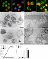
Disintegration of the nucleus and granules allows the formation of NETs. Transmission electron microscopy (a–h) and confocal immunomicroscopy (i–l) of neutrophils. Cells were incubated for 180 min in medium without stimulation (a, e, and i) or activated with 20 nM PMA for 60 (b, f, and j), 120 (c, g, and k), or 180 min (d, h, and l; all incubated for a total of 180 min). e–h are higher magnifications of a–d. (a and e) Naive neutrophils show a lobulated nucleus with clearly defined eu- and heterochromatin and numerous cytoplasmic vacuoles, even after 180 min of incubation. (i) Immunofluorescence staining for nuclear (red, histone–DNA complex antibodies) and granular components (green, neutrophil elastase antibodies) is clearly separated in naive neutrophils. Granules are distributed through the globular cell and exhibit a patchy cytoplasmic pattern in this projection of a confocal z stack. (b) After 60 min of stimulation, nuclei are less lobulated. (f) The space between inner and outer nuclear membrane is dilated. (j) Nuclei and granules are now individually visible because the cell has flattened out. (c) After 120 min, most nuclei no longer show separation of eu- and heterochromatin, and in some cells the nuclear envelope starts to disintegrate into a chain of vesicles surrounding the DNA (arrows). (g) DNA and cytoplasm are no longer separated by a membrane (arrows). (k) Cells lose their distinct granule pattern and show colocalization (yellow) of neutrophil elastase (green) and chromatin (red), especially at the nuclear periphery (arrows). (d) Most neutrophils stimulated for 180 min are nearly entirely filled with decondensed chromatin. (h) Small areas are occupied with residual granules and numerous vesicles. (l) Some cells already released NETs. (m) Extracellular neutrophil elastase was measured as described in Materials and methods. By 180 min, most of the neutrophil elastase was bound to chromatin (gray). The timecourse of release correlates with NET-DNA release (black). The experiment was repeated at least three times with neutrophils from independent donors, with similar results. The data shown is a representative triplicate experiment and presented as a mean value ± the SD. Bars: (a–d) 2 μm; (e–h) 1 μm; (i–l) 10 μm.
Figure 3.
Figure 3.
The nuclear membrane vesiculates after activation. Ultrathin cryosections of neutrophils stimulated with 20 nM PMA for 60 (a) and 180 (d) min and labeled with an antibody against the nuclear envelope–specific lamin B receptor. Bound antibody was detected with a gold-conjugated secondary antibody. In 60-min–stimulated neutrophils, the lamin B receptor was found on the inner nuclear membrane, as expected (a, arrows). 180 min after stimulation, the lamin B receptor was found in vesicles (d, arrows) in the cytoplasm. Nuclear material (d, arrowheads) is decondensed and not enclosed by a membrane. (b) 60 min after activation, neutrophils have flattened out and exhibit a lobulated nucleus and numerous granules. (c) Immunofluorescence staining for the nuclear membrane delineates the nuclear lobules (green) and is in continuity with the endoplasmic reticulum (red). (e and f) After 180 min of stimulation, neutrophil nuclei are inflated and fill nearly the entire cell. Some remaining granules are found in the periphery of the cell, while remnants of the nuclear membrane and the endoplasmic reticulum are restricted to a small central area surrounded by nuclear material (arrows). Bars: (a and d) 500 nm; (b, c, e, and d) 10 μm.
Figure 4.
Figure 4.
Neither apoptosis nor necrosis induce NETs. Neutrophils were stimulated with 20 ng/ml anti-Fas antibodies for 18 h to induce apoptosis, incubated with 25 μg/ml of pore-forming secreted toxins from S. aureus for 15 min to induce necrosis, or activated with 10 nM PMA for 4 h to induce NETs. (a–c) Transmission electron micrographs. (a) Neutrophil treated with anti-Fas antibodies show characteristic apoptotic morphology, including nuclear condensation and fragmentation as well as cytoplasmic vacuolization. (b) Features of necrosis in neutrophils treated with toxins are the loss of segregation into eu- and heterochromatin and of the nuclear lobules. The nuclear envelope, as well as the granules, remains intact. (c) Neutrophils undergoing NET-forming active cell death exhibit a morphology clearly different from both apoptosis and necrosis. The nuclear membranes are entirely fragmented while most of the granules are dissolved, allowing direct contact and mixing of nuclear, cytoplasmic, and granular components. Bars, 2 μm. (d) TUNEL analysis of different forms of cell death reveals that 60% of the anti-Fas–treated neutrophils have fragmented DNA, whereas toxin- and PMA-treated neutrophils do not. n = 200 cells/condition. (e) Neither apoptosis nor necrosis lead to NET formation, as revealed by quantifying extracellular DNA. The experiment was repeated at least three times with neutrophils from independent donors with similar results. The data shown is a representative triplicate experiment and presented as a mean value ± the SD.
Figure 5.
Figure 5.
NET-forming cell death depends on ROS production. (a) NET-DNA quantification of neutrophils stimulated with S. aureus (multiplicity of infection [MOI] 20) in the absence or presence of 10 μM of the NADPH oxidase inhibitor DPI, as indicated in the figure. (b) Quantification of NET formation of PMA-stimulated (10 nM) neutrophils in the absence (closed bars) or presence (open bars) of DPI. (c) ROS production 20 min after activation with S. aureus or PMA or 10 min after stimulation with 100 mU/ml GO was comparable (closed bars). After pharmacological inhibition of NADPH oxidase with DPI (open bars), ROS production was blocked when stimulated with S. aureus or PMA, but not with GO-treated cells. (d) Neutrophil cell death is dependent on ROS production. Neutrophils were stimulated with S. aureus for 180 min and PMA or GO for 240 min, and cell death was determined by vital dye exclusion. Cell death was abrogated by DPI in S. aureus– and PMA-, but not GO-, stimulated neutrophils. (e) Neutrophils incubated for 180 min with the indicated concentrations of GO in the absence (closed bars) or presence (open bars) of DPI. NET formation was induced by GO in a dose-dependent manner and was not dependent on NADPH oxidase activity. (f) GO treatment did induce NET formation in neutrophils, but not PBMCs. Neutrophils (closed bars) and PBMCs (open bars) were stimulated with GO for indicated periods and NET formation was quantified. NET-DNA release became detectable 90 min after activation only from neutrophils. (g) Neutrophils were activated with PMA in the presence of exogenous catalases (open bars), an inhibitor of endogenous catalases (AT, closed bars) or in the absence of these components (gray bars). (h) Stimulation of neutrophils with GO in the presence of exogenous catalases (open bars, 100 mU/ml), an inhibitor of endogenous catalases (AT, closed bars, 1 mM) or in the absence of these components (gray bars). In response to both stimuli, PMA and GO exogenous catalases reduced NET formation whereas NET formation was enhanced after inhibition of endogenous catalases as quantified by isolating NET – DNA. (i–l) Immunostaining of NET components (green, neutrophil elastase; red, histone–DNA complex). Bars, 10 μm. (i) Unstimulated neutrophils incubated for 180 min do not show NETs. Neutrophils stimulated with S. aureus (j, blue) for 120 min or PMA (k) for 180 min showed NETs. (l) Stimulation with GO for 180 min induced NET formation. The experiment was repeated at least 5 times with neutrophils from independent donors with similar results. The data shown is a representative triplicate experiment and presented as a mean value ± the SD.
Figure 6.
Figure 6.
NADPH oxidase is required to make NETs. (a–d) Immunostaining for NET components (green, neutrophil elastase; red, histone–DNA complex) of neutrophils isolated from a CGD patient. (a) Unstimulated neutrophils showed a typical lobulated nucleus and a distinct pattern of cytoplasmic granules. After S. aureus (b, blue; MOI 20) and 10 nM PMA (c) stimulation, we observed no substantial morphological changes. (d) Stimulation with 100 mU/ml GO elicited NET formation in neutrophils from CGD donors. (e–h) Transmission electron micrographs. Neutrophils isolated from CGD patients 3 h after PMA activation (e and f) showed no morphological signs of NET formation. The nuclei are still lobulated and granules are clearly visible. (g and h) GO-stimulated neutrophils isolated from CGD patients at the moment of NET formation. The plasma membrane ruptured, allowing the release of NETs. The nuclear membrane disintegrated, allowing the mixing of nuclear and granular components. (i) ROS production of PMA-activated neutrophils obtained from healthy donors in the absence or presence of 10 μM DPI and of PMA-activated neutrophils from CGD donors. ROS production was completely blocked by DPI to basal levels, as with neutrophils from CGD donors. (j) Quantification of NET-DNA of normal neutrophils and neutrophils from CGD patients. CGD neutrophils did not make NETs after S. aureus or PMA activation, but stimulation with GO elicited NETs to normal levels. The experiment was repeated at least five times with neutrophils from independent donors, with similar results. The data shown is a representative triplicate experiment and presented as a mean value ± the SD. Bars: (a–e and g) 10 μm; (f and h) 1 μm.
Figure 7.
Figure 7.
Neutrophils kill S. aureus first by phagocytosis, and then through NETs. Neutrophils were prestimulated with 10 nM PMA (a) or 100 mU/ml GO (b) for the indicated time or left unstimulated (a and b, 0 min). Before infection with S. aureus (MOI 1), the cells were treated with DNase or not (untreated). In the absence of DNase (open bars, untreated), neutrophils kill bacteria by both phagocytosis and NETs. In the presence of DNase, the NETs are dismantled and bacteria are killed only by phagocytosis. The differences in bacterial killing in cultures with and without DNase reflect the antimicrobial activity of NETs. Neutrophils that were not prestimulated killed S. aureus efficiently exclusively by phagocytosis because treatment with DNase did not affect the killing. However, cells prestimulated for 60 min showed decreased bacterial killing. Under both conditions, DNase treatment had no effect on killing the bacteria, as NETs had not yet emerged. After 120 min stimulation, when NETs are formed, DNase reduced killing of S. aureus. 3–4 h after stimulation, neutrophils killed S. aureus as efficiently as unstimulated cells. This killing was completely caused by NETs, as it was affected by the DNase treatment. (c) Unstimulated and PMA-activated (240 min) neutrophils of patients with CGD showed impaired killing of S. aureus. Their defective bacterial killing was circumvented when NETs were induced with GO (for 240 min). The experiment was repeated at least five times with neutrophils from independent donors with similar results. The data shown is a representative triplicate experiment and presented as a mean value ± the SD.

Similar articles

Cited by

References

    1. Aga, E., D.M. Katschinski, G. van Zandbergen, H. Laufs, B. Hansen, K. Muller, W. Solbach, and T. Laskay. 2002. Inhibition of the spontaneous apoptosis of neutrophil granulocytes by the intracellular parasite Leishmania major. J. Immunol. 169:898–905. - PubMed
    1. Beiter, K., F. Wartha, B. Albiger, S. Normark, A. Zychlinsky, and B. Henriques-Normark. 2006. An endonuclease allows Streptococcus pneumoniae to escape from neutrophil extracellular traps. Curr. Biol. 16:401–407. - PubMed
    1. Borregaard, N., and J.B. Cowland. 1997. Granules of the human neutrophilic polymorphonuclear leukocyte. Blood. 89:3503–3521. - PubMed
    1. Brinkmann, V., U. Reichard, C. Goosmann, B. Fauler, Y. Uhlemann, D.S. Weiss, Y. Weinrauch, and A. Zychlinsky. 2004. Neutrophil extracellular traps kill bacteria. Science. 303:1532–1535. - PubMed
    1. Buchanan, J.T., A.J. Simpson, R.K. Aziz, G.Y. Liu, S.A. Kristian, M. Kotb, J. Feramisco, and V. Nizet. 2006. DNase expression allows the pathogen group A Streptococcus to escape killing in neutrophil extracellular traps. Curr. Biol. 16:396–400. - PubMed

Publication types

MeSH terms