

Deciphering the cross talk between hnRNP K and c-Src: the c-Src activation domain in hnRNP K is distinct from a second interaction site
- PMID: 17178840
- PMCID: PMC1820454
- DOI: 10.1128/MCB.02014-06
Deciphering the cross talk between hnRNP K and c-Src: the c-Src activation domain in hnRNP K is distinct from a second interaction site
Abstract
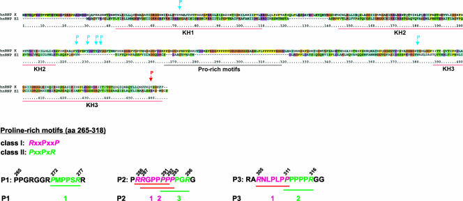
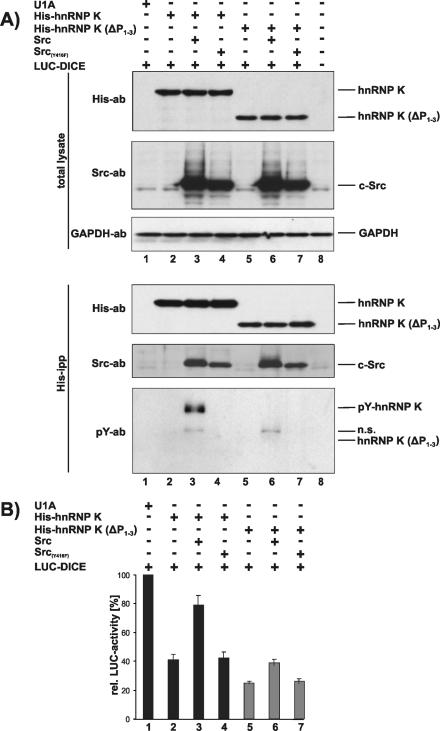
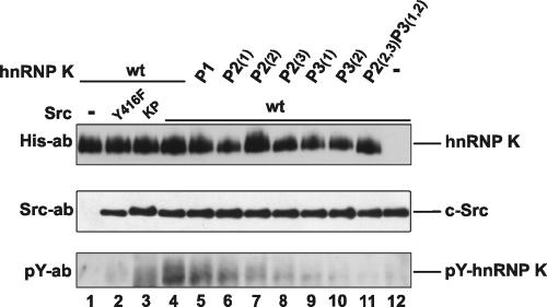
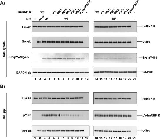
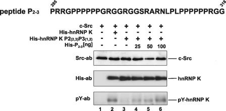
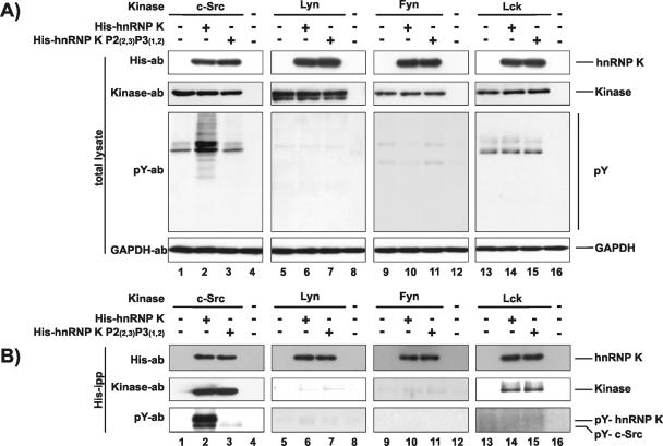
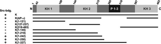
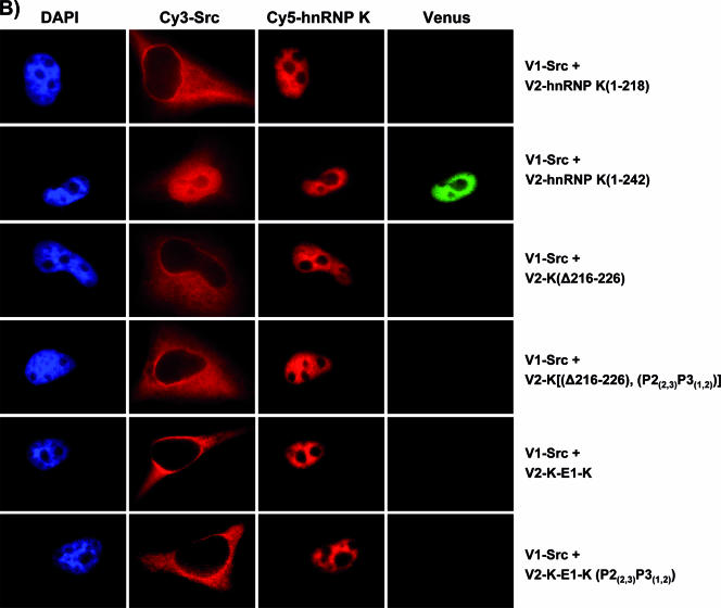
The protein tyrosine kinase c-Src is regulated by two intramolecular interactions. The repressed state is achieved through the interaction of the Src homology 2 (SH2) domain with the phosphorylated C-terminal tail and the association of the SH3 domain with a polyproline type II helix formed by the linker region between SH2 and the kinase domain. hnRNP K, the founding member of the KH domain protein family, is involved in chromatin remodeling, regulation of transcription, and translation of specific mRNAs and is a target in different signal transduction pathways. In particular, it functions as a specific activator and a substrate of the tyrosine kinase c-Src. Here we address the question how hnRNP K interacts with and activates c-Src. We define the proline residues in hnRNP K in the proline-rich motifs P2 (amino acids [aa] 285 to 297) and P3 (aa 303 to 318), which are necessary and sufficient for the specific activation of c-Src, and we dissect the amino acid sequence (aa 216 to 226) of hnRNP K that mediates a second interaction with c-Src. Our findings indicate that the interaction with c-Src and the activation of the kinase are separable functions of hnRNP K. hnRNP K acts as a scaffold protein that integrates signaling cascades by facilitating the cross talk between kinases and factors that mediate nucleic acid-directed processes.
Figures

Similar articles
-
Depressing time: Waiting, melancholia, and the psychoanalytic practice of care.In: Kirtsoglou E, Simpson B, editors. The Time of Anthropology: Studies of Contemporary Chronopolitics. Abingdon: Routledge; 2020. Chapter 5. In: Kirtsoglou E, Simpson B, editors. The Time of Anthropology: Studies of Contemporary Chronopolitics. Abingdon: Routledge; 2020. Chapter 5. PMID: 36137063 Free Books & Documents. Review.
-
The interplay between dysregulated metabolites and signaling pathway alterations involved in osteoarthritis: a systematic review.Ther Adv Musculoskelet Dis. 2024 Nov 25;16:1759720X241299535. doi: 10.1177/1759720X241299535. eCollection 2024. Ther Adv Musculoskelet Dis. 2024. PMID: 39600593 Free PMC article. Review.
-
Identification of heterogeneous nuclear ribonucleoprotein K as a transactivator for human low density lipoprotein receptor gene transcription.J Biol Chem. 2010 Jun 4;285(23):17789-97. doi: 10.1074/jbc.M109.082057. Epub 2010 Apr 6. J Biol Chem. 2010. PMID: 20371611 Free PMC article.
-
Qualitative evidence synthesis informing our understanding of people's perceptions and experiences of targeted digital communication.Cochrane Database Syst Rev. 2019 Oct 23;10(10):ED000141. doi: 10.1002/14651858.ED000141. Cochrane Database Syst Rev. 2019. PMID: 31643081 Free PMC article.
-
Metformin for endometrial hyperplasia.Cochrane Database Syst Rev. 2024 May 2;5(5):CD012214. doi: 10.1002/14651858.CD012214.pub3. Cochrane Database Syst Rev. 2024. PMID: 38695827 Review.
Cited by
-
SDCBP-AS1 destabilizes β-catenin by regulating ubiquitination and SUMOylation of hnRNP K to suppress gastric tumorigenicity and metastasis.Cancer Commun (Lond). 2022 Nov;42(11):1141-1161. doi: 10.1002/cac2.12367. Epub 2022 Oct 9. Cancer Commun (Lond). 2022. PMID: 36209503 Free PMC article.
-
Identification of RNA-binding Proteins in Macrophages by Interactome Capture.Mol Cell Proteomics. 2016 Aug;15(8):2699-714. doi: 10.1074/mcp.M115.056564. Epub 2016 Jun 8. Mol Cell Proteomics. 2016. PMID: 27281784 Free PMC article.
-
Heterogeneous nuclear ribonucleoprotein K promotes cap-independent translation initiation of retroviral mRNAs.Nucleic Acids Res. 2024 Mar 21;52(5):2625-2647. doi: 10.1093/nar/gkad1221. Nucleic Acids Res. 2024. PMID: 38165048 Free PMC article.
-
Downregulation of hnRNP K by RNAi inhibits growth of human lung carcinoma cells.Oncol Lett. 2014 Apr;7(4):1073-1077. doi: 10.3892/ol.2014.1832. Epub 2014 Jan 28. Oncol Lett. 2014. PMID: 24944671 Free PMC article.
-
Bimolecular fluorescence complementation (BiFC) analysis as a probe of protein interactions in living cells.Annu Rev Biophys. 2008;37:465-87. doi: 10.1146/annurev.biophys.37.032807.125842. Annu Rev Biophys. 2008. PMID: 18573091 Free PMC article. Review.
References
-
- Alexandropoulos, K., and D. Baltimore. 1996. Coordinate activation of c-Src by SH3- and SH2-binding sites on a novel, p130 Cas-related protein, Sin. Genes Dev. 10:1341-1355. - PubMed
-
- Alonso, G., M. Koegl, N. Mazurenko, and S. A. Courtneidge. 1995. Sequence requirements for binding of Src family tyrosine kinases to activated growth factor receptors. J. Biol. Chem. 270:9840-9848. - PubMed
-
- Ball, L. J., R. Kuhne, J. Schneider-Mergener, and H. Oschkinat. 2005. Recognition of proline-rich motifs by protein-protein-interaction domains. Angew. Chem. Int. Ed. Engl. 44:2852-2869. - PubMed
-
- Brown, M. T., and J. A. Cooper. 1996. Regulation, substrates and functions of Src. Biochim. Biophys. Acta 1287:121-149. - PubMed
Publication types
MeSH terms
Substances
LinkOut - more resources
Full Text Sources
Other Literature Sources
Molecular Biology Databases
Miscellaneous