. 2006 Dec;169(6):1925-38.
doi: 10.2353/ajpath.2006.060245.

Galectin-3 associates with the primary cilium and modulates cyst growth in congenital polycystic kidney disease

Affiliations

Galectin-3 associates with the primary cilium and modulates cyst growth in congenital polycystic kidney disease

Miliyun G Chiu et al. Am J Pathol. 2006 Dec.

Abstract

Several lines of evidence implicate the beta-galactoside-binding lectin galectin-3 in development and pathological processes in renal collecting ducts: galectin-3 is expressed in the ureteric bud/collecting duct lineage during nephrogenesis, modulates collecting duct growth/differentiation in vitro, and is expressed in human autosomal recessive polycystic kidney disease in cyst epithelia, almost all of which arise from collecting ducts. Moreover, exogenous galectin-3 restricts growth of cysts generated by Madin-Darby canine kidney collecting duct-derived cells in three-dimensional culture in collagen. Using the cpk mouse model of recessively inherited polycystic kidney disease, we observed widespread galectin-3 mRNA and protein in cyst epithelia. Exogenous galectin-3 reduced cyst formation in suspension culture, and mice-null mutant for galectin-3 had more extensive renal cysts in vivo. Galectin-3 was also detected for the first time in the centrosome/primary cilium, which has been implicated in diverse polycystic kidney disease. Cilia structure/number appeared normal in galectin-3-null mutants. Finally, paclitaxel, a therapy that retards polycystic kidney disease in cpk mice, increased extracellular galectin-3, in which the lectin could potentially interact with cilia. These data raise the possibility that galectin-3 may act as a natural brake on cystogenesis in cpk mice, perhaps via ciliary roles.


Figures
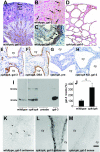
Figure 1
Figure 1
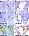
Renal galectin-3 protein and mRNA expression. All samples from 3-week-old mice. A–C, K: Wild-type kidneys; D–G, L, M: cpk/cpk cystic kidneys, with wild-type glactin-3; H: cpk/cpk cystic kidneys in galectin-3-null mutant (KO); A–F, H: galectin-3 immunohistochemistry; G: immunohistochemistry with preimmune serum; F: DBA-labeled; K–M: in situ hybridization with galectin-3 probes as labeled. A: Low-power view demonstrated galectin-3-positive collecting ducts in cortex (one prominent duct in longitudinal section is arrowed as an example) and medulla. B: Higher power view of cortex demonstrated galectin-3 expression in ducts (arrows) between PAS-positive (but galectin-3-negative) proximal tubules. Using adjacent sections, these galectin-3-labeled ducts also bound DBA, confirming that they were collecting ducts (data not shown). C: Strongly positive galectin-3 immunohistochemistry in the urothelium (arrows). D: Galectin-3-positive cysts throughout cortex and medulla of cystic kidney. E and F: Prominent galectin-3 expression in cyst (cy) epithelia, which also bound DBA, hence confirming collecting duct origin. G and H: Positive signal was not detected in cystic epithelia using either preimmune serum (G) or in galectin-3-null mutant mice (H). I: Galectin-3 Western blot from wild-type and cpk/cpk kidneys demonstrated a 30-kd major and a 60-kd minor band consistent with monomers and dimers, which was abolished by preabsorption with excess recombinant galectin-3 protein (preabs). Recombinant galectin-3 was used as a positive control (gal-3). J: Band densitometry confirmed a significantly increased amount of galectin-3 in cystic versus wild-type kidneys (n = 5; *P < 0.05). K: Anti-sense galectin-3 probe revealed a weak signal in subset of tubules (arrows). L: Prominent signal in cystic epithelia (cy). M: No significant signal in cystic epithelia using the sense probe (cy). Scale bars: 200 μm (A, D); 30 μm (B, C); 50 μm (E–H); 20 μm (K–M).
Figure 2
Figure 2
Galectin-3 and collecting duct proteins in wild-type and cystic kidneys. All samples from 3-week-old mice. A–C: Wild-type kidneys; D–F: cpk/cpk kidneys. Immunostaining was for galectin-3 (gal-3), aquaporin-2 (aq-2), and calbindin (calb). All counterstained with PAS. A and B: Similar distribution of immunoreactive galectin-3 and aquaporin-2 in collecting ducts (arrows) in normal kidneys; C: more widespread calbindin expression in distal tubules as well as collecting ducts. D: Prominent cytoplasmic galectin-3 expression in collecting ducts/cysts of all sizes, from small undilated tubules through to large cysts. E and F: Aquaporin-2 and calbindin expression was variable: immunoreactivity was observed in some but not all cysts, aquaporin-2 distribution was often heterogeneous within individual cysts, and there was intense calbindin immunostaining of tubules and small cysts but reduced intensity in larger cysts (*). Scale bar = 50 μm.
Figure 3
Figure 3
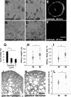
Exogenous galectin-3 reduced cyst growth in vitro, whereas the lack of the lectin accelerated cystogenesis on two genetic backgrounds in vivo. A–G: Suspension culture experiments; samples from mice on pure C57BL-6J background; G–J: cpk and galectin-3 mutant breeding results. A and B: Representative images of suspension cystogenesis in dissociated wild-type kidneys at the start of culture and after 24 hours. Single cells, aggregates, and partially digested tubules were observed at both times; cysts were very rarely seen after culture of these normal kidneys. C and D: Dissociated cpk/cpk kidneys appeared similar to those from wild-type littermates at the start of culture but multiple cysts were visible by 24 hours. E: Prominent cytoplasmic galectin-3 expression in cysts (L indicates lumen) as assessed by confocal microscopy; F: preimmune serum. G: Ten and thirty μmol/L galectin-3 caused decreased cyst numbers versus control (0) cultures after 24 hours (n = 5, *P < 0.05); there was no significant difference between the two doses. H and I: Initial experiments compared wet kidney weight or kidney to body weight (K/B) ratio in cpk/cpk mice on a mixed C57BL-6J/129Sv background with either wild-type (W) or null mutant (M) galectin-3 genotypes: both kidney weight and K/B ratio were larger in mice lacking galectin-3; mean kidney weight was 65 mg in galectin-3 mutants versus 46 mg in wild-type mice (*P < 0.03), whereas K/B ratio was 1.72% for mutants and 1.15% for galectin-3 wild-type mice (*P < 0.02). J and K: DBA-labeled sections of galectin-3 wild-type and null mutant cystic sections on the pure C57BL-6J background: cysts were consistently larger in the galectin-3-null mutants, bound DBA (ie, still derived from collecting ducts) and extended further into the cortex, often reaching the outer rim. (Note J was assessed as a cortical cystic index of two and K of three). L: Cortical cyst score was significantly increased in 1-week-old cpk/cpk galectin-3-null mutant mice on a pure C57BL-6J background (mean 2.83 versus 2.01 for galectin-3 wild type; *P = 0.01). Scale bars: 80 μm (A–D); 20 μm (E, F); and 200 μm (J, K).
Figure 4
Figure 4
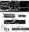
Effects of paclitaxel in vivo and in vitro; all samples are from cpk cystic kidneys. A–D: Galectin-3 confocal immunohistochemistry on control, vehicle-treated mice (A, C, D, F); or mice treated with paclitaxel (B, D); C and F: preimmune serum replaced anti-galectin-3 antibody; scanning voltage/offset was equalized to allow clearer comparison of these sections. Paclitaxel-treated kidneys contained fewer smaller cysts and renal architecture was better preserved. Galectin-3 distribution was predominantly cytoplasmic in control cyst (cy) epithelia (A and C); expression persisted in cysts and tubules (t) from paclitaxel-treated animals (B and D), although signal intensity appeared reduced compared with vehicle-treated kidneys and some cells had more prominent apical (arrowheads) rather than cytoplasmic (arrows) immunolocalization. Low levels of background signal were observed in sections exposed to preimmune serum. G: Western blot demonstrated less galectin-3 in whole kidney homogenates from paclitaxel-treated cystic mice versus vehicle-treated mice at 3 weeks, but this result may be biased by the decreased percentage of cystic epithelia in the paclitaxel group. H: Galectin-3 bands appeared stronger in cyst fluid from paclitaxel mice; one potential explanation is that this agent may cause galectin-3 secretion into the cysts. I and J: Digitally magnified images of galectin-3 immunocytochemistry on cysts grown in control medium (I) or with 50 μmol/L paclitaxel (J); prominent cytoplasmic galectin-3 immunoreactivity was observed in controls whereas positive signal was more prominent on the external surface of paclitaxel-treated cysts. Cilia were detected on the external surface of some cysts but could not be visualized at sufficient resolution to determine cilial galectin-3 expression; cy indicates the cyst lumen, arrows indicate the internal surface, and arrowheads the external surface. K: 35 and 50 μmol/L paclitaxel significantly reduced both cyst number and mean cyst area compared with DMSO (vehicle) control cultures (n = 4; *P < 0.05; **P < 0.02). L: Representative Western blot of galectin-3 protein in the cell-associated fraction (cells) or paired conditioned media (medium) from cultures treated with vehicle (0) or 50 μmol/L paclitaxel (50), ie, lane 1 of the cell blot came from the same culture as lane 1 of the medium blot and so forth. Galectin-3 was prominent in the cell preparation but barely detected in the media in control cultures, whereas the lectin was more prominent in the medium when exposed to 50 μmol/L paclitaxel. Scale bars: 20 μm (A–C); 5 μm (D–F, and I, J).
Figure 5
Figure 5
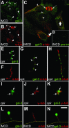
Galectin-3 is expressed in cilia and centrosomes in vitro. Confocal immunocytochemistry of IMCD (A–D, L–N) and cpk (E–K) epithelial monolayers. Galectin-3 is FITC (green)-labeled, and co-stained with TRITC (red)-labeled acetylated α-tubulin (A–G, K–M) or γ-tubulin (J and K). Note parallel cultures stained with preimmune and preabsorbed galectin-3 were completely negative (not shown). A–C: Diffuse cytoplasmic galectin-3, plus co-localization with acetylated α-tubulin; appearances consistent with centrosomes (arrowheads), mitotic spindle poles (narrow arrows), and basal bodies (thick arrows). D: Control sections (inset) using preimmune serum demonstrated low levels of background fluorescence, and cilia were not visible. E–G: Primary cilia appeared structurally normal and galectin-3 expression was detected in/on them (arrowheads) in a discontinuous pattern. H: Further punctate galectin-3 in a long cilium. I–K: Galectin-3 in the centrosome, co-localized with γ-tubulin (arrows). L–N: Galectin-3 immunoreactivity was still detected even when immunostained before permeabilization, suggesting that some galectin-3 protein is extracellular; yellow line indicates edge of cell. Acetylated α-tubulin staining was completely negative without permeabilization (data not shown). Scale bars: 10 μm (A, B, D); 5 μm (C); 0.5 μm (E, F); 0.25 μm (G, H, L, M); and 0.1 μm (I–K).
Figure 6
Figure 6
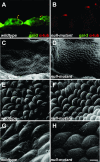
Cilia in cpk mice with wild-type and null mutant galectin-3. All images from cystic mice. Left column depicts sections from mice with wild-type galectin-3 genotype and right column from galectin-3-null mutant mice on the mixed C57BL-6J/129Sv background. A and B: Galectin-3 FITC (green); acetylated α-tubulin TRITC (red). Similar size and distribution of cilia observed using confocal microscopy; galectin-3 immunoreactivity was not detected in sections from galectin-3-null mutants. C–H: Scanning electron microscopy demonstrated cilia arising from more than 95% of cells lining the cysts. Gross differences in cilia size and appearance were not observed between the different genotypes and background strains. Scale bars: 2 μm (A, B); 80 μm (C, D); 8 μm (E, F); and 4 μm (G, H).

Similar articles

Cited by

References

    1. Barondes SH, Castronovo V, Cooper DN, Cummings RD, Drickamer K, Feizi T, Gitt MA, Hirabayashi J, Hughes C, Kasai K, Leffler H, Liu FT, Lotan R, Mercurio AM, Monsigny M, Pillai S, Poirier F, Raz A, Rigby PWJ, Rini JM. Galectins: a family of animal β-galactoside binding lectins. Cell. 1994;76:597–598. - PubMed
    1. Leffler H, Barondes SH. Specificity of binding of three soluble rat lung lectins to substituted and unsubstituted mammalian β-galactosides. J Biol Chem. 1986;261:10119–10126. - PubMed
    1. Winyard PJ, Bao Q, Hughes RC, Woolf AS. Epithelial galectin-3 during human nephrogenesis and childhood cystic diseases. J Am Soc Nephrol. 1997;8:1647–1657. - PubMed
    1. Bichara M, Attmane-Elakeb A, Brown D, Essig M, Karim Z, Muffat-Joly M, Micheli L, Eude-le-Parco I, Cluzeaud F, Peuchmaur M, Bonvalet JP, Poirier F, Farman N. Exploring the role of galectin-3 in kidney function: a genetic approach. Glycobiology. 2006;16:36–45. - PubMed
    1. Bullock SL, Johnson TM, Bao Q, Hughes RC, Winyard PJ, Woolf AS. Galectin-3 modulates ureteric bud branching in organ culture of the developing mouse kidney. J Am Soc Nephrol. 2001;12:515–523. - PubMed

Publication types

LinkOut - more resources