. 2006 Nov;18(11):3182-200.
doi: 10.1105/tpc.105.036400. Epub 2006 Nov 30.

Plant N-glycan processing enzymes employ different targeting mechanisms for their spatial arrangement along the secretory pathway

Affiliations

Plant N-glycan processing enzymes employ different targeting mechanisms for their spatial arrangement along the secretory pathway

Claude Saint-Jore-Dupas et al. Plant Cell. 2006 Nov.

Abstract

The processing of N-linked oligosaccharides in the secretory pathway requires the sequential action of a number of glycosidases and glycosyltransferases. We studied the spatial distribution of several type II membrane-bound enzymes from Glycine max, Arabidopsis thaliana, and Nicotiana tabacum. Glucosidase I (GCSI) localized to the endoplasmic reticulum (ER), alpha-1,2 mannosidase I (ManI) and N-acetylglucosaminyltransferase I (GNTI) both targeted to the ER and Golgi, and beta-1,2 xylosyltransferase localized exclusively to Golgi stacks, corresponding to the order of expected function. ManI deletion constructs revealed that the ManI transmembrane domain (TMD) contains all necessary targeting information. Likewise, GNTI truncations showed that this could apply to other type II enzymes. A green fluorescent protein chimera with ManI TMD, lengthened by duplicating its last seven amino acids, localized exclusively to the Golgi and colocalized with a trans-Golgi marker (ST52-mRFP), suggesting roles for protein-lipid interactions in ManI targeting. However, the TMD lengths of other plant glycosylation enzymes indicate that this mechanism cannot apply to all enzymes in the pathway. In fact, removal of the first 11 amino acids of the GCSI cytoplasmic tail resulted in relocalization from the ER to the Golgi, suggesting a targeting mechanism relying on protein-protein interactions. We conclude that the localization of N-glycan processing enzymes corresponds to an assembly line in the early secretory pathway and depends on both TMD length and signals in the cytoplasmic tail.


Figures

Figure 1.
Figure 1.
Schematic Representation of Fusion Proteins Analyzed in This Study. GCSI-GFP: full-length Arabidopsis GCSI fused to GFP. GCS90-GFP: the first 90 N-terminal amino acids of GCSI fused to GFP. Δ13GCS90-GFP: GCS90-GFP minus the first 13 N-terminal amino acids. ManI-GFP: full-length G. max ManI fused to GFP. Δ19Man-GFP: ManI-GFP minus the first 19 N-terminal amino acids. Man99-GFP and Man49-GFP: the first 99 and 49 amino acids of ManI, respectively, fused to GFP. Δ19Man49-GFP: Man49-GFP minus the first 19 N-terminal amino acids. ΔCTMan49-GFP: the whole CT was deleted from Man49-GFP. MAAAMan49-GFP: the CT of Man49-GFP was replaced by an artificial CT containing three Ala residues. ManTMD23-GFP and Man99TMD23-GFP: ManI-GFP and Man99-GFP, respectively, where the TMD was lengthened from 16 to 23 amino acids. GNTI-GFP: full-length N. tabacum GNTI fused to GFP. GNT38-GFP: the first 38 N-terminal amino acids of GNTI fused to GFP. XylT-GFP: full-length Arabidopsis XYLT fused to GFP. XylT35-GFP: the first 35 amino acids of XYLT fused to GFP. ST52-GFP/mRFP: the first 52 amino acids of a rat α-2,6-ST fused to mRFP. GFP-HDEL: a GFP version containing the sporamine signal peptide and a C-terminal HDEL ER retention sequence.
Figure 2.
Figure 2.
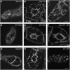
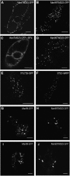
ManI-GFP and GNTI-GFP Are Located to the Golgi and to the ER, whereas GCSI-GFP Is Exclusively Accumulated in the ER and XYLT-GFP in the Golgi. Transgenic BY-2 tobacco cell lines were analyzed 3 to 4 d after subculturing. Bars = 8 μm. (A) and (B) ManI-GFP is located to the Golgi and to the ER ([A], cortical view; [B], cross section where ER labeling around the nucleus and in the vacuolar strands is characteristic). (C) After a 2-h treatment with the protein synthesis inhibitor cycloheximide, ER and Golgi labeling observed with ManI-GFP fusion remained unchanged, showing that the steady state localization of ManI-GFP is the Golgi and the ER. (D) and (E) GFP-HDEL highlights the ER. (F) After a 2-h treatment with BFA (50 mg·mL−1), fluorescent Golgi stacks have disappeared, while ER fluorescence is increased due to the relocation of ManI-GFP. (G) GCSI-GFP is an ER resident membrane protein and shows a similar fluorescence pattern as GFP-HDEL (D). (H) GNTI-GFP is targeted to the Golgi and to the ER as observed for ManI-GFP (B). (I) XYLT-GFP accumulates exclusively in Golgi stacks.
Figure 3.
Figure 3.
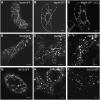
Luminal Domains Are Not Necessary to Target ManI and GNTI to the Golgi and the ER. (A) and (B) Cells expressing Man99-GFP display a punctuate and network pattern of GFP fluorescence typical of the organization of Golgi stacks and cortical ER in BY-2 cells and similar to the labeling observed for ManI-GFP. (C) The ER and Golgi labeling remains unchanged when cells expressing Man99-GFP were incubated with the protein synthesis inhibitor cycloheximide for 2 h. (D) Man49-GFP was located to the Golgi and to the ER in BY-2 suspension-cultured cells. (E) and (F) Leaf epidermal cells expressing Man99-GFP (E) and Man49-GFP (F) display the same labeling pattern 5 d after agroinfiltration as BY-2 cells expressing the same proteins. (G) and (H) As observed for the truncated forms of ManI fused to GFP, GNT38-GFP was located to the Golgi and to the ER in BY-2 suspension-cultured cells (G) and in Nicotiana leaf epidermal cells (H). (I) XYLT35-GFP highlights only the Golgi in Nicotiana leaf epidermal cells. Bars = 8 μm.
Figure 4.
Figure 4.
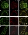
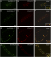
ManI and GNTI Fusions Accumulate into the Early Golgi. Transgenic cell lines coexpressing ST52-mRFP ([B], [E], [H], and [K]) and ManI-GFP (A), Man99-GFP (D), Man49-GFP (G), or GNT38-GFP (J) in BY-2 suspension-cultured cells and corresponding merged images ([C], [F], [I], and [L]). Insets: magnification of selected Golgi stacks (2.2× zoom). Note that stacks often appear tricolored (arrows in [F] and [I]) with the GFP fusions on one side (green), the ST52-RFP on the other side (red), and a region of overlap between them (yellow). This suggests that all the GFP constructs localize to the cis-Golgi as had been shown previously for ManI-GFP (Nebenführ et al., 1999), whereas ST52-RFP is in the trans-Golgi (Boevink et al., 1998). Bars = 8 μm in (A) to (I) and 16 μm in (J) to (L).
Figure 5.
Figure 5.
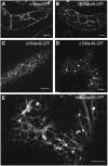
The CT Is Not Needed for ManI Localization to the Golgi and the ER. (A) to (C) Δ19Man-GFP (A) and Δ19Man49-GFP ([B] and [C]) are located to the Golgi and to the ER in BY-2 suspension-cultured cells. Bars = 16 μm. (D) Δ19Man49-GFP also highlights the Golgi and the ER in Nicotiana leaf epidermal cells. (E) A similar distribution pattern was observed for MAAAMan49-GFP after expression of this fusion in leaf epidermal cells. Bars = 8 μm.
Figure 6.
Figure 6.
An Increase in TMD Length Displaces ManI from the ER to the Golgi. Modification of TMD length in ManTMD23-GFP (A) and Man99TMD23-GFP (B) leads to the accumulation of fusion proteins in the Golgi of BY-2 tobacco cells, as observed for XYLT35-GFP (E) and ST52-mRFP (F). No ER labeling was observed in cells expressing these fusion proteins. The same results were obtained after transient expression in Nicotiana (D), G. max ([G] and [H]), or S. lycopersicum ([I] and [J]) leaf epidermal cells. ManTMD23-GFP is redistributed to the ER after a 2-h treatment with BFA (C). Bars = 16 μm.
Figure 7.
Figure 7.
An increase in TMD Length Displaces ManI and Man99 to Late Golgi Compartments: Evidence from Coexpression with the trans-Golgi Marker ST52-mRFP. (A) to (C) When ManTMD23-GFP (A) and ST52-mRFP (B) were coexpressed in BY-2 suspension-cultured tobacco cells, they both targeted to the Golgi and perfectly colocalized, as illustrated in the zoomed insets (2.2×) where the spots are yellow. (D) to (I) When Man99TMD23-GFP and ST52-mRFP were coexpressed in BY-2 suspension-cultured cells, they both targeted to the Golgi and perfectly colocalized (2.2× zoomed insets). In the cross sections ([G] to [I]), some red fluorescence can be distinguished from the yellow, whereas it is almost impossible in the cortical section, suggesting that the Golgi stacks are oriented toward the plasma membrane in the cortical cytoplasm. (J) to (L) XYLT35-GFP and ST52-mRFP were coexpressed in BY-2 suspension-cultured cells. Both fusion proteins targeted to the Golgi, but some green fluorescence can be distinguished from the yellow in some cases, consistent with their localization mainly in the medial Golgi (XYLT35-GFP) or in the trans-Golgi (ST52-mRFP). Bars = 8 μm.
Figure 8.
Figure 8.
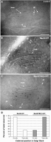
Subcompartmentation of Man99-GFP and Man99TMD23-GFP in the Golgi Apparatus. Electron micrographs of Golgi stacks were realized from suspension-cultured wild-type BY-2 tobacco cells (A) or from BY-2 cells expressing Man99-GFP (B) or Man99TMD23-GFP (C). Immunogold labeling was performed with anti-GFP antibodies. Distribution of fusion proteins in the Golgi was expressed as a percentage of label observed for 15 individual stacks analyzed in different cells (D), with counts of gold particles on the cis-side and trans-side after having divided Golgi in two domains. SV, secretory vesicle.
Figure 9.
Figure 9.
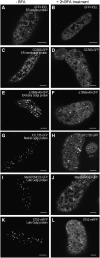
Effects of BFA on ER and/or Golgi Proteins in BY-2 Cells. BY-2 cells expressing a soluble ER marker (GFP-HDEL; [A] and [B]) or a membrane ER marker (GCS90-GFP; [C] and [D]) display a typical fluorescence of the ER in the presence or absence of BFA. An ER and early Golgi marker (Δ19Man49-GFP; [E] and [F]), a medial Golgi marker (XYLT35-GFP; [G] and [H]), or late Golgi markers (Man99TMD23-GFP and ST52-GFP; [I] to [L]) highlight the ER and aggregates assimilated to Golgi clusters in the presence of BFA. Note that in all cases, the ER network often turns into fenestrated sheets of fluorescence. Bars = 8 μm.
Figure 10.
Figure 10.
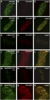
BFA Induces the Simultaneous Redistribution of Both Early and Late Golgi Markers into the ER and Golgi Clusters. BY-2 cells coexpressing GFP-HDEL ([A] to [F]), Δ19Man49-GFP ([G] to [L]), or Man99TMD23-GFP ([M] to [R]) with ST52-mRFP were analyzed in the absence ([A] to [C], [G], [H], and [M] to [O]) or presence ([D] to [F], [J] to [L], and [P] to [R]) of BFA (50 μg·mL−1).
Figure 11.
Figure 11.
Comparison of CT and TMD Length for Plant N-Glycosylation Enzymes. (A) Processing of N-linked glycans in the ER and Golgi apparatus of plant cells. A precursor oligosaccharide (Glc3Man9GlcNAc2) assembled onto a lipid carrier is transferred on specific Asn residues of the nascent polypeptide (P). This precursor is then modified by glycosidases and glycosyltransferases mainly in the ER and the Golgi apparatus during the transport of the glycoprotein downstream the secretory pathway. Glycosidases and glycosyltransferases responsible for plant N-glycan maturation and their localization in the biochemical processing pathway are indicated. N-glycan processing enzymes whose intracellular localization has been studied to date (in boldface characters; this study; Pagny et al., 2003; Strasser et al., 2006; C. Saint-Jore-Dupas, M.C. Kiefer-Meyer, and V. Gomord, unpublished data) confirm the assembly line concept in that their position within the organelles of the secretory pathway (at the right) mirrors their position in the biochemical pathway (at the left). (B) CT and TMD length of N-glycan processing enzymes that have been cloned from different plant species. Accession numbers are indicated at the right of each schematic representation. For each membrane protein, the position and the size of the TMD were estimated from TmHMM_v2 software (http://www.cbs.dtu.dk/services/TMHMM/). For some of the proteins, the probability to define the position of the TMD is below 50% (//). Boxes outlined in bold correspond to N-glycan processing enzymes whose intracellular localization has been studied to date by confocal and/or electronic microscopy. GNTII, N-acetylglucosaminyltransferase II; β1,2XYLT, β1,2 xylosyltransferase; α1,3 FucT, α1,3 fucosyltransferase; α1,4 FucT, α1,4 fucosyltransferase; β1,3 GalT, β1,3 galactosyltransferase.
Figure 12.
Figure 12.
The TMD Does Not Always Carry the Major Targeting Information: Evidence from GCSI Targeting to the ER. (A) to (C) In BY-2 cells, GCS90-GFP (A) highlighted the ER like the full-length GFP fusion GCSI-GFP (Figure 2G) and the ER marker GFP-HDEL (Figures 2D and 2E). In Nicotiana leaf epidermal cells, GCS90-GFP (B) also shows the same network pattern as GFP-HDEL (C). Bars = 8 μm. (D) to (F) After deletion of the first 13 amino acids of the CT of GCS90-GFP, Δ13GCS90-GFP localized in the Golgi in BY-2 cells (D) and Nicotiana leaf epidermal cells (E) as observed for XYLT or ST52-GFP/mRFP (Figures 6E and 6F, respectively) . Bars = 16 μm in (D) and (E) and 8 μm in (F).

Similar articles

Cited by

References

    1. Andreeva, A.V., Kutuzov, M.A., Evans, D.E., and Hawes, C. (1998). The structure of the Golgi apparatus: A hundred year of questions. J. Exp. Bot. 49 1281–1291.
    1. Ben-Tekaya, H., Miura, K., Pepperkok, R., and Hauri, H.P. (2004). Live imaging of bidirectional traffic from the ERGIC. J. Cell Sci. 118 357–367. - PubMed
    1. Boevink, P., Oparka, K., Santa Cruz, S., Martin, B., Betteridge, A., and Hawes, C. (1998). Stacks on tracks: The plant Golgi apparatus traffics on an actin/ER network. Plant J. 15 441–447. - PubMed
    1. Boisson, M., Gomord, V., Audran, C., Berger, N., Dubreucq, B., Granier, F., Lerouge, P., Faye, L., Caboche, M., and Lepiniec, L. (2001). Arabidopsis glucosidase I mutants reveal a critical role of N-glycan trimming in seed development. EMBO J. 20 1010–1019. - PMC - PubMed
    1. Brandizzi, F., Frangne, N., Marc-Martin, S., Hawes, C., Neuhaus, J.-M., and Paris, N. (2002. a). The destination for single-pass membrane proteins is influenced markedly by the length of the hydrophobic domain. Plant Cell 14 1077–1092. - PMC - PubMed

Publication types

MeSH terms