. 2007 Jan;151(1):28-40.
doi: 10.1016/j.molbiopara.2006.10.002. Epub 2006 Oct 19.

Characterization of protein kinase CK2 from Trypanosoma brucei

Affiliations

Characterization of protein kinase CK2 from Trypanosoma brucei

Bryan C Jensen et al. Mol Biochem Parasitol. 2007 Jan.

Abstract

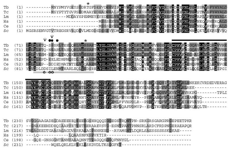
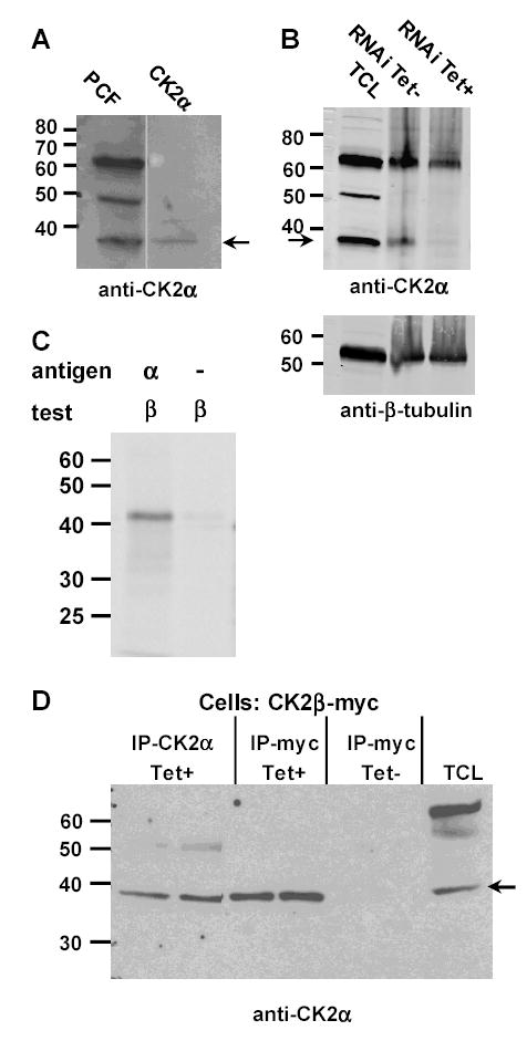
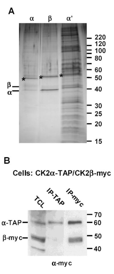
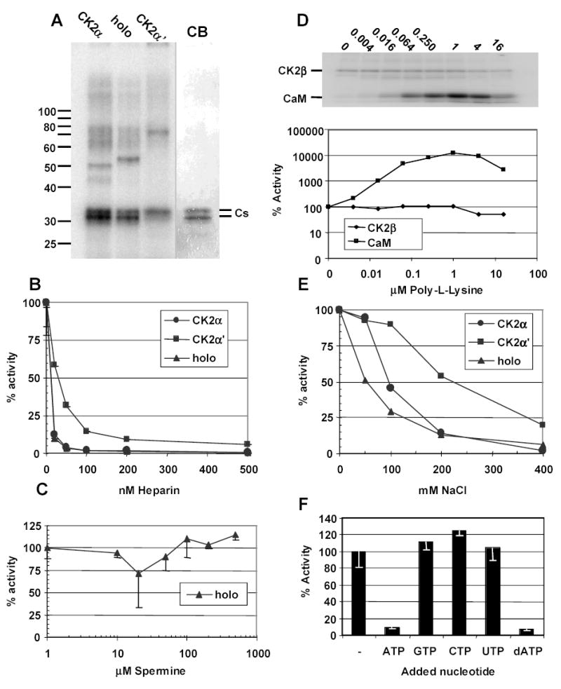
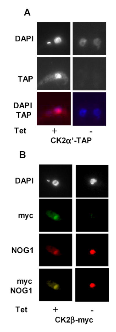
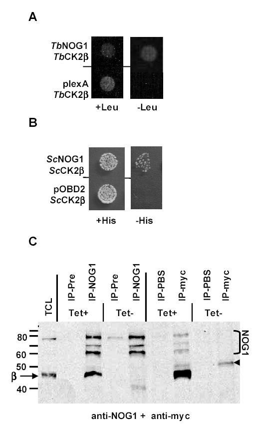
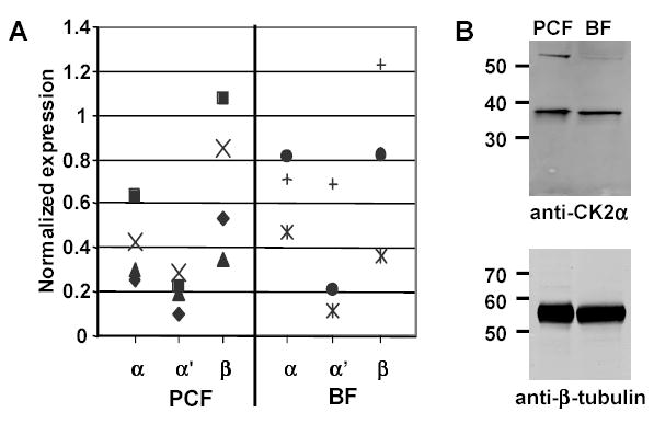
CK2 is a ubiquitous but enigmatic kinase. The difficulty in assigning a role to CK2 centers on the fact that, to date, no biologically relevant modulator of its function has been identified. One common theme revolves around a constellation of known substrates involved in growth control, compatible with its concentration in the nucleus and nucleolus. We had previously described the identification of two catalytic subunits of CK2 in Trypanosoma brucei and characterized one of them. Here we report the characterization of the second catalytic subunit, CK2alpha', and the identification and characterization of the regulatory subunit CK2beta. All three subunits are primarily localized to the nucleolus in T. brucei. We also show that CK2beta interacts with the nucleolar protein NOG1, adding to the interaction map which previously linked CK2alpha to the nucleolar protein NOPP44/46, which in turn associates with the rRNA binding protein p37. CK2 activity has four distinctive features: near equal affinity for GTP and ATP, heparin sensitivity, and stimulation by polyamines and polybasic peptides. Sequence comparison shows that the parasite orthologues have mutations in residues previously mapped as important in specifying affinity for GTP and stimulation by both polyamines and polybasic peptides. Studies of the enzymatic activity of the T. brucei CK2s show that both the affinity for GTP and stimulation by polyamines have been lost and only the features of heparin inhibition and stimulation by polybasic peptides are conserved.


Figures

Figure 1
Figure 1. Alignment of CK2β
Alignment of CK2β from T. brucei (Tb11.01.2590), T. cruzi (Tc00.1047053503757.40), L. major (LmjF36.5090), H. sapiens (AAA52123), C. elegans (AAA27983), and S. cerevisiae (CKB2, P38930). Residues conserved in all shown CK2β sequences are highlighted in black. Dark grey residues are conserved in three of the sequences while light grey are residues similar in at least three sequences. The black line above the sequence is the cysteine Zn++ binding motif. The acidic binding region in HsCK2β implicated in both poly-L-lysine and polyamine stimulation is indicated by the grey bar below the sequence. The two arrows denote the location of acidic residues in the human sequence suggested to mediate interactions between tetramers under low salt conditions via binding to basic residues in the active site of CK2α [52]. The filled circles mark residues, which when jointly mutated in Drosophila CK2β, result in decreased stimulation by polyamines [50]. Open circles mark three residues which when jointly mutated in human CK2β abrogated the response to poly-L-lysine [53]. Asterisks denote potential CK2 phosphorylation sites in the T. brucei protein.
Figure 2
Figure 2. Interaction of CK2α and CK2β
A. Analysis of anti-CK2α antiserum. Full-length untagged CK2α was generated in an in vitro coupled transcription and translation system. The in vitro generated protein (CK2α) was separated by SDS-PAGE, along with a protein lysate from 29-13 procyclic form cells (PCF). After transfer to nitrocellulose, blots were probed with anti-CK2α antiserum. The in vitro translated CK2α comigrates with the 38 kDa band detected by the anti-CK2a antiserum. The two lanes shown were from the same gel, separated by one lane. Arrow points to 38kDa band comigrating with CK2α. B. Disappearance of 38 kDa band after depletion of CK2α. Protein lysates generated from cells depleted for CK2α via RNAi (Tet+), see below Figure 7, were run on a gel along with protein lysates from untreated cells (Tet-) and a total cell lysate (TCL) from untransfected cells. Blots were imaged by IR scanning. Arrow points to CK2α. Lower panel shows loading control. C. In vitro translated CK2α and CK2β interact. CK2α (α) and 35S-methionine-labeled CK2β (β) were generated in an in vitro coupled transcription and translation system. The labeled CK2β either mixed with CK2α or on its own was immunoprecipitated with anti-CK2α antiserum. D. Coimmunoprecipitation of CK2α and CK2β-myc. Protein lysates made from either induced (Tet+) or uninduced (Tet-) cells were prepared from procyclic cells harboring a Tet-regulated, myc-tagged CK2β. Either CK2α (IP-CK2α) or CK2β-myc (IP-myc) were immunoprecipitated. Duplicate immunoprecipitates and the total cell lysate (TCL) were separated on an SDS-PAGE gel. Blots of the gel were probed with anti-CK2α antiserum. The location of CK2α is indicated by an arrow. The faint 50 kDa band in the CK2α immunoprecipitate is cross-reacting heavy chain IgG.
Figure 3
Figure 3. CK2 TAP complexes
A. Silver stained gel of samples from TAP purifications. Aliquots of the samples after TAP affinity purification procedure were separated on adjacent lanes of an 8-18% SDS-PAGE and silver stained. The location of the TAP-tagged protein in each lane is marked with an asterisk. Also indicated is the location of the native CK2α and CK2β proteins. B. Coprecipitation of CK2α-TAP and CK2β-myc. Protein lysates were made from procyclic cells expressing both CK2α-TAP and CK2β-myc. CK2α-TAP or CK2β-myc were precipitated and samples separated by SDS-PAGE along with the total cell lysate (TCL). The blot of the gel was probed with anti-myc mAb. The locations of the CK2α-TAP and CK2β-myc protein are indicated. Note the absence of CK2β-myc in the TAP purification. Due to the protein A portion of the TAP tag, CK2α-TAP is unavoidably bound by secondary antibodies and hence is observed in both the TCL and in the anti-myc immunoprecipitates.
Figure 4
Figure 4. Characterization of the kinase activity of CK2 preparations
A. TAP purified proteins have protein kinase activity. Aliquots of the CK2α (α), CK2 holoenzyme (holo) and CK2α’ (α’) preparations were used in a kinase reaction with casein as the substrate. Reactions were electrophoresed on an SDS-PAGE and stained. After drying the gels were exposed to phosphorimaging screen that was subsequently scanned. For comparison, a portion of the Coomassie blue stained gel is shown (CB). The location of the kinase substrate casein (Cs) is indicated. B. Heparin inhibition of CK2 activity. The activity of the purified proteins was assessed in the presence of increasing concentrations of heparin. Activities of the various enzymes were normalized to assays done in the absence of heparin. C. Spermine does not stimulate CK2 holoenzyme. The stimulatory effect of the polyamine spermine was tested in increasing concentrations with the holoenzyme. D. Poly-L-lysine stimulation of CK2 holoenzyme. The activity of the holoenzyme was tested with increasing concentrations of poly-L-lysine with calmodulin used as a substrate. The activity toward both calmodulin (CaM) and CK2β-TAP (CK2β) are shown normalized to untreated samples. E. Effect of NaCl on CK2 activity. Increasing concentrations of NaCl were added to reactions and normalized against reactions with no added salt. F. Nucleotide specificity of CK2α’. The ability of the CK2α’ catalytic subunit to utilize other nucleotides was tested. Unlabeled nucleotides (1mM) were added to the reactions to determine if they could compete for ATP binding
Figure 5
Figure 5. Localization of CK2α’ and CK2β to the nucleolus
A. Localization of CK2α’-TAP. CK2α’-TAP was stained in either induced (Tet+) or uninduced (Tet-) with rabbit anti-HRP, which binds the protein A part of the TAP tag. The stained protein was detected with Texas Red conjugated goat anti-rabbit Ig. The nucleus was stained with DAPI. The images were then overlayed, with red pseudocolor corresponding to TAP and blue to DAPI. B. Localization of CK2β-myc. CK2β-myc was stained in either induced (Tet+) or uninduced (Tet-) cells with anti-myc mAb and detected with FITC conjugated goat anti-mouse Ig. The nucleolar protein NOG1 was localized with anti-NOG1 antibodies and detected with Texas Red conjugated goat anti-rabbit Ig. The nucleus was stained with DAPI. The NOG1 and CK2β-myc images were overlayed.
Figure 6
Figure 6. CK2β interacts with NOG1
A. Yeast two-hybrid interaction between T. brucei proteins CK2β and NOG1. A yeast strain harboring plasmids that allow the expression of both the T. brucei NOG1 gene fused to LexA [30] and CK2β gene fused to the Gal4 activation domain was grown in liquid media. The culture was spotted onto media that contained leucine (+Leu) or lacked leucine (-Leu). Growth on the media without leucine indicates that the LEU2 reporter gene was expressed. B. Yeast two-hybrid interaction between S. cerevisiae proteins CK2β and NOG1. Similar to the above experiment only the HIS3 reporter gene was used. Shown is the data using the CKB2 gene fused to the region encoding the Gal4 activation domain constructed by Uetz et al [35]. Similar data was found using the second CK2β gene CKB1 (data not shown). C. Coimmunoprecipitation of CK2β and NOG1. Protein lysates were made from T. brucei transfectants either induced (Tet+) for expression of CK2β-myc or uninduced (Tet-). Proteins were immunoprecipitated with anti-NOG1 antiserum, the preimmune serum (Pre), anti-myc mAb, or PBS. Immunoprecipitates were separated by SDS-PAGE and proteins transferred to nitrocellulose. The membrane was probed simultaneously with anti-NOG1 and anti-myc reagents. The total cell lysate (TCL) was from induced cells. The faint band denoted by the arrowhead is IgG heavy chain.
Figure 7
Figure 7. Expression of CK2 subunits during the T. brucei life cycle
A. Analysis of transcript abundance. RNA from the two proliferative life stages, 29–13 procyclic cells (PCF) and single marker bloodform cells (BF) was isolated and the transcript abundance of CK2α, CK2α’, and CK2β quantified using real-time PCR. Quantities were normalized against α-tubulin transcript abundance. Shown is the data from four independent procyclic and three independent bloodform RNA samples. Each RNA preparation is marked by a different symbol so that the relationships of the subunits in individual samples can be seen. B. Analysis of CK2α protein abundance. Protein lysates from procyclic 29–13 (PCF) or single marker bloodform cells (BF) were loaded on an SDS-PAGE and transferred to nitrocellulose. Following detection of CK2α, the blot was stripped and reprobed with anti-β-tubulin mAb. Blots were imaged by IR scanning.
Figure 8
Figure 8. Effect of CK2 depletion on cell growth
A. Effect on growth for depletion for both CK2α and CK2α’. Growth of procyclic 29–13 cells harboring an inducible RNAi construct targeting the degradation of both CK2α and CK2α’ RNA was monitored after induction. Shown is the cumulative cell count for both induced (Tet+) and uninduced (Tet-) cultures. B. Northern analysis. RNA was extracted from either induced (Tet+) or uninduced (Tet-) cells at either day 2 or day 7 post induction. Northern blots were probed concurrently for CK2α and CK2α’. The location of the transcripts is indicated along with the double-stranded RNA (ds). Below the figure is shown the relative level of abundance of the transcripts after induction of RNAi as normalized to α-tubulin.

Similar articles

Cited by

References

    1. Dobrowolska G, Lozeman FJ, Li D, Krebs EG. CK2, a protein kinase of the next millennium. Mol Cell Biochem. 1999;191:3–12. - PubMed
    1. Glover CV. A filamentous form of Drosophila casein kinase II. J Biol Chem. 1986;261:14349–54. - PubMed
    1. Valero E, De BS, Filhol O, Wade RH, Langowski J, Chambaz EM, Cochet C. Quaternary structure of casein kinase 2. Characterization of multiple oligomeric states and relation with its catalytic activity. J Biol Chem. 1995;270:8345–52. - PubMed
    1. Chen M, Li D, Krebs EG, Cooper JA. The casein kinase II beta subunit binds to Mos and inhibits Mos activity. Mol Cell Biol. 1997;17:1904–12. - PMC - PubMed
    1. Abramczyk O, Zien P, Zielinski R, Pilecki M, Hellman U, Szyszka R. The protein kinase 60S is a free catalytic CK2alpha' subunit and forms an inactive complex with superoxide dismutase SOD1. Biochem Biophys Res Commun. 2003;307:31–40. - PubMed

Publication types

MeSH terms

LinkOut - more resources