. 2007 Feb;81(3):1274-87.
doi: 10.1128/JVI.00803-06. Epub 2006 Nov 8.

A hypervariable region within the 3' cis-acting element of the murine coronavirus genome is nonessential for RNA synthesis but affects pathogenesis

Affiliations

A hypervariable region within the 3' cis-acting element of the murine coronavirus genome is nonessential for RNA synthesis but affects pathogenesis

Scott J Goebel et al. J Virol. 2007 Feb.

Abstract

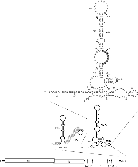
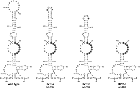
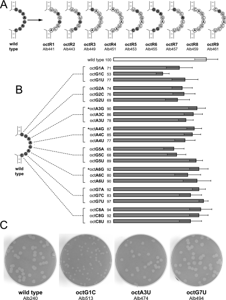
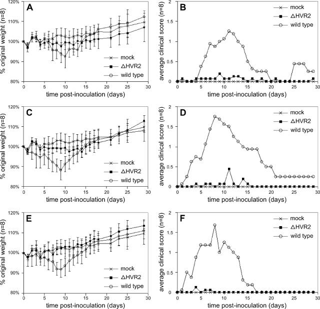
The 3' cis-acting element for mouse hepatitis virus (MHV) RNA synthesis resides entirely within the 301-nucleotide 3' untranslated region (3' UTR) of the viral genome and consists of three regions. Encompassing the upstream end of the 3' UTR are a bulged stem-loop and an overlapping RNA pseudoknot, both of which are essential to MHV and common to all group 2 coronaviruses. At the downstream end of the genome is the minimal signal for initiation of negative-strand RNA synthesis. Between these two ends is a hypervariable region (HVR) that is only poorly conserved between MHV and other group 2 coronaviruses. Paradoxically, buried within the HVR is an octanucleotide motif (oct), 5'-GGAAGAGC-3', which is almost universally conserved in coronaviruses and is therefore assumed to have a critical biological function. We conducted an extensive mutational analysis of the HVR. Surprisingly, this region tolerated numerous deletions, rearrangements, and point mutations. Most striking, a mutant deleted of the entire HVR was only minimally impaired in tissue culture relative to the wild type. By contrast, the HVR deletion mutant was highly attenuated in mice, causing no signs of clinical disease and minimal weight loss compared to wild-type virus. Correspondingly, replication of the HVR deletion mutant in the brains of mice was greatly reduced compared to that of the wild type. Our results show that neither the HVR nor oct is essential for the basic mechanism of MHV RNA synthesis in tissue culture. However, the HVR appears to play a significant role in viral pathogenesis.


Figures

FIG. 1.
FIG. 1.
Landscape of the MHV 3′ UTR. At the bottom is shown the organization of the 31.3-kb MHV genome. Above this is an expanded view of the secondary structure of the 301-nt 3′ UTR. The 3′ UTR comprises the bulged stem-loop (BSL) (nt 234 through 301) (11, 15, 16) and the overlapping pseudoknot (PK) (nt 185 through 238) (11, 55), the HVR (nt 46 through 156) (30), and the minimal element for the initiation of negative-strand RNA synthesis (MIN) (nt 1 through 45) (29). At the top is a detailed view of the structure of the downstream end of the 3′ UTR, as reported by Liu and coworkers (30), beginning at the nucleotide following the pseudoknot. The oct motif, 5′-GGAAGAGC-3′, is highlighted in gray. Stem segments A, B, and C of the HVR are those designated previously (30). Nucleotides are numbered from the first base at the 3′ end of the genome, excluding poly(A), and the N gene stop codon is boxed.
FIG. 2.
FIG. 2.
Selection of MHV 3′ UTR mutations by targeted RNA recombination. (A) Transcription vector pSG6 (1), the precursor of the plasmids used for synthesis of donor RNA for most recombinants, was derived from pMH54 (24) and contains a 5′ segment of the MHV genome (denoted by [1]) fused to a partial HE gene and all genes downstream of HE. The region between the 5′ segment and the N gene is not shown. The T7 RNA polymerase start site is indicated by an arrow. Particular 3′ UTR mutations, represented by a star, were constructed by splicing overlap extension-PCR and transferred into pSG6 by exchange of the BspEI-BclI restriction fragment. Donor RNAs were in vitro transcribed from PacI-linearized vectors. (B) For random mutagenesis of the oct motif, pTM11 was constructed from pSG6 to contain a fragment of the 3′ UTR preceding a site for BsmBI, which cuts at a distance from its recognition sequence. A PCR product was also generated to contain the remainder of the 3′ UTR, including a complementary BsmBI site in the opposite orientation and a randomized oct motif. BsmBI-linearized pTM11 was ligated to the BsmBI-restricted PCR fragment, and T7 transcripts were synthesized from the resulting template mixture. (C) MHV recombinants containing mutations in the 3′ UTR (solid rectangle) were generated by transfection of fMHV.v2-infected feline cells with synthetic donor RNA. The interspecies chimeric coronavirus fMHV.v2 (11) contains the ectodomain-encoding region of the feline infectious peritonitis virus (FIPV) S gene (gray rectangle), which renders it able to grow in feline cells but not in murine cells. Additionally, the order of the MHV structural protein genes downstream of S has been rearranged in fMHV.v2 to eliminate the possibility of unwanted secondary crossover events. Recombinants were selected as progeny that had regained the ability to grow in murine cells.
FIG. 3.
FIG. 3.
HVR truncation mutants. For each mutant, circled nucleotides are those that were changed from the wild-type sequence. The oct motif is highlighted in gray. Nucleotides are numbered according to the wild-type sequence, starting from the first base at the 3′ end of the genome, excluding poly(A).
FIG. 4.
FIG. 4.
HVR mutants that alter the accessibility of the oct motif. For each mutant, circled nucleotides are those that were changed from the wild-type sequence. The oct motif is highlighted in gray. Nucleotides are numbered according to the wild-type sequence, starting from the first base at the 3′ end of the genome, excluding poly(A).
FIG. 5.
FIG. 5.
oct motif point mutants. (A) Random, multiple-point mutants of oct. For each mutant, circled nucleotides are those that were changed from the wild-type sequence. Unchanged oct nucleotides are highlighted in gray. (B) Defined point mutants of oct. Each nucleotide in oct was changed to each of the three possible alternatives. Relative plaque sizes (diameters) of mutants are depicted as the percentage of the average diameter of wild-type plaques (± standard deviation). Mutants marked with an asterisk are those for which one naturally occurring variant coronavirus oct sequence has been reported. (C) Representative plaques for the three size classes of oct single-point mutants, compared to the wild type. Plaque titrations were performed on mouse L2 cells at 37°C. Monolayers were stained with neutral red at 48 h postinfection and were photographed 18 h later.
FIG. 6.
FIG. 6.
Mutants containing deletions encompassing the oct motif and the HVR. (A) At the right is the wild-type 3′ UTR, beginning at the nucleotide following the pseudoknot; the oct motif is highlighted in gray. Nucleotides in all structures are numbered according to the wild-type sequence, starting from the first base at the 3′ end of the genome, excluding poly(A). The Δoct mutant contains a deletion of the oct motif, nt 74 through 81, as well as the internal loop opposite oct, nt 130 through 136. The ΔHVR1 mutant contains a deletion of the region from nt 47 through 181, and the ΔHVR2 mutant contains a deletion of the region from nt 47 through 155. The Δ(3′ UTR) mutant contains a deletion of the entire 301-nt 3′ UTR, directly adjoining the N gene stop codon (boxed) to the poly(A) tail. (B) Plaques of the two viable deletion mutants, ΔHVR2 and Δoct, compared to those of an isogenic wild-type control. Plaque titrations were performed on mouse L2 cells at 37°C. Monolayers were stained with neutral red at 48 h postinfection and were photographed 18 h later.
FIG. 7.
FIG. 7.
Growth of the ΔHVR2 and Δoct mutants in tissue culture. (A) Single-step growth kinetics. Confluent monolayers of 17Cl1 cells were infected with wild-type, ΔHVR2, or Δoct viruses at a multiplicity of infection (moi) of 4.0 PFU per cell. At the indicated times postinfection, aliquots of medium were removed and infectious titers were determined on mouse L2 cells. Open and solid symbols represent results from two independent experiments. (B) Growth following a low multiplicity of infection. Confluent monolayers of 17Cl1 cells were infected with wild-type or ΔHVR2 viruses at a multiplicity of 0.01 PFU per cell. At the indicated times postinfection, aliquots of medium were removed and infectious titers were determined on mouse L2 cells. Open and solid symbols represent results from two independent experiments.
FIG. 8.
FIG. 8.
RNA synthesis by the ΔHVR2 and Δoct mutants. Infected or mock-infected 17Cl1 cells were metabolically labeled with [33P]orthophosphate in the presence of actinomycin D, and RNA was isolated and electrophoretically analyzed in 1% agarose containing formaldehyde as described in Materials and Methods.
FIG. 9.
FIG. 9.
Reduced virulence of the ΔHVR2 mutant. Six-week-old, female BALB/c mice, in groups of eight, were inoculated intranasally with 5 × 10³ PFU (A and B), 5 × 10⁴ PFU (C and D), or 5 × 10⁵ PFU (E and F) of wild-type or ΔHVR2 virus. A group of four mock-inoculated mice was inoculated with tissue culture supernatant. Mice were weighed and observed for clinical signs daily. (A, C, and E) Percentage of original weight (± standard deviation). (B, D, and F) Average clinical disease scores, as defined in Materials and Methods. The same mock-inoculated group is shown in all panels for comparison to each of the viral dose groups.
FIG. 10.
FIG. 10.
Reduced in vivo replication of the ΔHVR2 mutant. Six-week-old, female BALB/c mice, in two groups of 20, were inoculated intranasally with 5 × 10⁴ PFU of wild-type or ΔHVR2 virus (the dose shown in Fig. 9C and D). Four mice in each group were sacrificed at 1, 3, 5, 7, and 9 days p.i., and tissues were harvested. Viral titers in nasal washes (A) and brains (B) were determined by plaque assay in L2 cells, as described in Materials and Methods. Titers for individual mice are represented by circles or squares. Geometric means of titers for each set of four mice are connected by solid lines or broken lines. The thicker, horizontal broken line indicates the limit of detection for each set of assays (10 PFU/ml for nasal washes and 50 PFU/g for brains).

Similar articles

Cited by

References

    1. Boursnell, M. E. G., M. M. Binns, I. J. Foulds, and T. D. K. Brown. 1985. Sequences of the nucleocapsid genes from two strains of avian infectious bronchitis virus. J. Gen. Virol. 66:573-580. - PubMed
    1. Brian, D. A., and R. S. Baric. 2005. Coronavirus genome structure and replication. Curr. Top. Microbiol. Immunol. 287:1-30. - PMC - PubMed
    1. Brown, D. M., C. T. Cornell, G. P. Tran, J. H. Nguyen, and B. L. Semler. 2005. An authentic 3′ noncoding region is necessary for efficient poliovirus replication. J. Virol. 79:11962-11973. - PMC - PubMed
    1. Brown, D. M., S. E. Kauder, C. T. Cornell, G. M. Jang, V. R. Racaniello, and B. L. Semler. 2004. Cell-dependent role for the poliovirus 3′ noncoding region in positive-strand RNA synthesis. J. Virol. 78:1344-13451. - PMC - PubMed
    1. Chua, M. M., K. C. MacNamara, L. San Mateo, H. Shen, and S. R. Weiss. 2004. Effects of an epitope-specific CD8+ T-cell response on murine coronavirus central nervous system disease: protection from virus replication and antigen spread and selection of epitope escape mutants. J. Virol. 78:1150-1159. - PMC - PubMed

Publication types

LinkOut - more resources