. 2006 Nov 3;24(3):355-66.
doi: 10.1016/j.molcel.2006.09.007.

Phosphorylation-dependent ubiquitination of cyclin D1 by the SCF(FBX4-alphaB crystallin) complex

Affiliations

Phosphorylation-dependent ubiquitination of cyclin D1 by the SCF(FBX4-alphaB crystallin) complex

Douglas I Lin et al. Mol Cell. .

Abstract

Growth factor-dependent accumulation of the cyclin D1 proto-oncogene is balanced by its rapid phosphorylation-dependent proteolysis. Degradation is triggered by threonine 286 phosphorylation, which promotes its ubiquitination by an unknown E3 ligase. We demonstrate that Thr286-phosphorylated cyclin D1 is recognized by a Skp1-Cul1-F box (SCF) ubiquitin ligase where FBX4 and alphaB crystallin govern substrate specificity. Overexpression of FBX4 and alphaB crystallin triggered cyclin D1 ubiquitination and increased cyclin D1 turnover. Impairment of SCF(FBX4-alphaB crystallin) function attenuated cyclin D1 ubiquitination, promoting cyclin D1 overexpression and accelerated cell-cycle progression. Purified SCF(FBX4-alphaB crystallin) catalyzed polyubiquitination of cyclin D1 in vitro. Consistent with a putative role for a cyclin D1 E3 ligase in tumorigenesis, FBX4 and alphaB crystallin expression was reduced in tumor-derived cell lines and a subset of primary human cancers that overexpress cyclin D1. We conclude that SCF(FBX4-alphaB crystallin) is an E3 ubiquitin ligase that promotes ubiquitin-dependent degradation of Thr286-phosphorylated cyclin D1.


Figures

Fig. 1
Fig. 1. Identification of αB crystallin as a cyclin D1-associated protein
(A) Silver stain of affinity-purified cyclin D1 complexes prepared from parental NIH3T3 cells or Flag-D1 3T3 cell lines treated with the proteasome inhibitor, LLnL (leucyl-leucyl-norleucinal). Cyclin D1-interacting proteins were identified by mass spectrometry. (B) Western blot of endogenous cyclin D1 precipitates prepared from proliferating NIH3T3 cells treated with DMSO, LLM (N-acetyl-leucinyl-leucinyl-methioninal) or LLnL. Normal rabbit serum (NRS) precipitates served as a negative control. (C) The cyclin D1-αB crystallin interaction is dependent on cyclin D1 phosphorylation at Thr286. NIH3T3 cells expressing either wild-type cyclin D1 or the D1T286A mutant were treated with vehicle, LLM or LLnL for 6hrs and cell lysates prepared from these cells were precipitated with a cyclin D1 specific antibody (13-17G) and probed for cyclin D1 and αB crystallin.
Fig. 2
Fig. 2. Phosphorylation-dependent interaction between cyclin D1 and the SCFFBX4-αB crystallin complex
(A) Endogenous FBX4, cyclin D1 and αB crystallin associate in vivo. Cell lysates prepared from proliferating NIH3T3 cells treated with DMSO or MG132 were precipitated with antibodies directed against FBX4 or normal rabbit serum (NRS) and subjected to immunoblot with the indicated antibodies. (B) Phospho-T286-dependent binding of cyclin D1 to SCFFBX4-αB crystallin in vitro. Cyclin D1 or phosphorylation-deficient derivatives, D1T286A and D1P287A, were mixed with SCFFBX4-αB crystallin complexes and purified by immuno-affinity chromatography from 293T cells; where indicated, FBX4 and αB crystallin were omitted. (C) Cyclin D1-FBX4 binding is dependent on αB crystallin and Thr286 phosphorylation. 293T lysates containing the indicated proteins were incubated with beads coupled to the phosphorylated cyclin D1 peptide or to unphosphorylated cyclin D1 peptide. Bound proteins were detected by immunoblot. (D) Phospho-MYC and -IκKα do not interact with SCFFBX4-αB crystallin. Beads coupled to the indicated peptides were incubated with 293T lysates expressing SCFFBX4-αB crystallin complexes as in (C) and bound FBX4 was detected by immunoblot. (E) Beads coupled to the indicated peptides were incubated with 293T lysates harboring either SCFFBX4-αB crystallin or SCFFBW2 complexes and bound FBX4 or FBW2 were detected by anti-FLAG western blot. Uncoupled beads served as negative (background) control.
Fig. 3
Fig. 3. The SCFFBX4-αB crystallin ubiquitin ligase regulates cyclin D1 stability and protein levels in vivo.
(A) NIH3T3 cells expressing empty vector or (ΔF)FBX4 were pulse-labeled with 35S methionine/cysteine and “chased” for the indicated time periods. Cyclin D1 was precipitated and visualized by autoradiography. (B) Western analysis of whole cell extracts prepared from 293T cells transfected with cyclin D1, CDK4, (ΔF) FBX4 and αB crystallin. (C) (D) NIH3T3 cells expressing either control, αB crystallin (C) or FBX4 shRNAs (D) were treated with 100 μg/mL of cycloheximide for the indicated time periods, and cyclin D1 half-life was determined by western blotting. (E) Expression of FBX4 and αB crystallin promotes cyclin D1 proteolysis. 293T cells were transfected with cyclin D1, CDK4, αB crystallin, FBX4 and GFP. Cell lysates were prepared and immunoblotted for cyclin D1, FBX4 and GFP. (F) Proteasome inhibition inhibits FBX4-dependent cyclin D1 proteolysis.
Fig. 4
Fig. 4. The SCFFBX4-αB crystallin complex catalyzes cyclin D1 ubiquitination in vivo and in vitro.
(A) NIH3T3 cells expressing Flag-cyclin D1 were transfected with HA-ubiquitin, (ΔF) FBX4 and αB crystallin and treated with 20 μM MG132 for 0, 2 and 4 hours. Cell lysates were subjected to precipitation with either normal rabbit serum (NRS) or with a cyclin D1 specific antibody and ubiquitinated proteins were visualized by anti-HA immunoblot. (B) Same as in (A) except that cyclin D1 immunoprecipitates were probed with a cyclin D1 antibody. (C) Human U2OS cells were co-transfected with HA-tagged ubiquitin, cyclin D1 and shRNAs specific for firefly luciferase or FBX4. Cyclin D1 ubiquitination was detected by immunoprecipitation with an anti-flag antibody followed by anti-HA western blotting. (D) The SCFFBX4-αB crystallin complex catalyzes ubiquitination of cyclin D1 in vitro. Ubiquitination of in vitro transcribed and translated cyclin D1 was assessed by mixing cyclin D1 with GSK3β ATP, ubiquitin, E1, E2, and a complete SCFFBX4 αBcrystallin complex (lane 2). The reactions were also carried out omitting the following: the entire E3 ligase (lane1), FBX4 (lane 3), Cul1, Skp1, Roc 1(lane 4) or E1, E2 (lane 5). (E) The F-box of FBX4 and αB crystallin are required for cyclin D1 polyubiquitination in vitro. Ubiquitination reactions were initiated by addition of an ATP, ubiquitin, E1 and E2 enzymes. Where indicated wild-type FBX4 was replaced with (ΔF)FBX4 (lane 2), αB crystallin was omitted from the reaction (lane 3). (F) Phosphorylation deficient cyclin D1-T286A is refractory to SCFFBX4-αB crystallin-dependent ubiquitination. Cyclin D1 ubiquitination D-F was assessed by autoradiography (upper panels) and input is shown (lower panels).
Fig. 5
Fig. 5. Cytoplasmic localization of FBX4 and αB crystallin
(A) Immunofluorescence of asynchronously growing NIH3T3 and U2OS cells with N-terminus (top) or C-terminus (middle and bottom panels) FBX4-specific antibodies. (B) Immunofluorescence of asynchronously growing mouse fibroblasts stably expressing FBX4-specific hairpins (F-3T3-2, F-3T3-4). (C) Western analysis confirming knockdown of FBX4 in NIH3T3 cells. Asterisk indicates a nonspecific band detected by the secondary antibody. (D) Stable NIH3T3 cells expressing empty vector or Flag-tagged FBX4 were fractionated into cytoplasmic (C), nuclear (N) and total (T) extracts. Fractions were used for immunoprecipitation with an anti-flag antibody or for direct western blots as indicated.
Fig. 6
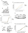
Fig. 6. The SCFFBX4-αB crystallin ubiquitin ligase regulates cell cycle progression
(A) Cell cycle dependent binding between FBX4 and cyclin D1. NIH3T3 cell were released from a G0 (serum starvation) block and binding between cyclin D1 and FBX4 was assessed by IP-western. (B) Lysates prepared from NIH-3T3 cells transfected with GFP or GSK3β kinase-dead (KD) constructs and treated with MG132 were precipitated with FBX4 antiserum or rabbit anti-mouse IgG and subjected to immunoblot with indicated antibodies. (C) Kinetics of cell cycle re-entry from G0 in NIH3T3 cells shRNAs against FBX4 or αB crystallin (D) NIH3T3 cells expressing shRNAs against FBX4/αB crystallin were released from a nocodazole (G2/M) block in media containing BrDU and percentage of cells in S phase assessed by FACS. (E) Immunoblot (cyclin D1) of NIH3T3 cells expressing shRNAs against FBX4/αB crystallin released from a nocodazole (G2/M) block for the indicated time. (F) Knockdown of FBX4/αB crystallin in D1−/− fibroblasts (right panel); control blot of D1 and Skp1 (left panel). (G) D1−/− cells expressing shRNAs against FBX4/αB crystallin were released from a nocodazole and S-phase entry was assessed as in D.
Fig. 7
Fig. 7. FBX4 and αB crystallin levels are altered in human cancers
(A) Quantification of FBX4 and αB crystallin mRNA levels in a tumor vs. matching normal tissue in an mRNA array. (B) Expression of αB crystallin, cyclin D1 and FBX4 in normal human mammary epithelial (HMEC), non-tumorigenic breast MCF10A cells and tumorigenic breast cancer cell lines (MCF7, MDA-MB-231, BT474). (C) MCF10A, MCF7 or MCF7 cells infected with a retrovirus encoding αB crystallin were treated with cycloheximide for the indicated intervals and levels of cyclin D1, αB crystallin and actin were assessed by immunoblot.

Similar articles

Cited by

References

    1. Alt JR, Cleveland JL, Hannink M, Diehl JA. Phosphorylation-dependent regulation of cyclin D1 nuclear export and cyclin D1-dependent cellular transformation. Genes Dev. 2000;14:3102–3114. - PMC - PubMed
    1. Alt JR, Gladden AB, Diehl JA. p21(Cip1) Promotes cyclin D1 nuclear accumulation via direct inhibition of nuclear export. J Biol Chem. 2002;277:8517–8523. - PubMed
    1. Bai F, Xi JH, Wawrousek EF, Fleming TP, Andley UP. Hyperproliferation and p53 status of lens epithelial cells derived from alphaB-crystallin knockout mice. J Biol Chem. 2003;278:36876–36886. - PubMed
    1. Bashir T, Dorrello NV, Amador V, Guardavaccaro D, Pagano M. Control of the SCF(Skp2-Cks1) ubiquitin ligase by the APC/C(Cdh1) ubiquitin ligase. Nature. 2004;428:190–193. - PubMed
    1. Bax B, Carter PS, Lewis C, Guy AR, Bridges A, Tanner R, Pettman G, Mannix C, Culbert AA, Brown MJ, et al. The structure of phosphorylated GSK-3beta complexed with a peptide, FRATtide, that inhibits beta-catenin phosphorylation. Structure (Camb) 2001;9:1143–1152. - PubMed

Publication types

MeSH terms