. 2007 Jan 19;365(3):835-55.
doi: 10.1016/j.jmb.2006.09.083. Epub 2006 Oct 3.

Involvement of DEAD-box proteins in group I and group II intron splicing. Biochemical characterization of Mss116p, ATP hydrolysis-dependent and -independent mechanisms, and general RNA chaperone activity

Affiliations

Involvement of DEAD-box proteins in group I and group II intron splicing. Biochemical characterization of Mss116p, ATP hydrolysis-dependent and -independent mechanisms, and general RNA chaperone activity

Coralie Halls et al. J Mol Biol. .

Abstract

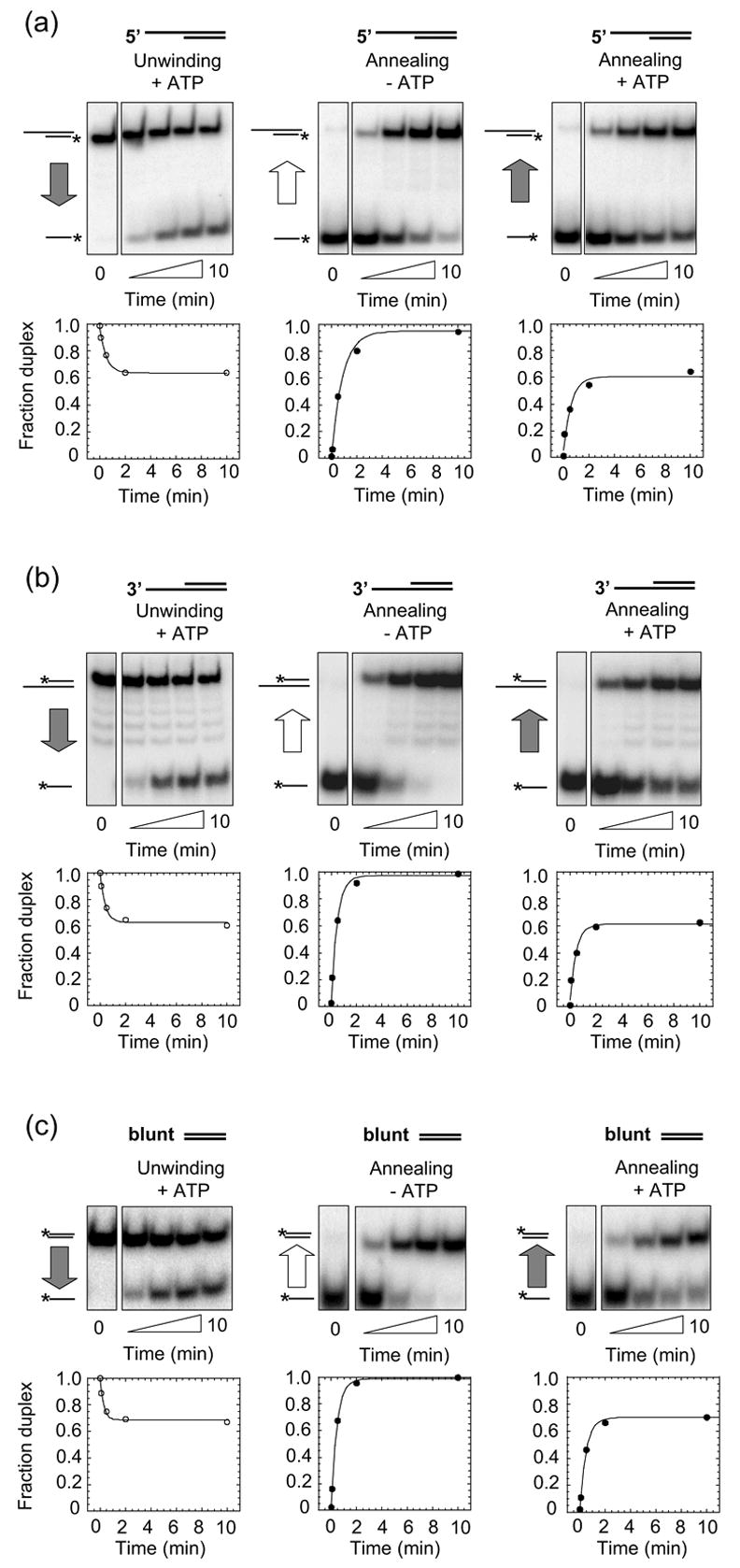
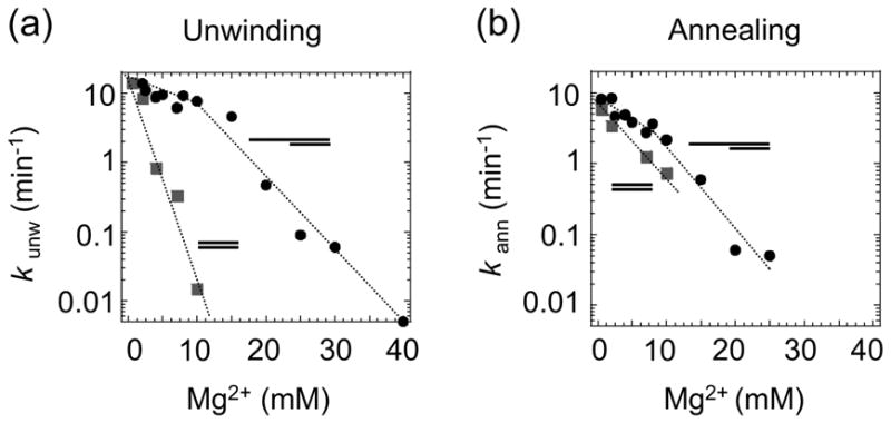
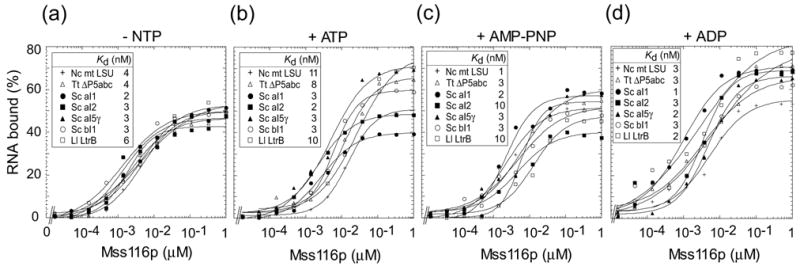
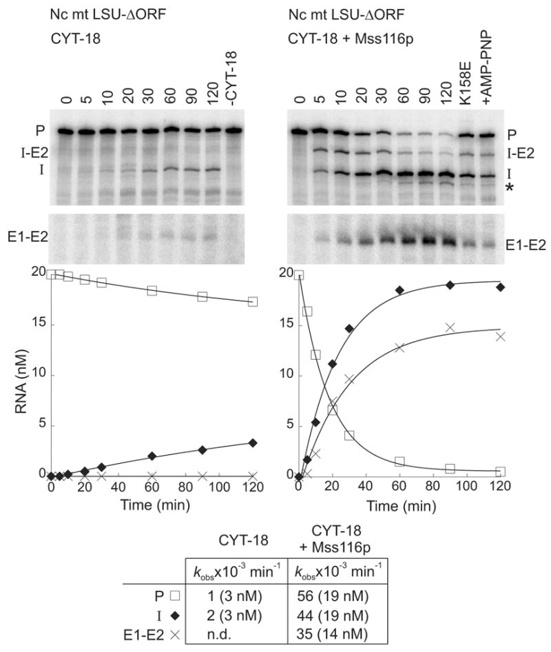
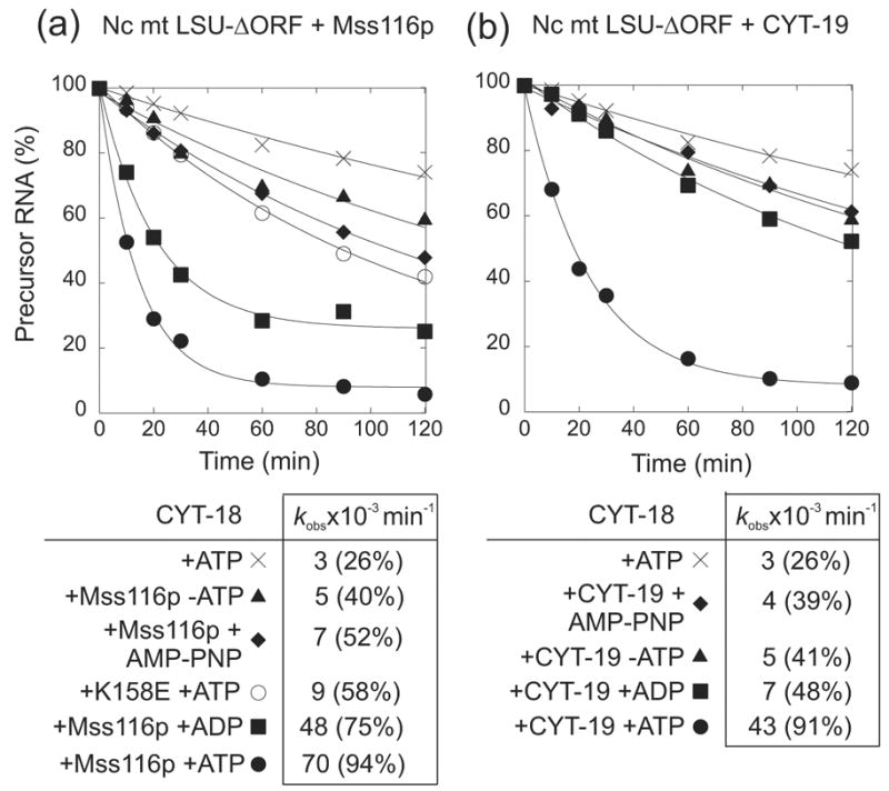
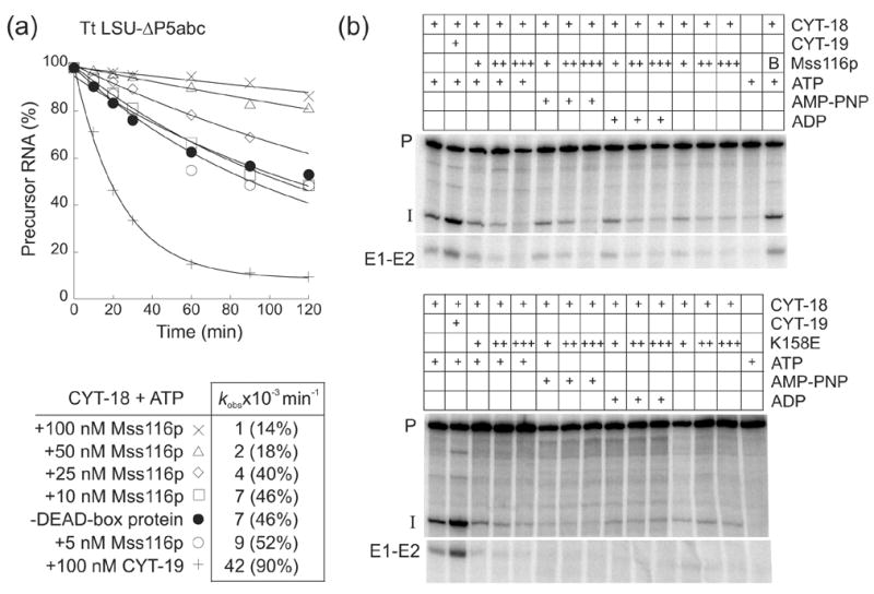
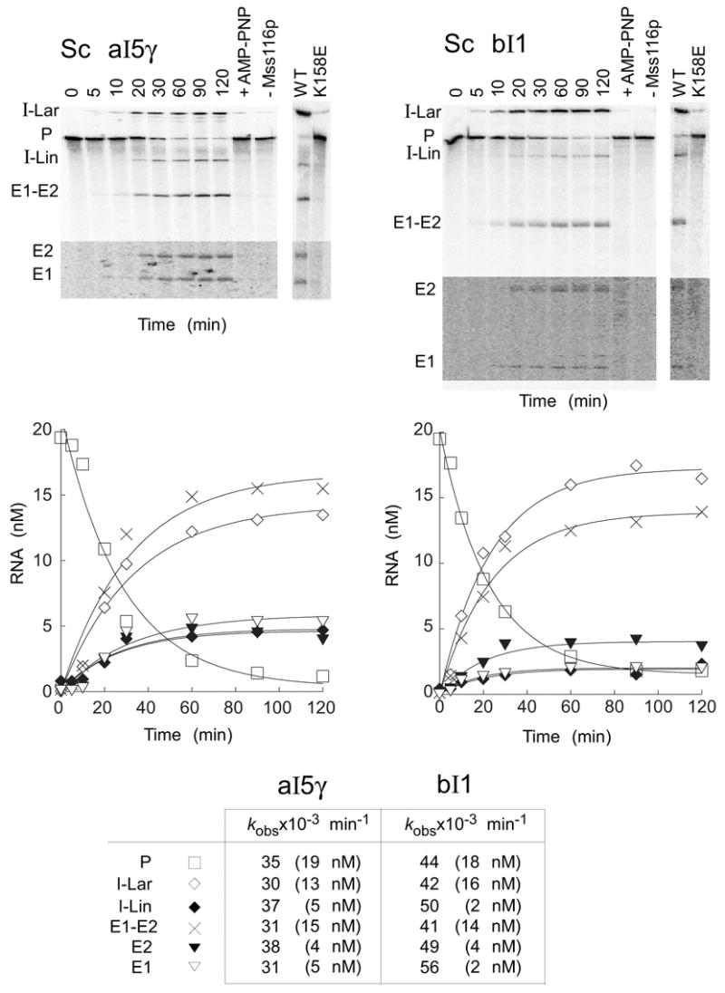
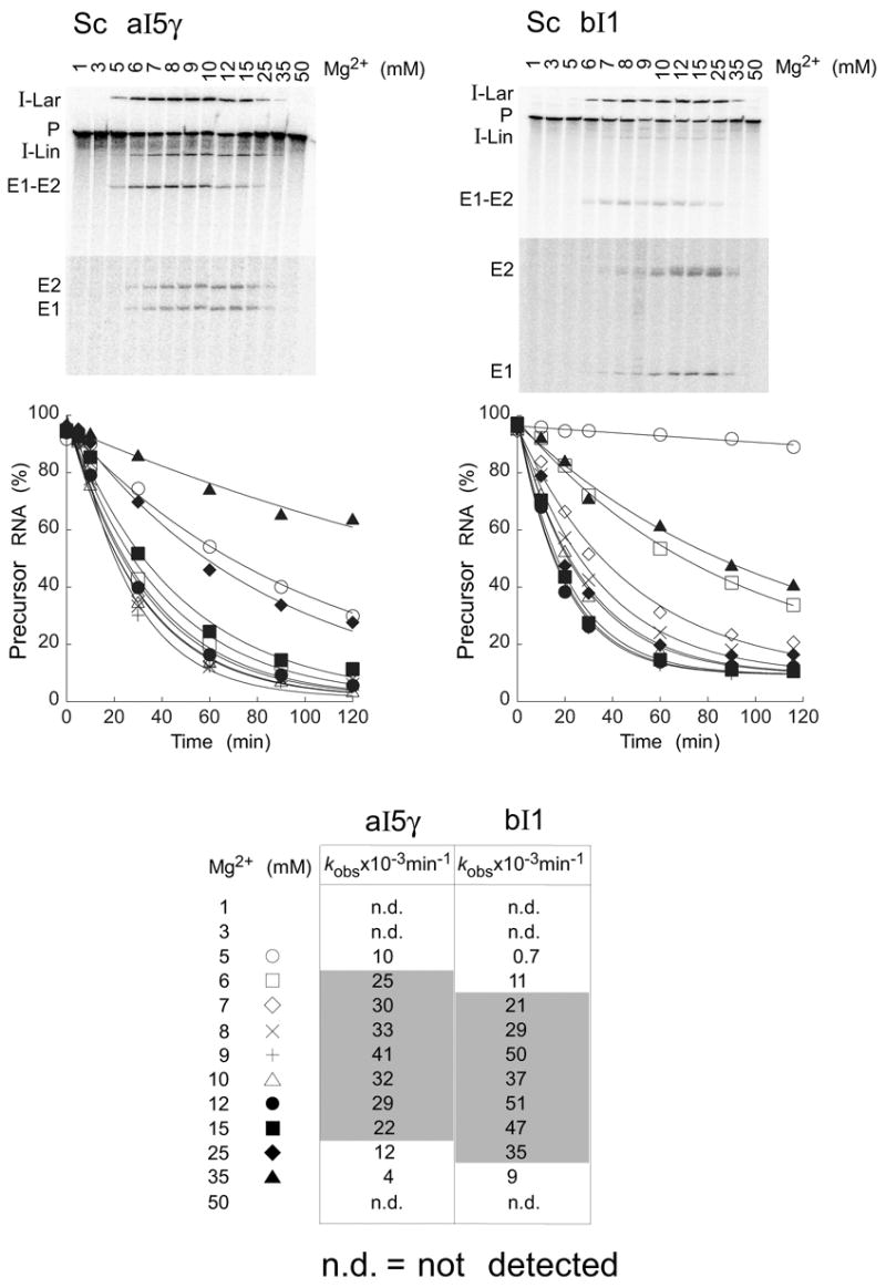
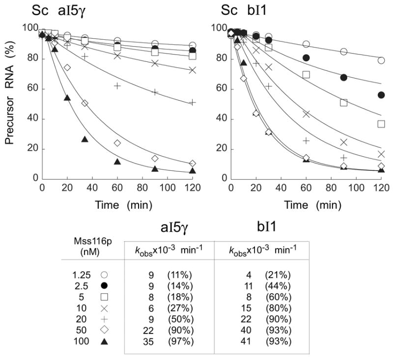
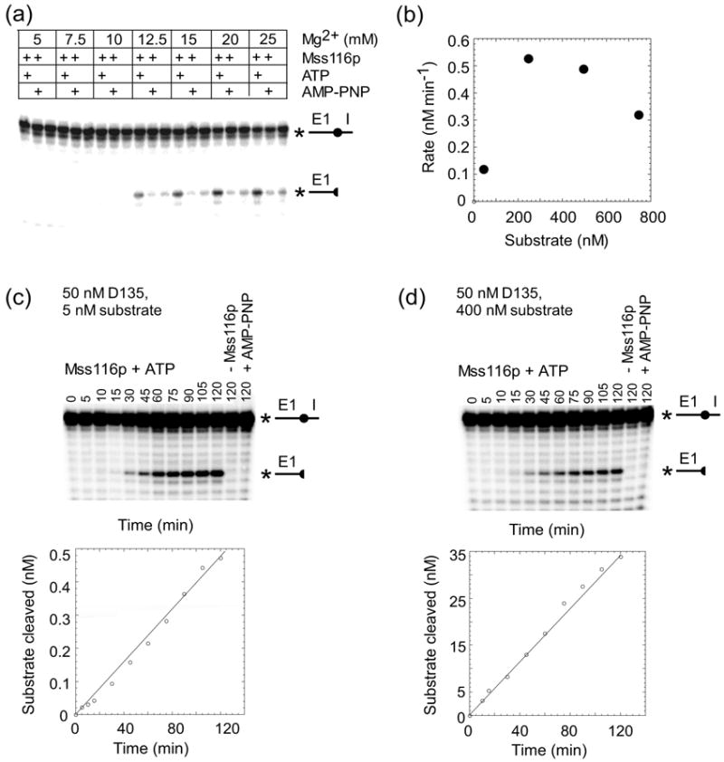
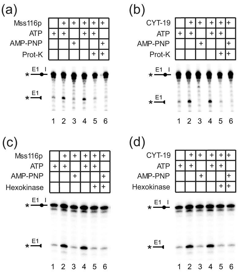
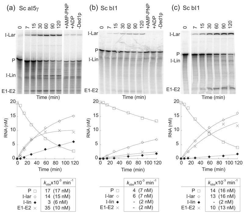
The RNA-catalyzed splicing of group I and group II introns is facilitated by proteins that stabilize the active RNA structure or act as RNA chaperones to disrupt stable inactive structures that are kinetic traps in RNA folding. In Neurospora crassa and Saccharomyces cerevisiae, the latter function is fulfilled by specific DEAD-box proteins, denoted CYT-19 and Mss116p, respectively. Previous studies showed that purified CYT-19 stimulates the in vitro splicing of structurally diverse group I and group II introns, and uses the energy of ATP binding or hydrolysis to resolve kinetic traps. Here, we purified Mss116p and show that it has RNA-dependent ATPase activity, unwinds RNA duplexes in a non-polar fashion, and promotes ATP-independent strand-annealing. Further, we show that Mss116p binds RNA non-specifically and promotes in vitro splicing of both group I and group II intron RNAs, as well as RNA cleavage by the aI5gamma-derived D135 ribozyme. However, Mss116p also has ATP hydrolysis-independent effects on some of these reactions, which are not shared by CYT-19 and may reflect differences in its RNA-binding properties. We also show that a non-mitochondrial DEAD-box protein, yeast Ded1p, can function almost as efficiently as CYT-19 and Mss116p in splicing the yeast aI5gamma group II intron and less efficiently in splicing the bI1 group II intron. Together, our results show that Mss116p, like CYT-19, can act broadly as an RNA chaperone to stimulate the splicing of diverse group I and group II introns, and that Ded1p also has an RNA chaperone activity that can be assayed by its effect on splicing mitochondrial introns. Nevertheless, these DEAD-box protein RNA chaperones are not completely interchangeable and appear to function in somewhat different ways, using biochemical activities that have likely been tuned by coevolution to function optimally on specific RNA substrates.


Figures

Figure 1
Figure 1
Mss116p duplex-unwinding and strand-annealing activities. Reactions were done with RNA oligonucleotides that form a 16-bp duplex with (a) a 25-nt 5′ overhang; (b) a 25-nt 3′ overhang; or (c) blunt-ends. For each substrate, the figure shows representative duplex- unwinding reactions with 2 mM ATP (left panels), strand-annealing reactions without ATP (middle panels), and strand-annealing reactions with 2 mM ATP (right panels). Plots beneath the gels show fraction duplex versus reaction time fitted to the integrated form of a homogenous first-order rate law. Substrates and products are depicted schematically to the left of the gel, with the asterisk indicating the radiolabel and the vertical arrow indicating the direction of the reaction. Gray arrows indicate presence and white arrows indicate absence of ATP in the reactions. The zero time points for the duplex-unwinding reactions were recorded before ATP addition, and the zero time points for the strand-annealing reactions were recorded before Mss116p addition. Strand annealing in the absence of Mss116p was not significant (<5% of the RNA spontaneously annealed within 10 min; data not shown). Rate constants calculated from at least two independent time courses for each reaction are summarized in Table 3.
Figure 2
Figure 2
Mg2+-concentration dependence of the Mss116p duplex-unwinding and strand- annealing reactions. The Mg2+-concentration dependence of the rate constants for (a) duplex unwinding (kunw), and (b) strand annealing (kann) of RNA oligonucleotides containing a 13-bp duplex region with a 5′ overhang (filled circles) or blunt-ends (filled squares). Reactions were done in the presence of 1 mM ATP.
Figure 3
Figure 3
Equilibrium-binding of Mss116p to group I and group II intron RNAs. 32P-labeled RNAs (5 pM) were incubated with increasing concentrations of Mss116p in reaction medium containing 100 mM KCl/8 mM MgCl2 for 90 min at 30°C (a) without nucleotide (-NTP), or with 1 mM (b) ATP, (c) AMP-PNP, or (d) ADP. RNAs were group I introns N. crassa mt LSU-ΔORF and T. thermophila LSU-ΔP5abc, and group II introns S. cerevisiae aI1-ΔORF, aI2-ΔORF, aI5γ, and bI1 and L. lactis Ll.LtrB-ΔORF. The plots show the percent input RNA bound versus Mss116p concentration. Essentially identical binding curves for each condition were obtained when the RNA and protein were incubated for 10 or 120 min before filtration through nitrocellulose, or in reaction medium containing 100 mM KCl/5 mM MgCl2 (data not shown).
Figure 4
Figure 4
Mss116p promotes splicing of the N. crassa mt LSU-ΔORF group I intron. Splicing time courses for CYT-18 alone (left) and CYT-18 plus Mss116p (right) were done by incubating 32P-RNA substrate (20 nM) with one or both proteins (each at 100 nM) in reaction medium containing 100 mM KCl, 5 mM MgCl2, and 1 mM ATP at 25°C. The splicing products were analyzed in a denaturing 4% polyacrylamide gel, which was dried and quantified with a phosphorimager. In controls (right lanes), RNA was incubated for 120 min with 1 mM ATP without CYT-18 (-CYT-18), with mutant Mss116p-K158E plus 1 mM ATP, or with wild-type Mss116p plus 1 mM AMP-PNP (+AMP-PNP). The plots beneath the gel show the disappearance of precursor RNA and the appearance of products as a function of time, with the data fit to a single exponential. The table at the bottom summarizes kobs values, with the numbers in parentheses indicating the concentration of RNA (nM) reacted or produced after 120 min. Abbreviations: E1–E2, ligated exons; I, intron; I-E2, intermediate after the first step of splicing; P, precursor RNA; *, slightly shorter intron RNA resulting from use of an upstream cryptic 3′-splice site; n.d., not detected.
Figure 5
Figure 5
Mss116p promotes CYT-18-dependent splicing of the N. crassa mt LSU-ΔORF group I intron by both ATP-hydrolysis-dependent and ATP-hydrolysis independent mechanisms. Splicing time courses were done with 20 nM 32P-labeled precursor RNA, 100 nM CYT-18 dimer, and (a) 100 nM Mss116p or (b) 100 nM CYT-19 in reaction medium containing 100 mM KCl and 5 mM MgCl2 at 25°C in the presence or absence of 1 mM ATP, AMP-PNP, and ADP. The time course for Mss116p-K158E in (a) was done in the presence of 1 mM ATP. The tables below the plots summarize kobs values for the disappearance of precursor RNA, with the numbers in parentheses indicating the percent of precursor RNA reacted after 120 min.
Figure 6
Figure 6
Mss116p inhibits CYT-18-dependent splicing of the T. thermophila LSU-ΔP5abc group I intron. (a) Splicing time courses were done with 50 nM 32P-labeled precursor RNA, 50 nM CYT-18 dimer, and 5 to 100 nM Mss116p or 100 nM CYT-19 in reaction medium containing 100 mM KCl and 5 mM MgCl2 at 30°C. The products were analyzed in a denaturing 4% polyacrylamide gel, which was dried and scanned with a phosphorimager. The table below the plots summarizes kobs values for the disappearance of precursor RNA, with the numbers in parentheses indicating the percent of precursor RNA reacted after 120 min. (b) Splicing reactions were carried out as above for 30 min with different concentrations of wild-type Mss116p or Mss116p-K158E in the presence or absence of 1 mM ATP, AMP-PNP, or ADP. In panel (a) reactions were initiated by adding CYT-18; in panel (b), precursor RNA was preincubated with 50 nM CYT-18 for 10 min at 30°C, and the reaction was initiated by adding the DEAD-box protein plus 1 mM GTP. Concentrations of wild-type Mss116p and Mss116p-K158E were 25 nM (+), 50 nM (++), and 100 nM (+++). In the top gel, the far right lane shows a control reaction in which 50 nM wild-type Mss116p was boiled (B) for 5 min prior to the start of the splicing reaction. Abbreviations are as in Figure 4.
Figure 7
Figure 7
Mss116p-promoted splicing of group II introns aI5γ and bI1. Splicing time courses for aI5γ (left) and bI1 (right) were done by incubating 20 nM 32P-labeled RNA substrate with 100 nM Mss116p protein and 1 mM ATP in reaction medium containing 100 mM KCl and 8 mM MgCl2 at 30°C. Splicing products were analyzed in a denaturing 4% polyacrylamide gel, which was dried and quantified with a phosphorimager. The bottom segments of the gels are darker exposures to show E1 and E2, which result form SER. Control lanes show RNA substrate incubated for 120 min with Mss116p plus 1 mM AMP-PNP (+AMP-PNP) or without Mss116p (-Mss116p). The additional lanes (far right) show precursor RNAs incubated with wild-type (WT) Mss116p and mutant Mss116p-K158E under the same conditions for 120 min. The plots beneath the gels show disappearance of precursor RNA and appearance of products as a function of time, with the data fit to a single exponential. The table at the bottom summarizes kobs values, with the numbers in parentheses indicating the concentration of RNA (nM) reacted or produced after 120 min. The I-Lin band may contain a mixture of linear intron and broken lariat RNA. Abbreviations: E1, 5′ exon; E2, 3′ exon; E1–E2, ligated exons; I-Lar, intron lariat; I-Lin, linear intron; P, precursor RNA.
Figure 8
Figure 8
Mg2+-concentration dependence of Mss116p-promoted splicing of aI5γ and bI1. Splicing time courses for aI5γ (left) and bI1 (right) were done by incubating 20 nM 32P-labeled precursor RNAs with 100 nM Mss116p plus 1 mM ATP in reaction media containing 100 mM KCl and the indicated Mg2+ concentrations at 30°C. Products were analyzed and quantified, as in Figure 7. The gels show the extent of splicing after 20 min at different Mg2+ concentrations, and the plots show disappearance of precursor RNA as a function of time, with the data fit to a single exponential. The bottom segments of the gels are darker exposures to show E1 and E2, which result form SER. The table at the bottom summarizes kobs values for aI5γ and bI1 precursor RNA disappearance at different Mg2+ concentrations. The shaded area highlights the broad Mg2+ optimum. Abbreviations are as in Figure 7.
Figure 9
Figure 9
Mss116p-concentration dependence of aI5γ and bI1 group II intron splicing. Splicing time courses for aI5γ (left) and bI1 (right), respectively, were done with 20 nM 32P-labeled precursor RNA and 1.25 to 100 nM Mss116p in reaction media containing 100 mM KCl and 8 mM MgCl2 at 30°C. The plots show disappearance of precursor RNA as a function of time, with data fit to a single exponential. The table at the bottom summarizes kobs values and amplitudes (%) at 120 min. Abbreviations are as in Figure 7.
Figure 10
Figure 10
Mss116p promotes RNA-oligomer cleavage by the D135 ribozyme. (a) Mg2+-concentration dependence. D135 RNA (50 nM) was pre-incubated at 25°C with Mss116p (50 nM) for 5 min in reaction medium containing 100 mM KCl and the indicated Mg2+ concentrations in the presence or absence of 1 mM ATP or AMP-PNP. The reaction was initiated by adding 5′-32P-labeled RNA-oligomer substrate (5 mM), incubated for 90 min at 25°C, and terminated by adding 100 mM EDTA, followed by phenol-CIA extraction. The products were analyzed in a denaturing 20% polyacrylamide gel, which was dried and quantified with a phosphorimager. (b) Substrate-concentration dependence. For each substrate concentration (25, 250, 500, and 750 nM), a 2 h-time course were done as above with 50 nM D135 ribozyme and 50 nM Mss116p plus 1 mM ATP in reaction medium containing 100 mM KCl and 25 mM MgCl2 at 25°C. The plot shows the rate of cleavage as a function of RNA substrate concentration. (c) and (d) Time courses of D135-catalyzed RNA-cleavage with substoichiometric and saturating concentrations of RNA-oligomer substrate, respectively. 50 nM D135 RNA was pre-incubated with 50 nM Mss116p and 1 mM ATP in reaction medium containing 100 mM KCl and 25 mM Mg2+ for 5 min at 25°C, and the reaction was initiated by adding 5 or 400 nM RNA substrate in (c) or (d), respectively. The right-hand lanes show control reactions with the same amount of RNA substrate incubated for 2 h at 25°C in the absence of Mss116p or with 1 mM AMP-PNP instead of ATP. The plots beneath the gels show the appearance of product as a function of time. The 5′-32P-labeled RNA oligomer substrate and labeled cleavage product are depicted schematically to the right of the gels, with the circle indicating the 5′-exon-intron junction, and the star indicating the location of the label.
Figure 11
Figure 11
Protease sensitivity of DEAD-box protein-promoted D135-ribozyme cleavage reactions and requirement for ATP-hydrolysis after substrate binding. (a) and (b), protease sensitivity of D135 ribozyme cleavage promoted by (a) Mss116p or (b) CYT-19. Mss116p (100 nM) or CYT-19 (500 nM) were incubated with D135 RNA (100 nM) and RNA-oligomer substrate (5 nM) without or with 1 mM ATP or AMP-PNP in reaction medium containing 100 mM KCl and 25 mM MgCl2 at 25°C. Lanes 1-3, D135 RNA was pre-incubated in reaction medium for 90 min with ATP (lane 1), DEAD-box protein plus ATP (lane 2), or DEAD-box protein plus AMP-PNP (lane 3). Lanes 4 and 5, D135 RNA was pre-incubated for 60 min with DEAD-box protein plus ATP or AMP-PNP, respectively, then split with halves incubated for 30 min without (lane 4) or with 20 mg/ml proteinase K (prot-K) and 0.5% SDS (lane 5). Lane 6, D135 RNA was pre-incubated with DEAD-box protein for 60 min, then for 30 min with proteinase K plus SDS. After the pre-incubations, reactions were initiated by adding 5′-32P-labeled RNA oligomer, incubated for 30 min, then terminated, and products analyzed in a denaturing 20% polyacrylamide gel. (c) and (d), effect of ATP-depletion prior to substrate addition. Reactions were done as above, with D135 RNA pre-incubated with the indicated additions for 30 min, and then for an additional 15 min without or with addition of hexokinase plus glucose. Reactions were initiated by adding RNA-oligomer substrate, incubated for 120 min, and products analyzed as above.
Figure 12
Figure 12
Ded1p-promoted splicing of group II introns aI5γ and bI1. Splicing time courses for (a) aI5γ with 300 nM Ded1p, and (b) and (c), bI1 with 300 and 600 nM Ded1p, respectively, were done using 20 nM 32P-labeled precursor RNA and 1 mM ATP in reaction medium containing 100 mM KCl and 8 mM MgCl2 at 30°C. Splicing products were analyzed in a denaturing 4% polyacrylamide gel, which was dried and quantified with a phosphorimager. Control lanes show precursor RNA incubated for 120 min without Ded1p (-Ded1p) or with Ded1p plus 1 mM AMP-PNP (+AMP-PNP) or 1 mM ADP (+ADP). The plots beneath the gels show disappearance of precursor RNA and appearance of products as a function of time, with the data fit to a single exponential. The table at the bottom summarizes kobs values, with the numbers in parentheses indicating the concentration of RNA (nM) reacted or produced after 120 min. kobss could not be determined for the low amounts of I-Lin and E1–E2 produced in panel (b) and I-Lin in panel (c) due to high errors in the curve fitting. The I-Lin band may contain a mixture of linear intron and broken lariat RNA. Abbreviations are as in Figure 7.

Similar articles

Cited by

References

    1. Lambowitz AM, Caprara MG, Zimmerly S, Perlman PS. Group I and group II ribozymes as RNPs: clues to the past and guides to the future. In: Gesteland RF, Cech TR, Atkins JF, editors. The RNA World. 2. Cold Spring Harbor Laboratory Press: Plainview, NY; 1999. pp. 451–485.
    1. Lehmann K, Schmidt U. Group II introns: structure and catalytic versatility of large natural ribozymes. Crit Rev Biochem Mol Biol. 2003;38:249–303. - PubMed
    1. Pyle AM, Lambowitz AM. Group II introns: ribozymes that splice RNA and invade DNA. In: Gesteland RF, Cech TR, Atkins JF, editors. The RNA World. 3. Cold Spring Harbor Laboratory Press; Plainview, NY: 2006. pp. 469–505.
    1. Caprara MG, Nilsen TW. RNA: versatility in form and function. Nat Struct Biol. 2000;7:831–833. - PubMed
    1. Weeks KM. Protein-facilitated RNA folding. Curr Opin Struct Biol. 1997;7:336–342. - PubMed

Publication types

MeSH terms