. 2007 Jan 15;55(2):165-77.
doi: 10.1002/glia.20445.

S100B expression defines a state in which GFAP-expressing cells lose their neural stem cell potential and acquire a more mature developmental stage

Affiliations

S100B expression defines a state in which GFAP-expressing cells lose their neural stem cell potential and acquire a more mature developmental stage

Eric Raponi et al. Glia. .

Abstract

During the postnatal development, astrocytic cells in the neocortex progressively lose their neural stem cell (NSC) potential, whereas this peculiar attribute is preserved in the adult subventricular zone (SVZ). To understand this fundamental difference, many reports suggest that adult subventricular GFAP-expressing cells might be maintained in immature developmental stage. Here, we show that S100B, a marker of glial cells, is absent from GFAP-expressing cells of the SVZ and that its onset of expression characterizes a terminal maturation stage of cortical astrocytic cells. Nevertheless, when cultured in vitro, SVZ astrocytic cells developed as S100B expressing cells, as do cortical astrocytic cells, suggesting that SVZ microenvironment represses S100B expression. Using transgenic s100b-EGFP cells, we then demonstrated that S100B expression coincides with the loss of neurosphere forming abilities of GFAP expressing cells. By doing grafting experiments with cells derived from beta-actin-GFP mice, we next found that S100B expression in astrocytic cells is repressed in the SVZ, but not in the striatal parenchyma. Furthermore, we showed that treatment with epidermal growth factor represses S100B expression in GFAP-expressing cells in vitro as well as in vivo. Altogether, our results indicate that the S100B expression defines a late developmental stage after which GFAP-expressing cells lose their NSC potential and suggest that S100B expression is repressed by adult SVZ microenvironment.


Figures

Figure 1
Figure 1
S100B is expressed long after the gain of GFAP and the loss of RC2 and characterizes a mature developmental stage of the astrocytic lineage. A–E, coronal sections of developing post-natal telencephalon of mice at P2, P8, P14 and P60 were subjected to double immunolabeling as indicated. A–B, at P2, RC2+ transforming RGCs do not yet express S100B (A, arrowheads) but start to express GFAP in their radial processes (B, arrows). In B, arrowheads point to RC2+ RGC bodies which are still negative for GFAP staining. C–D, at P7, RG transforming cells are more stellate and express GFAP in their cell bodies (C and D, arrowheads). Some of these cells start to express S100B (C, arrow). In D, arrows point to a radial process with residual RC2 staining. E, at P14, most of GFAP expressing cells express S100B (arrows). The arrow points to a GFAP+ cell that does not yet express S100B. F, at P60, all GFAP+ cells express S100B (arrows). In A, C, E and F, asterisks indicate S100B expressing cells that do not express RC2 or GFAP. These cells likely are oligodendroglial cells as indicated in supplemental figure 1. G, Western blot analysis of GFAP and S100B expression during the forebrain postnatal development. α-tubulin (αTub) was used as loading control. H, quantitative analysis of GFAP+ cells expressing S100B in different telencephal regions including the cortex, the corpus callosum (CC) and the striatum during postnatal development. 3 mice were analyzed at P8 and P14 and 5 at P60. Errors are ± SD. ** p<0.001, * p<0,05. Scale bars: 20 μm.
Figure 2
Figure 2
S100B expression pattern in developing and adult germinal zones A–E, coronal sections of developing and adult SVZ (A–C and E) and adult hippocampus (D) were subjected to double immunolabeling as indicated. A, at P8, RC2+ RG transforming cells express GFAP in radial processes. B–C, cell bodies (arrowhead) and radial process (arrows) of transforming astrocytic cells at P7 (B) and P14 (C) express GFAP but are devoid of S100B staining. In developing SVZ, S100B is expressed in ependymocytes (asterisks). D, in adult dentate gyrus, radial GFAP+ cells of the subgranular layer never express S100B (arrowheads). Arrows point to stellate GFAP+/S100B+ cells. E, in adult SVZ, S100B expression is restricted to ependymal cells. The subependymal GFAP+ cells with bipolar morphology are devoid of S100B staining (arrowheads). lv, lateral ventricle; St, striatum; gl, granular layer. Scale bars: 20 μm.
Figure 3
Figure 3
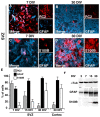
Cultured subventricular and cortical astrocytes mature into S100B+ cells. Astrocytic cultures derived from the SVZ (A–D) and the neocortex (not shown) of newborn mice were immunolabeled at 7, 15 and 30 DIV as indicated. The percentages of RC2+, GFAP+ and S100B+ cells in the SVZ and the cortical cultures are represented in E. Western blot analysis of GFAP and S100B expression during astrocytic development in culture is presented in F. α-tubulin (αTub) was used as loading control. Each bar represents the average of 2 to 4 independent cultures. Errors are ± SD. DIV, days in vitro. Scale bars: 20 μm.
Figure 4
Figure 4
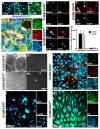
In astrocytes, activation of s100b gene is linked to the loss of NSC potential. A and B, cultured astrocytic cells were obtained from whole forebrains of newborn transgenic s100b-EGFP mice and were characterized at 7, 15 and 30 DIV as indicated. A–A2, EGFP expression increases with the time spent in culture. B, at 30 DIV, nearly all EGFP+ cells express both endogenous S100B and GFAP. C–E, EGFP+ and EGFP cells derived from 15 and 30 DIV cultures were sorted by FACS and double immunolabeled as indicated. Most sorted EGFP cells are in a RC2+/GFAP or a RC2+/GFAP+ developmental stage, all of them express GLAST cells (C,E), while EGFP+ cells are GFAP+/GLAST+ cells (D). F–H, unlike sorted EGFP cells, most sorted EGFP+ cells are unable to generate neurospheres (NSs) with a size higher than 50 μm (H). EGFP expression is maintained in small NSs derived from sorted EGFP+ cells even after 12 days of EGF/bFGF treatment (H1). In F, each bar represents the average of 4 independent cultures, each counted in triplicate. Errors are ± SD. I–K, NSs derived from EGFP cells are multipotent and give rise to β-tubulin III+ neuroblasts (I), O4+ oligodendroglial cells (J) and GFAP+ astrocytic cells (I and J), whereas the small NSs derived from sorted EGFP+ cells are mostly unipotent and give rise to GFAP+ astrocytes (K). A and G, Zeiss conventional microscopy. B–F and H–J, stack of 5 to 8 optical sections captured with Leica confocal microscope. Scale bars: 20 μm in A–E and H–J; 100 μm in G.
Figure 5
Figure 5
Maturation of grafted astrocytic cells is controlled by micro-environmental factors. A–C, 15 DIV cultured astrocytic cells derived from transgenic newborn mice expressing GFP under the ubiquitous β-actin promoter were grafted stereotaxically in the striatum (A–A1, B–B1) or in the SVZ (C–C1) of adult mice as indicated in D. GFP+ cells grafted in the striatum progressively expressed S100B after 1 and 4 weeks (A–A1 and B–B1 respectively) whereas most of GFAP+/GFP+ cells grafted in SVZ were maintained in S100B immature state even after 4 weeks (C–C1). Arrows and arrowheads point to GFP+/GFAP+/S100B+ and GFP+/GFAP+/S100B respectively. E, Quantitative analysis of GFP+/GAFP+ expressing S100B. At least 400 GFP+/GFAP+ cells were counted and n is the number of transplanted mice. Errors are ± SD. lv, lateral ventricle; St, striatum, WPG, Week Post Grafting. Scale bars are 50 μm in A, B and C and 20 μm in A1, B1 and C1.
Figure 6
Figure 6
EGF prevents the activation of the s100b gene and maintains astrocytic cells in S100B immature state in vitro and in vivo. A–D, sorted s100b-EGFP cells (EGFP) from 15 DIV primary astrocytic cultures were replated in medium with serum in the absence (−EGF) or in the presence of EGF (+EGF), and double-immununolabeled for the presence of RC2 and GFAP at 8 DIV and 15 DIV as indicated. EGF treatment stimulated the transition from RC2+/GFAP/EGFP and RC2+/GFAP+/EGFP stages to RC2/GFAP+/EGFP stage and maintained the cells in this stage (C–D). E, percentages of different cell populations. F, EGF treatment increases the number of NSs generated from cultured astrocytic cells after 15 DIV. NSs are generated from 15 DIV secondary astrocytic culture in the absence (−EGF) or in the presence of EGF (+EGF) and from sorted s100b-EGFP cells (Ctl) derived from 15 DIV primary culture. Each bar represents the average of 3 independent cultures, each counted in triplicate. Errors are ± SD, Scale bars are 50 μm in A and C and 20 μm in B and D. G–G1, 15 DIV cultured astrocytic cells derived from transgenic newborn mice expressing GFP under the ubiquitous β-actin promoter were grafted stereotaxically in the striatum of adult mice as described in figure 4. Multiple i.v. injections of EGF (400 ng/40 μl every second day for 4 weeks) prevent the expression of S100B in GFP+/GFAP+ grafted cells compared to control mice (see fig. 4B–B1). H, Quantitative analysis of GFP+/GAFP+ cells expressing S100B. At least 400 GFP+/GFAP+ cells were counted and n the number of transplanted mice. Errors are ± SD. Scale bars: 50 μm in G and 20 μm in G1.
Figure 7
Figure 7
Model of GFAP+ astrocytic development. The onset of GFAP expression in parallel with the progressive down-regulation of RC2 characterizes the stage I and the stage II respectively. Next, GFAP+ astrocytes continue their developmental program and the onset of S100B expression defines the stage III. This stage includes restricted astrocyte precursors and terminally differentiated astrocytes. EGF signaling exerts a double effect on GFAP+ astrocytic development. It activates the transition from stage I to the stage II and blocks the transition from stage II to stage III.

Similar articles

Cited by

References

    1. Ahn S, Joyner AL. In vivo analysis of quiescent adult neural stem cells responding to Sonic hedgehog. Nature. 2005;437:894–7. - PubMed
    1. Alvarez-Buylla A, Garcia-Verdugo JM. Neurogenesis in adult subventricular zone. J Neurosci. 2002;22:629–34. - PMC - PubMed
    1. Anthony TE, Klein C, Fishell G, Heintz N. Radial glia serve as neuronal progenitors in all regions of the central nervous system. Neuron. 2004;41:881–90. - PubMed
    1. Bonaguidi MA, McGuire T, Hu M, Kan L, Samanta J, Kessler JA. LIF and BMP signaling generate separate and discrete types of GFAP-expressing cells. Development. 2005;132:5503–14. - PubMed
    1. Burrows RC, Wancio D, Levitt P, Lillien L. Response diversity and the timing of progenitor cell maturation are regulated by developmental changes in EGFR expression in the cortex. Neuron. 1997;19:251–67. - PubMed

Publication types

MeSH terms