. 2006 Nov;25(5):783-93.
doi: 10.1016/j.immuni.2006.08.023. Epub 2006 Oct 19.

The adaptor molecule MyD88 activates PI-3 kinase signaling in CD4+ T cells and enables CpG oligodeoxynucleotide-mediated costimulation

Affiliations

The adaptor molecule MyD88 activates PI-3 kinase signaling in CD4+ T cells and enables CpG oligodeoxynucleotide-mediated costimulation

Andrew E Gelman et al. Immunity. 2006 Nov.

Abstract

While T cells respond directly to toll-like receptor (TLR) agonists, TLR-signaling pathways in T cells are poorly characterized. Here we demonstrate in CD4(+) T cells that CpG DNA directly enhances proliferation, prevents anergy, and augments humoral responses to a T cell-dependent antigen by a Myeloid differentiation primary-response protein 88 (MyD88) and Phosphatidylinositol 3-kinase (PI-3 kinase)-dependent pathway. PI-3 kinase activation required a putative Src-homology domain (SH2) binding motif in the MyD88 Toll-Like or IL-1 Receptor (TIR) domain. Reconstitution of MyD88-deficient primary T cells with a MyD88 transgene mutated in this motif abrogated association of PI-3 kinase with MyD88, phosphorylation of protein kinase B (Akt) and Glycogen Synthetase Kinase-3 (GSK-3), and interleukin-2 (IL-2) production. The MyD88 death domain, on the other hand, was required for NF-kB activation and survival. These studies identify a MyD88-dependent PI-3 kinase-signaling pathway in T cells that differentiates CpG DNA-mediated proliferation from survival and is required for an in vivo T cell-dependent immune response.


Figures

Figure 1
Figure 1. CpG DNA Directly Promotes CD4+ T Cell Proliferation in a MyD88-Dependent Manner
(A) Purified Myd88−/− or wild-type littermate control CD4+ T cells were stimulated with graded concentrations of CD3 mAb in the presence of irradiated T cell-depleted Myd88−/− splenocytes. Cultures were performed in the absence (open squares) or presence (closed squares) of 3.0 μM CpG ODN. Additionally, irradiated T cell-depleted Myd88−/− splenocytes were also first pretreated for 8 hr with 3 μM CpG ODN and then washed 3 times prior to culture (open circles). After 72 hr, cultures were pulsed with [3H]thymidine and then harvested 16 hr later. Results are indicated as mean ± SD and are representative of two independent experiments. (B) Purified Myd88−/− or wild-type (WT) littermate control CFSE-labeled CD4+ T cells were cultured with plate-bound CD3 mAb (0.3 μg/ml) in the absence or presence of CpG DNA (3.0 μM), LPS (100 ng/ml), or CD28 mAb (1 μg/ml) for 72 hr. Proliferation was assessed by FACS analysis and is represented by responder frequency (inset). Results are representative of five experiments. (C) CD4+ T cells were purified by FACS, labeled with CFSE, and stimulated with plate-bound CD3 mAb at a limiting concentration of 0.3 μg/ml. CpG ODN (1668), GpC ODN (1720), or a nonstimulatory ODN (AP1) were added to the cultures at varying concentrations as shown. LPS (100 ng/ml) was used as a negative control and to assess the purity of the preparation, because CD4+ T cells are not directly stimulated by LPS but receive costimulatory signals from LPS-activated APCs. Cells were cultured for 72 hr and harvested, and flow cytometry was performed to assess proliferation by CFSE dilution and viability by 7-AAD exclusion. The number above each inset represents the percentage of live CD4+ T cells that responded to stimulation by dividing (responder frequency). One experiment representative of three is shown.
Figure 2
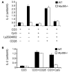
Figure 2. CpG DNA Promotes IL-2 Synthesis and Blocks Anergy Induction in a MyD88-Dependent Manner
(A) Purified Myd88−/− or wild-type control CD4+ T cells were activated with plate-bound CD3 (2.5 μg/ml) in the absence or presence of listed combinations of CpG DNA (3.0 μM), Ly294002 (10 μM), or plate-bound CD28 (2.5 μg/ml). IL-2 amounts were determined by ELISA. Results are indicated as mean ± SD and are representative of three independent experiments. (B) Purified Myd88−/− or wild-type CD4+ T cells were stimulated with plate-bound CD3 mAb (2.5 μg/ml) in the presence or absence of either CpG DNA (3.0 μM) or CD28 mAb for 24 hr, washed, rested for 48 hr, and restimulated with plate-bound CD3 and CD28 mAb (2.5 μg/ml each) for additional 18 hr, and supernatant was assessed for IL-2 by ELISA. Results are indicated as mean ± SD and are representative of three independent experiments.
Figure 3
Figure 3. CpG DNA Stimulates a MyD88-Dependent PI-3 Kinase Pathway
(A) Purified CD4+ T cells were activated with polystyrene beads coated with CD3 mAb (1 μg/ml) in the presence or absence of listed combinations of CpG DNA (3.3 μM), Ly294002 (10 μM), or CD28 mAb-coated polystyrene beads (1 μg/ml) for 30 min. Cells were then lysed and analyzed by immunoblot with antibodies specific for p-Akt or Akt. The result is representative of three experiments. (B) Purified Myd88−/− or wild-type control littermate CD4+ T cells were activated with polystyrene beads coated with CD3 mAb (1 μg/ml) in the presence or absence of CpG DNA (3.3 μM), CD28 mAb-coated polystyrene beads (1 μg/ml), or both for 30 min. Cells were then lysed and analyzed by immunoblot with antibodies specific for p-Akt, p-GSK-3β, or actin. The result is representative of three experiments. (C) Purified CD4+ T cells were stimulated with CD3 mAb (1.0 μg/ml)-coated beads in the presence or absence of 10 μM CpG DNA for 0, 30, or 60 min. Cells were then lysed and immunoprecipitated with rabbit antibodies specific for MyD88 and analyzed by immunoblot with antibodies specific for p85 (top) and goat antibodies specific for MyD88 (bottom). Results are representative of two experiments.
Figure 4
Figure 4. Y257 Is Required for the Association of the Regulatory Subunit of PI-3 Kinase p85 with MyD88 and for PI-3 Kinase-Generated Signals
(A) Alignment of the MyD88 TIR domains of human, mouse, and zebrafish. The consensus YKXM motif is denoted with a large box, and boxed amino acids denote differences between mouse with human or mouse with zebrafish. (B) Myd88−/− mice were lethally irradiated and reconstituted with syngeneic Myd88−/− bone marrow that had been retrovirally transduced with wild-type or mutant MyD88 constructs (mutation of Y → F mutation of Y257, truncated MyD88 TIR domain only, or both, as per Experimental Procedures). 3 months after reconstitution, purified NGFR+ CD4+ T cells were lysed and immunoblotted and probed with MyD88-specific antibodies. (C) Purified CD4+ T cells from chimeras as described in (B) were stimulated with CD3 mAb (1.0 μg/ml)-coated beads in the presence or absence of 10 μM CpG DNA for 0, 30, or 60 min. Cells were then lysed and immunoprecipitated with rabbit antibodies specific for MyD88 and analyzed by immunoblot with antibodies specific for p85 and goat antibodies specific for MyD88. Results are representative of two experiments. (D) Purified CD4+ T cells as in (B) were activated with polystyrene beads coated with CD3 mAb (1 μg/ml) in the presence or absence of listed combinations of CpG DNA (3.3 μM), Ly294002 (10 μM), or CD28 mAb-coated polystyrene beads (1 μg/ml) for 30 min. CD4+ T cells were then lysed and analyzed by immunoblot with antibodies specific for p-Akt or Akt. The result is representative of two experiments.
Figure 5
Figure 5. MyD88 Y257 and MyD88 Death Domain Are Both Required for CpG DNA-Mediated Proliferative Responses
(A) Purified NGFR+ CD4+ T cells coexpressing MyD88, MyD88 Y257, MyD88 TIR, or MyD88 TIRY257F transgenes isolated from radiation bone chimeras were stimulated with graded concentrations of CD3 mAb in the presence of irradiated T cell-depleted Myd88−/− splenocytes. Cultures were performed in the absence (open squares) or presence (closed squares) of 3.0 μM CpG ODN. Additionally, irradiated T cell-depleted Myd88−/− splenocytes were first pretreated for 8 hr with 3 μM CpG ODN and then washed three times prior to culture (open circles). After 72 hr, cultures were pulsed with 3H thymidine and then harvested 16 hr later. Results are indicated as mean ± SD and are representative of two experiments. (B) CSFE-labeled CD4+ T cells prepared as in (A) were cultured with plate-bound CD3 mAb (0.3 μg/ml) in the absence or presence of CpG ODN (3.0 μM) or CD28 (1 μg/ml) for 72 hr and then analyzed by FACS. Proliferation is represented by percent CSFE dilution (inset). Results are representative of two experiments. (C) Purified CD4+ T cells prepared as in (A) were activated with plate-bound CD3 (2.5 μg/ml) alone (open bars) or in the presence of either 3.0 μM CpG DNA (closed bars), 3.0 μM CpG DNA and Ly2924002 (horizontal striped bars), or 2.5 μg/ml of plate-bound CD28 (vertical striped bars) for 18 hr. IL-2 amounts were determined by bead ELISA. Results are indicated as a mean ± SD and are representative of two independent experiments.
Figure 6
Figure 6. MyD88 Death Domain but Not Y257 Is Required for CpG DNA-Mediated Activated CD4+ T Cell Survival, NF-kB Activation, and Bcl-xL Upregulation
(A) Purified NGFR+ CD4+ T cells coexpressing MyD88, MyD88 Y257F, MyD88 TIR, or MyD88 TIRY257F transgenes isolated from bone marrow chimeras were activated with plate-bound CD3 mAb (0.3 μg/ml) + CD28 mAbs (2.5 μg/ml) for 16 hr, washed, and then recultured in quadruplicate wells at 500,000 cells/well alone (open bars) or in the presence of either 10 μM CpG DNA (closed bars) or 30 μg/ml of poly I:C (shaded bars) for an additional 72 hr. Cells were then either assessed for viability by the exclusion of annexin staining (top) or live cell counts (bottom). Results are indicated as a mean ± SD and are representative of two independent experiments. (B) Purified NGFR+ CD4+ T cells coexpressing MyD88, MyD88 Y257, or MyD88 TIR transgenes isolated from bone marrow chimeras were activated with plate-bound CD3 mAb (2.5 μg/ml) + CD28 mAbs (2.5 μg/ml) for 8 hr, washed, rested for 24 hr, treated with 10 μM CpG DNA for indicated periods, lysed, and analyzed by immunoblot with antibodies specific for p-IκBα and total IκBα. (C) Purified NGFR+ CD4+ T cells prepared as in (B) were activated with plate-bound CD3 mAb (2.5 μg/ml) in the presence or absence of CD28 mAb (2.5 μg/ml) or CpG ODN (3.0 μM) for 48 hr, lysed, and probed by immunoblot with antibodies specific for Bcl-xL and β-actin.
Figure 7
Figure 7
Requirement for MyD88 on CD4+ T Cells for CpG DNA-Mediated Anti-IgG Responses to a T Cell-Dependent Antigen In Vivo Wild-type B cells alone or with either wild-type, Cd28−/−, Myd88−/−, NGFR only, MyD88 Y257, or MyD88 TIR CD4+ T cells were adoptively transferred into SCID mice. After a 6 week rest, reconstituted SCID mice were immunized with either 50 μg KLH in 200 μl PBS (open squares) or 50 μg KLH and 4 μM CpG DNA in 200 μl PBS (closed circles), and serum was assessed at day 21 for anti-KLH IgG by ELISA. Results are representative of two independent experiments.

Similar articles

Cited by

References

    1. Akira S, Yamamoto M, Takeda K. Role of adapters in Toll-like receptor signalling. Biochem. Soc. Trans. 2003;31:637–642. - PubMed
    1. Banchereau J, Steinman RM. Dendritic cells and the control of immunity. Nature. 1998;392:245–252. - PubMed
    1. Beals CR, Sheridan CM, Turck CW, Gardner P, Crabtree GR. Nuclear export of NF-ATc enhanced by glycogen synthase kinase-3. Science. 1997;275:1930–1934. - PubMed
    1. Bendigs S, Salzer U, Lipford GB, Wagner H, Heeg K. CpG-oligodeoxynucleotides co-stimulate primary T cells in the absence of antigen-presenting cells. Eur. J. Immunol. 1999;29:1209–1218. - PubMed
    1. Burr JS, Savage ND, Messah GE, Kimzey SL, Shaw AS, Arch RH, Green JM. Cutting edge: distinct motifs within CD28 regulate T cell proliferation and induction of Bcl-XL. J. Immunol. 2001;166:5331–5335. - PubMed

Publication types

MeSH terms

Substances