Review
. 2006 Dec;27(4):415-35.
doi: 10.1016/j.yfrne.2006.09.004. Epub 2006 Oct 20.

Estrogen and brain-derived neurotrophic factor (BDNF) in hippocampus: complexity of steroid hormone-growth factor interactions in the adult CNS

Affiliations
Review

Estrogen and brain-derived neurotrophic factor (BDNF) in hippocampus: complexity of steroid hormone-growth factor interactions in the adult CNS

Helen E Scharfman et al. Front Neuroendocrinol. 2006 Dec.

Abstract

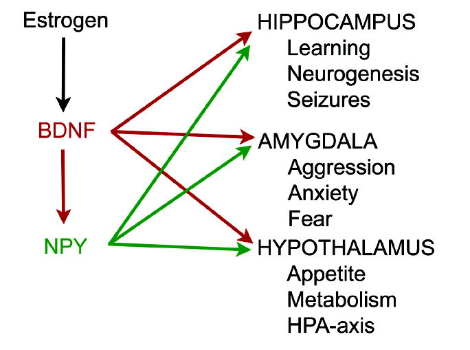
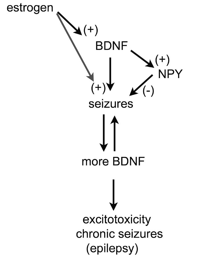
In the CNS, there are widespread and diverse interactions between growth factors and estrogen. Here we examine the interactions of estrogen and brain-derived neurotrophic factor (BDNF), two molecules that have historically been studied separately, despite the fact that they seem to share common targets, effects, and mechanisms of action. The demonstration of an estrogen-sensitive response element on the BDNF gene provided an impetus to explore a direct relationship between estrogen and BDNF, and predicted that the effects of estrogen, at least in part, might be due to the induction of BDNF. This hypothesis is discussed with respect to the hippocampus, where substantial evidence has accumulated in favor of it, but alternate hypotheses are also raised. It is suggested that some of the interactions between estrogen and BDNF, as well as the controversies and implications associated with their respective actions, may be best appreciated in light of the ability of BDNF to induce neuropeptide Y (NPY) synthesis in hippocampal neurons. Taken together, this tri-molecular cascade, estrogen-BDNF-NPY, may be important in understanding the hormonal regulation of hippocampal function. It may also be relevant to other regions of the CNS where estrogen is known to exert profound effects, such as amygdala and hypothalamus; and may provide greater insight into neurological disorders and psychiatric illness, including Alzheimer's disease, depression and epilepsy.


Figures

Figure 1
Figure 1
Estrogen-BDNF interactions: convergence vs. induction. A. A schematic illustrates the potential convergence of estrogen- and BDNF-induced signal transduction. Estrogen may act either on membrane or nuclear receptors, and possibly both. These receptors are not precisely defined, so their similarity in the diagram is for schematic purposes only. BDNF acts on either trkB or p75 receptors to activate similar signaling pathways as estrogen, and major targets include transcription factors that influence growth, survival, neural plasticity, and learning in hippocampus, as well as other effects. Estrogen and BDNF may not necessarily act on the same cell, as shown, but cells that are distinct, such as pyramidal cells and GABAergic interneurons, as well as glia. B. An alternative to the convergence shown in part A is that estrogen and BDNF interact directly, because estrogen induces BDNF gene expression, which in turn acts on trkB and p75 to exert its effects. Induction of this kind may occur by an estrogen-sensitive response element (ERE) on the BDNF gene or by estrogen-induced increase in neural activity that upregulates BDNF, because BDNF expression is regulated by activity (for further discussion, see text).
Figure 2
Figure 2
Neurotrophin receptors and targets of BDNF. A. The Neurotrophin family includes nerve growth factor (NGF), BDNF, and also neurotrophin -3 (NT-3) and neurotrophin-4 (NT-4). Receptor specificity exists for trkA, for which NGF is the specific ligand, and trkC (NT-3 is the ligand), whereas trkB has two potential ligands, BDNF and NT-4 act. All neurotrophins bind to p75. BDNF is considered the main ligand for trkB in hippocampus, but the effects of NT-4 may be underestimated because in forebrain BDNF knockout mice, trkB can still be phosphorylated [236]. B. A schematic illustration of target pathways and molecules of trkB for postsynaptic, presynaptic, and glial processes. From [237].
Figure 3
Figure 3
The expression of BDNF, its receptors, and estrogen receptors in the adult rat dentate gyrus. A. A schematic illustrates the fundamental circuitry of the dentate gyrus. From [238]. B. The expression of BDNF and its receptors is shown schematically for the granule cell of the dentate gyrus and neighboring glia. BDNF is thought to be cleaved from its precursor, proBDNF and then anterogradely transported [36] to dense core vesicles. It may also be located or trafficked to dendrites under some conditions [37]. ProBDNF may be released and act on p75 receptors [34]. For further discussion, see text. TrkB FL refers to the full-length receptor; there also is a truncated receptor (TrkB TK-) that is likely to be associated with glia and potentially scavenge BDNF. C. The expression of BDNF, its receptors, and the potential locations of estrogen receptors in a simplified circuit of granule cell and GABAergic interneurons are illustrated. The GABAergic neurons, which express estrogen receptors may innervate granule cells. This, and evidence from CA1 studies that estradiol can disinhibit pyramidal cells [49], has led to the suggestion that estrogen may disinhibit granule cells indirectly [27]. However, estrogen receptors may be on a subset of interneurons that do not control granule cell inhibition [26] and even if so, estrogen actions on those GABAergic neurons may not lead to granule cell disinhibition, because such studies have not been conducted specifically (see text for further discussion).
Figure 4
Figure 4
BDNF expression and physiological changes in hippocampal slices across the estrous cycle of the adult female rat. A. Immunocytochemistry using an antibody to BDNF illustrates increased immunoreactivity in the hilar mossy fibers (arrows) at proestrus and estrus relative to metestrus. B. Physiological recordings from the CA3 pyramidal cells, targets of the mossy fibers, at proestrus, estrus, metestrus, and after ovariectomy. The recordings represent responses to hilar stimuli (at the dots) triggered in pairs (40 msec interstimulus interval). After the 5th pair, multiple responses (population spikes) occurred in the animals examined on the morning of proestrus (arrows) or estrus, when BDNF in the mossy fibers was elevated. However, even 10 pairs of stimuli failed to evoke multiple population spikes in animals examined on the morning of metestrus or after ovariectomy, when BDNF levels were relatively low. From [86].
Figure 5
Figure 5
Neuropeptide Y (NPY) changes during the estrous cycle of the adult female rat. A. A representative section from an adult male rat stained with an antibody to NPY shows numerous immunoreactive neurons in the hilar region, and additional staining in the outer molecular layer, reflecting the major axonal projection of these cells. B. A schematic illustration of the normal location and axonal projections of GABAergic neurons of the dentate gyrus that co-express NPY. There is strong evidence for their molecular layer projection and hilar collaterals [29], but less evidence that they directly innervate the dentate gyrus progenitors (PRE) or mossy fibers. However, NPY exerts robust actions on progenitors and mossy fiber transmission (see text for further discussion). C-D. At higher magnification, a comparison is shown of NPY immunoreactivity in the dentate gyrus of a normal adult female rat that was euthanized on the morning of proestrus (C) relative to a section processed concurrently from another animal that was euthanized on the morning of metestrus (D). The arrows indicate increased immunoreactivity in the axon plexus of the hilar NPY-immunoreactive GABAergic interneurons at metestrus. E. A schematic illustrates a hypothesis for the changes in excitability of the CA3 pyramidal cells in response to hilar stimulation during the estrous cycle. Following the proestrus surge of estradiol, which appears to be followed by an increase in mossy fiber BDNF, which lasts for the duration of proestrus and at least the morning of estrus (Figure 4; [86]). There is a subsequent decline in BDNF and elevation in NPY in GABAergic neurons of the dentate hilus. It is suggested that the elevation in NPY in this interneuronal population limits the ability of hilar stimulation to produce repetitive activity in CA3 pyramidal cells on the morning of metestrus (Figure 4). This may be due not just to the hilar NPY neurons, but possibly also the NPY neurons of area CA3. Together, they potentially release NPY, which decreases glutamate release from mossy fibers [239, 240].
Figure 6
Figure 6
A schematic illustration of the potential for estrogen to exert actions via BDNF and NPY. Although estrogen potentially has direct effects on target structures like the hippocampus, as well as others such as the amygdala and hypothalamus, some of the effects previously attributed to direct actions could be due to the ability of estrogen to induce BDNF synthesis. Further modulation of the effects of BDNF could be due to subsequent induction of NPY expression by BDNF.
Figure 7
Figure 7
The effects of BDNF on seizure threshold and epilepsy. Given that BDNF can facilitate many different excitatory pathways that can contribute to seizures, and that other evidence suggests BDNF decreases seizure threshold and in fact can induce seizures, the diagram shown starts with the hypothesis that BDNF could be proconvulsant. If so, a positive feedback may develop, because seizures upregulate BDNF expression. If sufficient, such a positive feedback loop may lead to repeated seizures and seizure-induced excitotoxicity, the hallmark of temporal lobe epilepsy. In addition, estrogen may facilitate this, because estrogen induces BDNF gene expression, and this may have relevance to hormone-sensitive seizures in women with epilepsy (discussed further in the text). However, because BDNF can induce NPY synthesis, a natural brake on this process is present normally, and may serve to prevent epileptogenesis, or simply be responsible for the long interictal periods in many individuals with epilepsy. This hypothesis illustrates the interesting implications of steroid hormone-growth factor interactions, but also emphasizes their complexity.

Similar articles

Cited by

References

    1. Stancel GM, Gardner RM, Kirkland JL, Lin TH, Lingham RB, Loose-Mitchell DS, Mukku VR, Orengo CA, Verner G. Interactions between estrogen and EGF in uterine growth and function. Adv Exp Med Biol. 1987;230:99–118. - PubMed
    1. Ignar-Trowbridge DM, Pimental M, Teng CT, Korach KS, McLachlan JA. Cross talk between peptide growth factor and estrogen receptor signaling systems. Environ Health Perspect. 1995;103:35–38. - PMC - PubMed
    1. Mendez P, Cardona-Gomez GP, Garcia-Segura LM. Interactions of insulin-like growth factor-I and estrogen in the brain. Adv Exp Med Biol. 2005;567:285–303. - PubMed
    1. Toran-Allerand CD, Singh M, Setalo G. Novel mechanisms of estrogen action in the brain: new players in an old story. Front Neuroendocrinol. 1999;20:97–121. - PubMed
    1. Sandstrom NJ, Williams CL. Spatial memory retention is enhanced by acute and continuous estradiol replacement. Horm Behav. 2004;45:128–135. - PubMed

Publication types

MeSH terms

LinkOut - more resources