. 2006 Oct 31;103(44):16307-12.
doi: 10.1073/pnas.0605154103. Epub 2006 Oct 18.

Sog/Chordin is required for ventral-to-dorsal Dpp/BMP transport and head formation in a short germ insect

Affiliations

Sog/Chordin is required for ventral-to-dorsal Dpp/BMP transport and head formation in a short germ insect

Maurijn van der Zee et al. Proc Natl Acad Sci U S A. .

Abstract

Bone morphogenetic protein (BMP) signaling plays a major role in dorsoventral patterning in vertebrates and in Drosophila. Remarkably, in Tribolium, a beetle with an ancestral type of insect development, early BMP/dpp exhibits differential expression along the anteroposterior axis. However, the BMP/Dpp inhibitor Sog/chordin is expressed ventrally and establishes a dorsal domain of BMP/Dpp activity by transporting BMPs toward the dorsal side, like in Drosophila. Loss of Tribolium Sog not only abolishes dorsoventral polarity in the ectoderm, but also leads to the complete absence of the CNS. This phenotype suggests that sog is the main BMP antagonist in Tribolium, in contrast to vertebrates and Drosophila, which possess redundant antagonists. Surprisingly, Sog also is required for head formation in Tribolium, as are the BMP antagonists in vertebrates. Thus, in Tribolium, the system of BMP and its antagonists is less complex than in Drosophila or vertebrates and combines features from both, suggesting that it might represent an ancestral state.


Conflict of interest statement

The authors declare no conflict of interest.

Figures

Fig. 1.
Fig. 1.
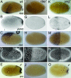
Expression of Tc-dpp compared with expression of Tc-sog. (B–D% and F–L%) In situ hybridizations with Tc-dpp (B, F, I, and K) and Tc-sog (C, D, G, H, J, and L). (A–D%) Uniform blastoderm stages. (E–H%) Differentiated blastoderm stages. (I and J) Extending germ-band embryos. (K and L) Cross-sections from the growth zone, taken at the position of the lines in I and J, respectively. Schematic drawings are shown at Right. Dashed lines indicate the future border between amnion and embryo proper (A) DAPI counterstaining of the embryo shown in B. The nuclei have a uniform distribution. (B) Lateral view. Tc-dpp is ubiquitously expressed, with stronger expression at the anterior pole. (C) Ventral view. Tc-sog is expressed in a broad ventral domain. (D) Lateral view of the embryo shown in C. (E) DAPI counterstaining of the embryo shown in F. The serosa can be recognized by big, widely spaced nuclei; the germ rudiment by smaller, dense nuclei. (F) Lateral view. Tc-dpp is expressed in a stripe along the germ rudiment/serosa border. (G) Ventral view. The Tc-sog expression domain becomes narrower. A gap is observed at the germ rudiment/serosa border (white arrowhead). (H) Lateral view of the embryo shown in G. (I) Tc-dpp is expressed along the dorsal borders of the embryo (arrows), except for the growth zone. (J) Tc-sog is expressed in a ventral, ectodermal domain. Except for the growth zone, Tc-sog is not expressed in the mesoderm. (K) Tc-dpp is strongly expressed in two stripes in the outer layer (OL) directly flanking the IL. Weak expression is found in the amnion. (L) Tc-sog is expressed in IL cells between the OL.
Fig. 2.
Fig. 2.
Tc-Sog transports Dpp toward the dorsal side. Lateral views of embryos at the differentiated blastoderm stage. (A–E%) Wild type (wt). (F–J%) Tc-dpp RNAi (dpp). (K–O%) Tc-sog RNAi (sog). (A, F, and K) Tc-dpp in situ hybridization. (B, G, and L) pMAD antibody staining. (C, H, and M) Tc-doc in situ hybridization. (D, I, and N) DAPI counterstaining of the embryos shown in C, H, and M, respectively. Serosal nuclei are bigger and wider spaced than those of the germ rudiment. (E, J, and O) Tc-pnr in situ hybridization. (A) Tc-dpp is expressed in a stripe along the border of the germ rudiment and serosa and in the primitive pit (p). (B) pMAD accumulates along the whole dorsal side of the embryo. See Results for details. Black lines indicate the germ rudiment/serosa border. (C) Tc-doc is transcribed in a subset of dorsal cells in the serosa. (D) The border of the germ rudiment and serosa is oblique (white lines indicate the dorsal and ventral point of the border). (E) Tc-pnr is expressed at the dorsal side of the germ rudiment (arrow) and in the primitive pit. (F) No Tc-dpp expression is detected. (G) pMAD could not be detected. (H) Tc-doc transcripts could not be detected. (I) The serosa/germ rudiment border is straight. (J) Tc-pnr transcripts could not be detected. (K) Tc-dpp is weakly expressed along the germ rudiment/serosa border. (L) pMAD is present in a band along the border of the germ rudiment and the serosa. Additional pMAD is found in the primitive pit. (M) Tc-doc is expressed in a DV symmetrical broad band in the serosa. (N) The germ rudiment/serosa border is straight. (O) Tc-pnr is expressed in a rim along the anterior of the germ rudiment and in the primitive pit but not at the dorsal side of the germ rudiment (arrow).
Fig. 3.
Fig. 3.
Tc-sog RNAi deletes the head; Tc-dpp RNAi enlarges the head. (A–F%) Differentiated blastoderm stages. (A–C%) Tc-Otd antibody stainings, lateral views. (D–F%) Tc-twist in situ hybridizations, differentiated blastoderm stages, ventral views. (G–I%) Tc-Otd antibody stainings; extended germ-band embryos. (A) WT. Tc-Otd is present in an anterior triangle. (B) Tc-dpp RNAi. Tc-Otd is present in a band in the anterior germ rudiment. (C) Tc-sog RNAi. Tc-Otd could not be detected. (D) WT. (E) Tc-dpp RNAi. The Tc-twist domain extends to a WT position along the AP axis but is slightly broader in the anterior half. (F) Tc-sog RNAi. The Tc-twi domain is only half as long as in the WT. (G) WT. Tc-Otd is found in the head lobes (open arrowhead) and along the ventral midline (filled arrowhead). (H) Tc-dpp RNAi, lateral view. Tc-Otd is detected along the ventral midline (filled arrowhead) and in an enlarged anterior domain (open arrowhead). (I) Tc-sog RNAi. Tc-Otd is detected only in some patches along the ventral midline (arrowhead).
Fig. 4.
Fig. 4.
Tc-sog RNAi leads to a complete loss of the neurogenic ectoderm; extending germ-band embryos. (A–C%) WT. (D and E) Tc-dpp RNAi. (F–H%) Tc-sog RNAi. (A, D, and F) Tc-achaete-scute in situ hybridizations. (B–H%) Tc-snail in situ hybridizations (blue) with engrailed antibody staining (brown). (A) Tc-ASH is expressed in cells of the CNS and in a transverse stripe at the anterior of each segment. (B) Tc-snail is expressed in cells of the CNS. Seventeen engrailed stripes could be counted. Segments are labeled as follows: I, intercallary; Md, mandibular; Mx, maxillary; Lb, labial; T, thoracic; A, abdominal. (C) Magnification of a part of the embryo boxed in B. Tc-snail is also detected in single clusters marking the peripheral neurons of the lateral ectoderm (arrows). (D) Tc-ASH transcripts can be detected throughout the embryo. (E) Tc-snail can be detected throughout the embryo. (F) Tc-ASH can be detected only in segmental stripes. (G) Tc-snail expression is found only in single clusters. Only 13 engrailed stripes were counted. (H) Magnification of segment A5 from the embryo shown in E. The arrow points at the peripheral neurons.
Fig. 5.
Fig. 5.
Tc-sog RNAi embryos show a double dorsal phenotype. (A–E%) WT. (F–J%) Tc-sog RNAi. (A and F) Tc-dpp in situ hybridizations. (B, C, G, and H) pMAD antibody staining. (D and I) Cross-sections with Tc-pnr in situ hybridization (blue) and Twist antibody staining marking the mesoderm (dark brown). (E and J) Tc-pnr in situ hybridizations. (A) Tc-dpp is expressed along the dorsal borders of the extending germ band and in two stripes in the growth zone. (B) pMAD is detected along the dorsal margins of the extended germ band. (C) Magnification of the area boxed in B. (D and E) Tc-pnr is expressed at the dorsal margins. (F) Tc-dpp is weakly expressed along the dorsal margins and in two strong ectopic stripes along the ventral midline. The stripes are continuous, with the stripes in the growth zone. (G) pMAD is detected along the dorsal margins of the germ band and in a strong ectopic domain along the ventral midline. (H) Magnification of the area boxed in D. (I and J) Tc-pnr is expressed along the dorsal margin and in a strong, ventral, ectopic stripe in the ectoderm.
Fig. 6.
Fig. 6.
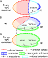
Schematic drawings of the Tc-sog and Tc-dpp RNAi phenotypes. (A) Tc-sog RNAi. The neurogenic ectoderm (green) is absent. Dorsal cell fates occupy domains along the AP axis: Dorsal serosal cells (red) are present in a broad band anterior to the germ rudiment, and amniotic cells (dark blue) are present along the anterior margin of the germ rudiment and in the primitive pit. Dorsal amnion is absent. The serosa/germ rudiment border is straight and is located at the position of the dorsal border in WT. The presumptive mesoderm (dotted line with arrow) is correspondingly shorter along the AP axis. (B) WT. Dorsal serosal cells and amniotic cells are localized to the dorsal side. Neurogenic ectoderm and mesoderm are present. The germ rudiment/serosa border is oblique and runs from a dorsal, posterior position to a ventral, anterior position. (C) Tc-dpp RNAi. Dorsal serosa and amnion are absent. Mesoderm and an anterior serosa (orange) are present. The germ rudiment/serosa border is straight and located at the position of the WT ventral border. The rest of the embryo consists of neurogenic ectoderm. pp, primitive pit.

Similar articles

Cited by

References

    1. De Robertis EM, Kuroda H. Annu Rev Cell Dev Biol. 2004;20:285–308. - PMC - PubMed
    1. Hammerschmidt M, Mullins MC. Results Probl Cell Differ. 2002;40:72–95. - PubMed
    1. Ferguson EL, Anderson KV. Cell. 1992;71:451–461. - PubMed
    1. Wharton KA, Ray RP, Gelbart WM. Development (Cambridge, UK) 1993;117:807–822. - PubMed
    1. Biehs B, Francois V, Bier E. Genes Dev. 1996;10:2922–2934. - PubMed

Publication types

MeSH terms

Associated data

LinkOut - more resources