. 2007 Jan;81(1):42-58.
doi: 10.1128/JVI.00648-06. Epub 2006 Oct 18.

Modulation of host gene expression by the K15 protein of Kaposi's sarcoma-associated herpesvirus

Affiliations

Modulation of host gene expression by the K15 protein of Kaposi's sarcoma-associated herpesvirus

Melanie M Brinkmann et al. J Virol. 2007 Jan.

Abstract

Kaposi's sarcoma-associated herpesvirus (KSHV) contains several open reading frames (ORFs) encoding proteins capable of initiating signal transduction pathways. Among them is the K15 ORF, which consists of eight exons encoding a protein with 12 predicted transmembrane domains and a cytoplasmic C terminus. When transiently expressed, the 8-exon K15 transcript gives rise to a protein with an apparent molecular mass of 45 kDa. K15 interacts with cellular proteins, TRAF (tumor necrosis factor receptor-associated factor) and Src kinases, and activates AP-1, NF-kappaB, and the mitogen-activated protein kinases (MAPKs) c-jun-N-terminal kinase and extracellular signal-regulated kinase. This signaling activity of K15 is related to phosphorylation of Y(481) of the K15 SH2-B motif Y(481)EEV. In this study we demonstrate the expression of an endogenous 45-kDa K15 protein in KSHV BAC36-infected epithelial cells. This endogenous K15 protein shows the same intracellular localization as transiently expressed K15, and expression kinetic studies suggest it to be a lytic gene. We have further determined the downstream target genes of K15 signaling using DNA oligonucleotide microarrays. We demonstrate that K15 is capable of inducing expression of multiple cytokines and chemokines, including interleukin-8 (IL-8), IL-6, CCL20, CCL2, CXCL3, and IL-1alpha/beta, as well as expression of Dscr1 and Cox-2. In epithelial cells, K15-induced upregulation of most genes was dependent on phosphorylation of Y(481), whereas in endothelial cells mutation of Y(481) did not result in a complete loss of Dscr1 and Cox-2 expression and NFAT-activity. Our study establishes K15 as one of the KSHV lytic genes that are inducing expression of multiple cytokines, which have been shown to play an important role in KSHV-associated pathogenesis.


Figures

FIG. 1.
FIG. 1.
A K15 protein of 45 kDa is expressed in 293 cells stably transfected with KSHV BAC36. (A) In 293 cells stably transfected with KSHV BAC36, a 45-kDa protein was detected with the K15 polyclonal antibody (right blot, indicated by arrows), which was not present in 293 cells (left blot). The K15 polyclonal antibody detects an unspecific band of approximately 48 kDa in lysates of 293 and 293 KSHV BAC36. For induction of the lytic viral life cycle, cells were either treated with butyrate or infected with baculovirus coding for the KSHV RTA/ORF50 (regulator of transcriptional activation) protein, or both. At 48 h postinduction, cells were lysed and analyzed for K15 protein expression by Western blotting with the K15 polyclonal antibody. 293 cells were treated in a similar manner. (B) 293-T cells were transiently transfected with empty expression vector pFJ-EA (mock) or a full-length K15 cDNA construct encompassing exons 1 to 8 (aa 1 to 489). At 6 h prior to DNA transfection, cells were transfected with a K15 siRNA directed against exon 8, which codes for the C-terminal cytoplasmic domain of K15 (+, K15 siRNA transfected; −, not K15 siRNA transfected). At 36 h posttransfection, cells were lysed and analyzed by Western blotting with the polyclonal K15 antibody. (C) The 293 KSHV BAC36 cell line was transfected with K15 siRNA as described in Material and Methods. Twenty hours after siRNA transfection, either cells were left untreated (uninduced) or the lytic viral life cycle was induced by addition of butyrate and baculovirus RTA/ORF50. Cells were lysed 24 h postinduction, and lysates were analyzed by Western blotting with the polyclonal K15 antibody. WB, Western blotting.
FIG. 2.
FIG. 2.
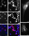
Subcellular localization of K15 in 293 KSHV BAC36 cells and in transiently transfected PtK2 and SLK cells. (A) PtK2 cells were transiently transfected with the eight-exon K15 expression construct. At 48 h posttransfection, cells were fixed and labeled with the K15 polyclonal antibody and anti-rabbit Cy3 as secondary antibody (A1) as described in Material and Methods. Nuclei were stained with Hoechst 33258 (A2). A3 shows an overlay of images A1 and A2. (B) 293 cells stably transfected with KSHV BAC36 were treated with 1 mM butyrate for 23 h, fixed, and labeled with the K15 polyclonal antibody (B1) as described in Material and Methods. The arrows indicate cells expressing K15 protein. Nuclei were stained with Hoechst 33258 (B2). B3 shows an overlay of images B1 and B2. (C) Image showing a single 293 KSHV BAC36 cell expressing endogenous K15. The lytic cycle was induced in 293 KSHV BAC36 cells by adding 1 mM butyrate and baculovirus coding for KSHV RTA/ORF50 to the cell culture medium. At 38 h postinduction, cells were fixed and labeled with the K15 polyclonal antibody as described in Material and Methods. (D) SLK endothelial cells were transiently transfected with the eight-exon K15 cDNA expression construct, fixed, labeled with the K15 polyclonal antibody, and analyzed by fluorescence microscopy.
FIG. 3.
FIG. 3.
Induction of cytokine and chemokine secretion by K15. (A) HeLa cells were transiently transfected with K15 expression constructs (K15 WT and K15 F481) or the empty vector pFJ-EA (mock). At 32 h posttransfection, the conditioned medium was collected and analyzed with a cytokine antibody array encompassing 42 different human cytokines spotted in duplicate onto a nitrocellulose membrane. The cytokine antibody array contains two different controls (c), one spotted in quadruplicate (upper left corner) and one in duplicate (lower right corner) and was performed as described in Materials and Methods. Arrays are labeled at left. The spots that were upregulated by K15 are marked as indicated on the figure. (B) Quantification of the cytokine antibody array shown in panel A. The intensity of the spots was measured by densitometry and normalized to the control spots (c). The signal reached with medium of cells that were mock-transfected was set at 1. Standard deviation is shown. (C) The ELISA was carried out according to the manufacturer's instructions with supernatant from transiently transfected HeLa cells as described in panel A. Shown is the mean value of two independent experiments. (D) After the conditioned medium was collected from transiently transfected HeLa cells for the cytokine antibody array (A) and ELISA (C), cells were lysed and analyzed by Western blotting for K15 protein expression with the K15 polyclonal antibody. Actin served as a loading control. (E) Real-time PCR analysis for IL-8 and VEGF was performed with 7 ng of cDNA from HeLa cells transiently transfected with K15 expression constructs (K15 WT and K15 F481) or pFJ-EA (mock). The graph shows the relative changes of mRNA expression after normalization against β-actin (ΔΔCT method; see Material and Methods).WB, Western blotting.
FIG. 4.
FIG. 4.
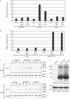
K15 activates the cox-2 and dscr1 promoter and the NFAT transcription factor in luciferase-based reporter assays in 293-T cells. 293-T cells were transiently transfected with 1 μg of K15 WT or K15 F481 and 100 ng of reporter plasmid per well of a six-well plate. The Dscr1 and Cox-2 reporter plasmids contain the promoter regions of the dscr1 and cox-2 genes, respectively, cloned in the promoterless pGL3b vector (A). The pTA-Luc reporter plasmid contains the minimal TA promoter upstream of the luciferase reporter gene. The pNFAT-TA-Luc reporter plasmid additionally contains three tandem copies of the NFAT transcription factor consensus sequence (B). At 20 h posttransfection, cells were serum starved by reducing the serum concentration from 10% to 1% (for dscr1 and cox-2) or 0% (for NFAT). At 29 h posttransfection, cells were lysed, and luciferase activities were determined. As a positive control for NFAT activation, mock-transfected 293-T cells were incubated with TPA and calcium ionophore (IO; both dissolved in DMSO) 4 h before cells were lysed, and treatment of mock-transfected cells with DMSO served as negative control (B). The luciferase activities are depicted as relative activity compared to mock transfections (luciferase activity of mock and pGL3b, Dscr1, and Cox-2 transfections in panel A or mock- and pTA- and pNFAT-TA-cotransfected cells treated with DMSO in panel B was set at 1). The data are representative of three independent experiments, each performed in duplicate. Standard deviations are shown. For both panels, lysates were analyzed by SDS-polyacrylamide gel electrophoresis and Western blotting (WB) with the K15 polyclonal antibody. Membranes were stripped and probed with an α-actin antibody as loading control.
FIG. 5.
FIG. 5.
K15 activates the cox-2 and dscr1 promoter and the NFAT transcription factor in SLK endothelial cells. SLK cells were transiently cotransfected with 3 μg of K15 WT, K15 F481, or pFJ-EA (empty vector, mock) and 1 μg of reporter plasmids pGL3b (negative control), Dscr1, or Cox-2 (A) or 1 μg of reporter plasmids pTA (negative control) or pNFAT-TA (B). At 21 h posttransfection, cells were serum starved by reducing the serum concentration from 10% to 0%. At 29 h posttransfection, cells were lysed, and luciferase activities were determined. The luciferase activities are depicted as relative activity compared to mock transfections. The data are from one experiment representative of three independent experiments, each performed in duplicate. Standard deviations are shown. Equal expression levels of K15 proteins were analyzed by Western blotting (data not shown). (C) Dscr1 and GAPDH semiquantitative RT-PCR. cDNA of SLK or HeLa cells transiently transfected with empty vector (mock) or K15 expression constructs K15 WT or K15 F481 was generated as described in Material and Methods. A semiquantitative PCR with 22, 23, or 24 PCR cycles was performed with dscr1-for and dscr1-rev primers amplifying a spliced transcript of 196 bp. The GAPDH PCR shows that equal amounts of cDNA were utilized for the PCR. (D) To control equal protein expression of K15 WT and K15 F481 in transiently transfected SLK cells, cells were lysed for Western blotting (WB) analysis in parallel to the cells being lysed for RNA preparation and subsequent RT-PCR analysis. K15 proteins were detected with the K15 polyclonal antibody. Blots were stripped and probed with an α-actin antibody as loading control. The transfection efficiency of SLK cells with K15 expression constructs was about 40% as judged by immunofluorescence (data not shown).

Similar articles

Cited by

References

    1. Abe, M., and Y. Sato. 2001. cDNA microarray analysis of the gene expression profile of VEGF-activated human umbilical vein endothelial cells. Angiogenesis 4:289-298. - PubMed
    1. Aoki, Y., E. S. Jaffe, Y. Chang, K. Jones, J. Teruya-Feldstein, P. S. Moore, and G. Tosato. 1999. Angiogenesis and hematopoiesis induced by Kaposi's sarcoma-associated herpesvirus-encoded interleukin-6. Blood 93:4034-4043. - PubMed
    1. Aoki, Y., and G. Tosato. 1999. Role of vascular endothelial growth factor/vascular permeability factor in the pathogenesis of Kaposi's sarcoma-associated herpesvirus-infected primary effusion lymphomas. Blood 94:4247-4254. - PubMed
    1. Armesilla, A. L., E. Lorenzo, d. A. Gomez, S. Martinez-Martinez, A. Alfranca, and J. M. Redondo. 1999. Vascular endothelial growth factor activates nuclear factor of activated T cells in human endothelial cells: a role for tissue factor gene expression. Mol. Cell. Biol. 19:2032-2043. - PMC - PubMed
    1. Asou, H., J. W. Said, R. Yang, R. Munker, D. J. Park, N. Kamada, and H. P. Koeffler. 1998. Mechanisms of growth control of Kaposi's sarcoma-associated herpes virus-associated primary effusion lymphoma cells. Blood 91:2475-2481. - PubMed

Publication types

MeSH terms