. 2006 Dec;142(4):1642-55.
doi: 10.1104/pp.106.088476. Epub 2006 Oct 13.

Geminivirus infection up-regulates the expression of two Arabidopsis protein kinases related to yeast SNF1- and mammalian AMPK-activating kinases

Affiliations

Geminivirus infection up-regulates the expression of two Arabidopsis protein kinases related to yeast SNF1- and mammalian AMPK-activating kinases

Wei Shen et al. Plant Physiol. 2006 Dec.

Abstract

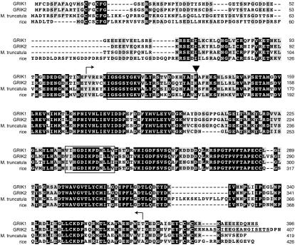
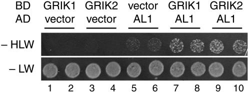
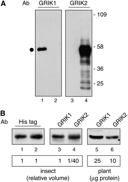
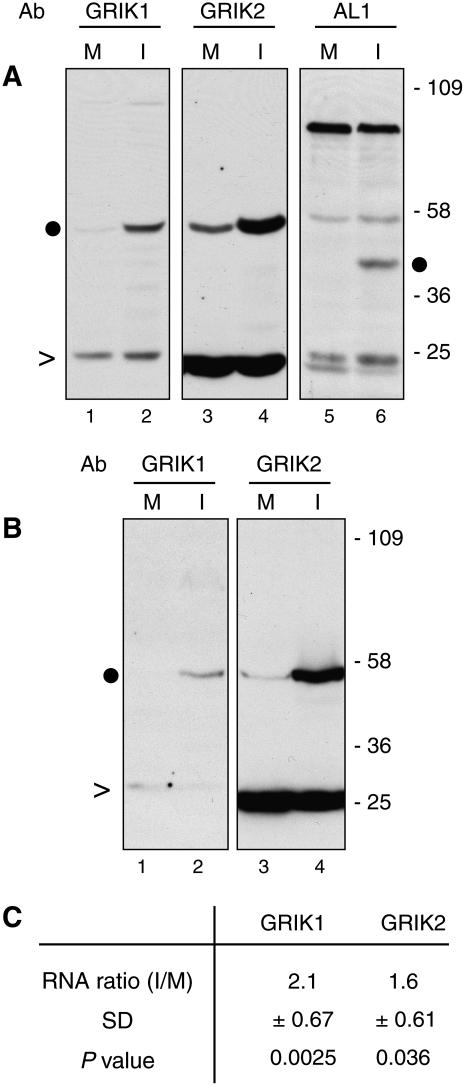
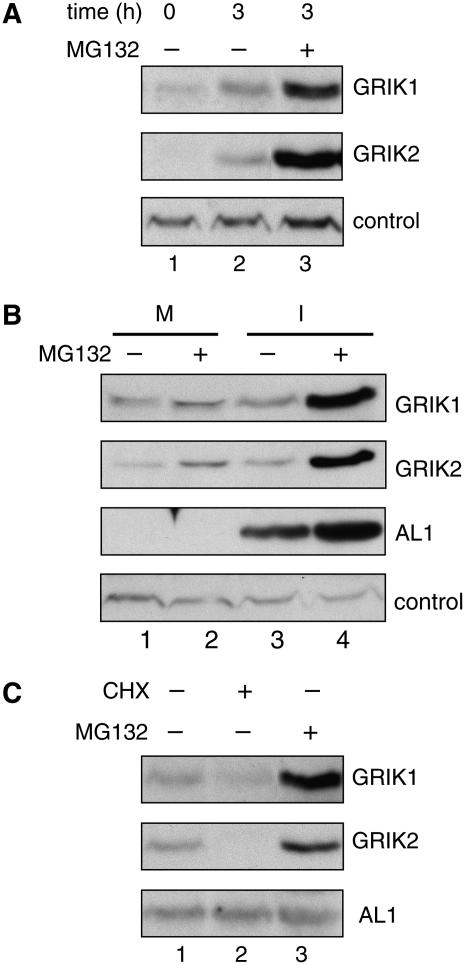
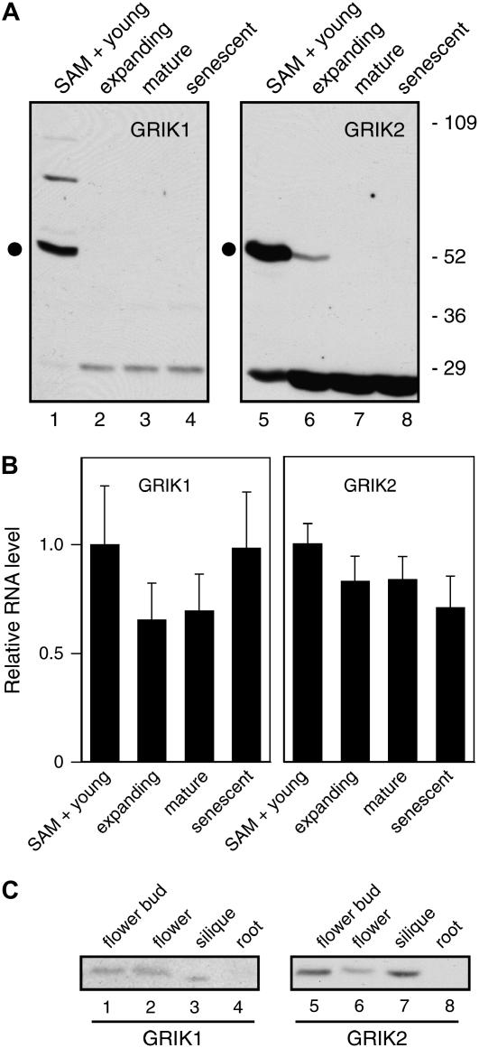
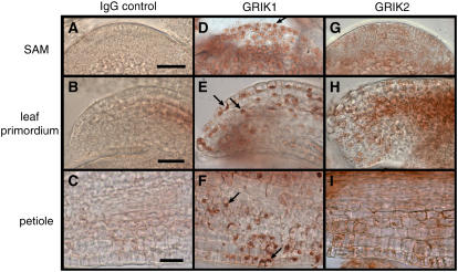
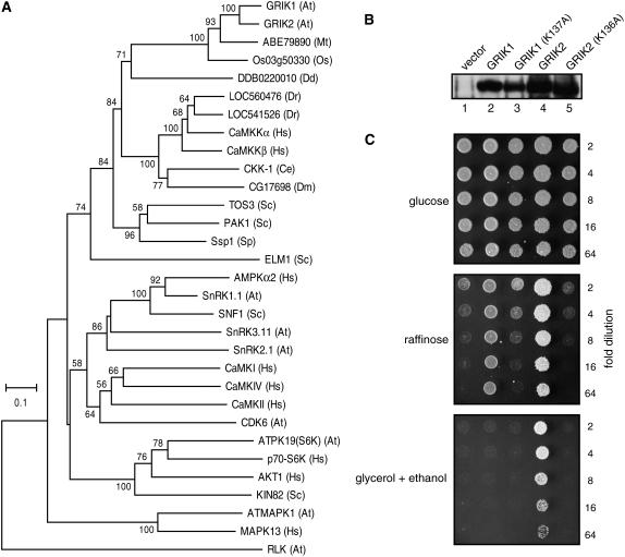
Geminivirus Rep-interacting kinase 1 (GRIK1) and GRIK2 constitute a small protein kinase family in Arabidopsis (Arabidopsis thaliana). An earlier study showed that a truncated version of GRIK1 binds to the geminivirus replication protein AL1. We show here both full-length GRIK1 and GRIK2 interact with AL1 in yeast two-hybrid studies. Using specific antibodies, we showed that both Arabidopsis kinases are elevated in infected leaves. Immunoblot analysis of healthy plants revealed that GRIK1 and GRIK2 are highest in young leaf and floral tissues and low or undetectable in mature tissues. Immunohistochemical staining showed that the kinases accumulate in the shoot apical meristem, leaf primordium, and emerging petiole. Unlike the protein patterns, GRIK1 and GRIK2 transcript levels only show a small increase during infection and do not change significantly during development. Treating healthy seedlings and infected leaves with the proteasome inhibitor MG132 resulted in higher GRIK1 and GRIK2 protein levels, whereas treatment with the translation inhibitor cycloheximide reduced both kinases, demonstrating that their accumulation is modulated by posttranscriptional processes. Phylogenetic comparisons indicated that GRIK1, GRIK2, and related kinases from Medicago truncatula and rice (Oryza sativa) are most similar to the yeast kinases PAK1, TOS3, and ELM1 and the mammalian kinase CaMKK, which activate the yeast kinase SNF1 and its mammalian homolog AMPK, respectively. Complementation studies using a PAK1/TOS3/ELM1 triple mutant showed that GRIK1 and GRIK2 can functionally replace the yeast kinases, suggesting that the Arabidopsis kinases mediate one or more processes during early plant development and geminivirus infection by activating SNF1-related kinases.


Figures

Figure 1.
Figure 1.
Alignment of Arabidopsis GRIK1, GRIK2, and their homologs from M. truncatula and rice. Identical residues are in black blocks and identical or similar residues from at least three proteins are highlighted in gray. The protein kinase catalytic domains are indicated by the regions bordered by the bent arrows. The upper boxed sequences indicate the conserved protein kinase ATP-binding region (PROSITE accession PS00107) and the lower boxed sequences are the Ser/Thr protein kinase catalytic loop (PROSITE accession PS00108). The triangle marks the conserved Lys residue that is required for ATP binding (Hanks and Hunter, 1995) and was altered to Ala in GRIK1(K137A) and GRIK2(K136A). Underlined sequences in GRIK1and GRIK2 correspond to the peptides used for antibody production.
Figure 2.
Figure 2.
Both GRIK proteins interact with a geminivirus replication protein. Full-length GRIK1 (spots 7 and 8) or GRIK2 (spots 9 and 10) fused with the GAL4 DNA binding domain (BD) was assayed for interaction with the TGMV AL1 protein fused with the GAL4 activation domain (AD) in the yeast two-hybrid system. The empty vector controls with GRIK1 (spots 1 and 2), GRIK2 (spots 3 and 4), and TGMV AL1 (spots 5 and 6) are shown. Protein interaction was scored as growth of the yeast transformants expressing the HIS3 reporter gene on the synthetic complete medium without His (−HLW). Equal inoculations are indicated by the growth on medium containing His (−LW). Two independent transformants of each combination are shown.
Figure 3.
Figure 3.
Specificity and relative sensitivity of the GRIK1 and GRIK2 peptide antibodies. A, Total protein extracts from insect cells expressing His-tagged GRIK1 (lanes 1 and 3) or His-tagged GRIK2 (lanes 2 and 4) were resolved by SDS-PAGE followed by immunoblotting with affinity-purified peptide antibodies against GRIK1 (lanes 1 and 2) or GRIK2 (lanes 3 and 4). The dot indicates the GRIK1 or GRIK2 band. Molecular mass markers (in kilodaltons) are indicated. B, Volumes of the insect cell protein extracts were adjusted to contain equal amounts of the His-tagged GRIK1 (lane 1) and His-tagged GRIK2 (lane 2) proteins as determined by immunoblotting with anti-His-tag antibodies. One volume of GRIK1 (lane 3) and 1/40 volume of GRIK2 (lane 4) proteins show similar intensities on immunoblots probed with their corresponding peptide antibodies at 1 μg/mL and 0.1 μg/mL, respectively. The same GRIK1 (lane 5) and GRIK2 (lane 6) antibody dilutions were used to probe a total protein extract (25 and 10 μg, respectively) from young leaves.
Figure 4.
Figure 4.
Geminivirus infection induces the accumulation of GRIK1 and GRIK2 in expanding leaves. A, Total proteins from mock-inoculated (M; lanes 1, 3, and 5) and CaLCuV-infected (I; lanes 2, 4, and 6) leaves (0.5–1.5 cm long) at 12 dpi were resolved by SDS-PAGE followed by immunoblotting with GRIK1 (lanes 1 and 2) and GRIK2 (lanes 3 and 4) peptide antibodies and a polyclonal antiserum against CaLCuV AL1 (lanes 5 and 6). Molecular mass markers (in kilodaltons) are indicated. This experiment was repeated twice with similar results. B, Proteins from mock-inoculated (M; lanes 1 and 3) and BCTV-infected (I; lanes 2 and 4) leaves (0.5–1.5 cm long) at 28 dpi were analyzed similarly using the GRIK1 (lanes 1 and 2) and GRIK2 (lanes 3 and 4) antibodies. In A and B, the dots indicate the GRIK1, GRIK2, or AL1 band, while the carats indicate nonspecific bands as internal controls. C, Real-time quantitative RT-PCR of GRIK1 and GRIK2 mRNAs in CaLCuV-infected leaves (0.5–1.5 cm long). The average ratios of mRNAs from CaLCuV-infected versus mock-inoculated leaves (I/M) were calculated from three independent experiments. The sd of the ratios and the P values from Z tests are given.
Figure 5.
Figure 5.
GRIK1 and GRIK2 are degraded by the proteasome. A, Two-week-old Arabidopsis seedlings were treated with MG132 (lane 3) or buffer (lane 2) for 3 h and total proteins were subjected to immunoblot analysis using GRIK1 or GRIK2 peptide antibodies. Seedlings collected immediately before treatment (time = 0) were tested in lane 1. A nonspecific band was used as an internal control. B, Mock-inoculated (M; lanes 1 and 2) or CaLCuV-infected (I; lanes 3 and 4) leaves were excised and treated with MG132 (lanes 2 and 4) or buffer only (lanes 1 and 3) for 3 h, and total proteins were analyzed by immunoblotting with antibodies against GRIK1, GRIK2, or CaLCuV AL1. A nonspecific band served as an internal control. C, CaLCuV-infected leaves were excised and treated for 3 h with cycloheximide (CHX; lane 2), MG132 (lane 3), or buffer (lane 1), and total proteins were analyzed by immunoblotting with antibodies against GRIK1, GRIK2, and CaLCuV AL1.
Figure 6.
Figure 6.
GRIK1 and GRIK2 protein and mRNA accumulate differentially during plant development. A, Leaves from 5-week-old plants were collected in fractions: young leaves (<0.5 cm) with SAM (lanes 1 and 5), expanding leaves (0.5–1.5 cm, lanes 2 and 6), fully expanded leaves (lanes 3 and 7), and senescent leaves (lanes 4 and 8). Total protein extracts were analyzed for GRIK1 (lanes 1–4) and GRIK2 (lanes 5–8) by immunoblotting with their respective peptide antibodies. The dot indicates the GRIK1 or GRIK2 band. Molecular mass markers (in kilodaltons) are indicated. B, Real-time quantitative RT-PCR of GRIK1 and GRIK2 mRNAs in the same leaf tissues. Relative mRNA levels are normalized to the levels in young leaves using Act2 mRNA as reference. The averages and sds of triplicate reactions are shown. This experiment was repeated once with similar results. C, Total proteins from flower buds (lanes 1 and 5), fully opened flowers (lanes 2 and 6), siliques (lanes 3 and 7), and roots (lanes 4 and 8) were analyzed by immunoblotting using antibodies to GRIK1 (lanes 1–4) or GRIK2 (lanes 5–8).
Figure 7.
Figure 7.
Immunolocalization of GRIK1 and GRIK2 in the SAM and young leaves. Sections from 5-week-old Arabidopsis SAM (A, D, and G), leaf primordium (B, E, and H), and emerging petiole (C, F, and I) were incubated with the GRIK1 (D, E, and F) or GRIK2 (G, H, and I) antibodies followed by visualization using a secondary antibody HRP-conjugate and the AEC substrate. Normal rabbit IgG (A–C) was used as a primary antibody control. Arrows indicate some of the stained nuclei. The bars indicate 100 nm.
Figure 8.
Figure 8.
GRIK1 and GRIK2 are related to activating kinases for the yeast SNF1 and mammalian AMPK. A, A phylogenetic tree containing GRIK1, GRIK2, their plant homologs, and related protein kinases from nonplant species. The protein kinase catalytic domains were aligned and used for building the tree with the neighbor-joining method. The numbers at branches indicate the percentage of bootstrap support (only those greater than 50% are shown). The two letter abbreviations in the brackets indicate the species sources of the kinases: At, Arabidopsis; Mt, M. truncatula; Os, rice; Dd, D. discoideum; Dr, zebrafish; Hs, human; Ce, C. elegans; Dm, D. melanogaster; Sc, S. cerevisiae; and Sp, S. pombe. An Arabidopsis RLK (At4g22730) was used as an outgroup control. B, Protein expression of GRIK1, GRIK2, and their mutant forms GRIK1(K137A) and GRIK2(K136A) in the S. cerevisiae pak1/tos3/elm1 mutant MCY5138. Total protein from yeast cells containing the expression plasmids or the empty vector were analyzed by immunoblotting with a mixture of GRIK1 and GRIK2 antibodies. C, Complementation of the S. cerevisiae pak1, tos3, and elm1 mutations by GRIK1, GRIK1(K137A), GRIK2, and GRIK2(K136A). Equal volumes of a 2-fold dilution series of the transformants containing the expression plasmids were spotted onto synthetic complete media containing either Glc, or raffinose, or glycerol and ethanol as carbon sources for further growth. Transformants carrying the empty expression vector was used as control.

Similar articles

Cited by

References

    1. Ach RA, Durfee T, Miller AB, Taranto P, Hanley-Bowdoin L, Zambryski PC, Gruissem W (1997) RRB1 and RRB2 encode maize retinoblastoma-related proteins that interact with a plant D-type cyclin and geminivirus replication protein. Mol Cell Biol 17: 5077–5086 - PMC - PubMed
    1. Bagewadi B, Chen S, Lal SK, Choudhury NR, Mukherjee SK (2004) PCNA interacts with Indian mung bean yellow mosaic virus rep and downregulates Rep activity. J Virol 78: 11890–11903 - PMC - PubMed
    1. Bisaro DM (2006) Silencing suppression by geminivirus proteins. Virology 344: 158–168 - PubMed
    1. Carvalho MF, Turgeon R, Lazarowitz SG (2006) The geminivirus nuclear shuttle protein NSP inhibits the activity of AtNSI, a vascular-expressed Arabidopsis acetyltransferase regulated with the sink-to-source transition. Plant Physiol 140: 1317–1330 - PMC - PubMed
    1. Castillo AG, Collinet D, Deret S, Kashoggi A, Bejarano ER (2003) Dual interaction of plant PCNA with geminivirus replication accessory protein (Ren) and viral replication protein (Rep). Virology 312: 381–394 - PubMed

Publication types

MeSH terms