. 2006 Dec;26(24):9387-401.
doi: 10.1128/MCB.01335-06. Epub 2006 Oct 9.

Smc5/6 is required for repair at collapsed replication forks

Affiliations

Smc5/6 is required for repair at collapsed replication forks

Eleni Ampatzidou et al. Mol Cell Biol. 2006 Dec.

Abstract

In eukaryotes, three pairs of structural-maintenance-of-chromosome (SMC) proteins are found in conserved multisubunit protein complexes required for chromosomal organization. Cohesin, the Smc1/3 complex, mediates sister chromatid cohesion while two condensin complexes containing Smc2/4 facilitate chromosome condensation. Smc5/6 scaffolds an essential complex required for homologous recombination repair. We have examined the response of smc6 mutants to the inhibition of DNA replication. We define homologous recombination-dependent and -independent functions for Smc6 during replication inhibition and provide evidence for a Rad60-independent function within S phase, in addition to a Rad60-dependent function following S phase. Both genetic and physical data show that when forks collapse (i.e., are not stabilized by the Cds1Chk2 checkpoint), Smc6 is required for the effective repair of resulting lesions but not for the recruitment of recombination proteins. We further demonstrate that when the Rad60-dependent, post-S-phase Smc6 function is compromised, the resulting recombination-dependent DNA intermediates that accumulate following release from replication arrest are not recognized by the G2/M checkpoint.


Figures
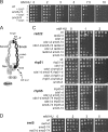
FIG. 1.
FIG. 1.
smc6 mutants are sensitive to HU. (A) Diagram of the Smc5/6 complex, showing the relative positions of the smc6-T2 (ts), smc6-X, and smc6-74 mutations. (B) smc6 mutants are sensitive to HU, and the sensitivity of smc6-74 but not smc6-X can be rescued by overexpression of brc1. Overexpression of brc1 slightly sensitizes smc6+ cells to HU. Row 1, smc6+ plus pREP41 (vector control); row 2, smc6+ plus pREP41brc1; row 3, smc6-74 plus pREP41; row 4, smc6-74 plus pREP41brc1; row 5, smc6-X plus pREP41; row 6, smc6-X plus pREP41brc1. (C) Epistasis analysis of smc6 mutants with cds1CHK2 and recombination null mutants in response to HU. Representative double mutants are shown. cds1CHK2 with recombination null double mutants are slightly more sensitive than cds1CHK2 null mutants (compare the samples in the third and sixth rows at 1 mM HU in each panel). smc6 cds1-d CHK2 double mutants are more sensitive than the parental strains. Middle panel, smc6-X cds1-d; top and bottom panels, smc6-74 cds1-d. rad22-dRAD52 smc6 double mutants are more sensitive than the parental strains. Top panel, rad22-d smc6-74. rhp51RAD51 or rhp55RAD55 rescues the sensitivities of cells carrying smc6 single and smc6 cds1CHK2 double mutations. Middle panel, rhp51-d smc6-X; bottom panel, rhp55-d smc6-74. (D) Epistasis analysis with swi5 shows that swi5 mutants are not sensitive at 5 mM HU and show no genetic interaction with smc6. wt, wild type; OP, overproduction.
FIG. 2.
FIG. 2.
smc6 mutants are defective in processing collapsed forks. rDNA replication intermediates were analyzed using 2-D gels. (A) The structure of the rDNA region. (B) The expected position of each intermediate. (C) Genomic DNA was prepared from smc6+ (wt), smc6-X (X), smc6-74 (74), the cds1CHK2 null mutant (cds1), and the indicated double mutants, digested with XhoI and KpnI, run in both dimensions, and probed with the XhoI and KpnI fragment (XK) spanning ars3001 and the RFB. Asynchronous cultures (−HU) or cultures treated with 10 mM HU for 2.5 h (+HU) were analyzed. The bubble arc is visible in the HU-treated cells. Asynchronous cultures have two pause sites on the large Y arc. Only one is seen in HU-arrested cells. In smc6 mutants, replication intermediates are similar to those in smc6+ with or without HU treatment. The X spike (X-shaped molecules, such as recombination intermediates) is slightly increased in smc6-X. No aberrant structures are seen in smc-74. In the HU-arrested cds1CHK2 null cells, no large Y or bubble arc structures are visible, consistent with fork collapse. In the cds1CHK2 smc6 double mutants, these replication intermediates are preserved and the X spike increases, consistent with an increase in recombination intermediates.
FIG. 3.
FIG. 3.
smc6-X and smc6-74 cells start to lose viability upon entry into mitosis. (A) Upon transient HU exposure, smc6 mutants start to lose viability after about 4 to 6 h. At 6 h, cells are overcoming the S-phase block and entering mitosis. smc6 cds1CHK2 double mutants lose viability with kinetics similar to that of cds1CHK2 null cells, dying in S phase. wt, wild type. (B) FACS analysis of smc6+, smc6-X, and smc6-74 cells in 10 mM HU. S. pombe is a haploid organism that spends ∼70% of its time in G2. In asynchronous cultures (time zero), there is a strong 2C peak. In an unperturbed cell cycle, G1 is very short and cytokinesis coincident with S phase, giving rise to the 2 to 2x2C shoulder. When cells are blocked in HU, cytokinesis proceeds as normal but S phase is slowed, leading to uncoupling of septation and replication and the appearance of a 1C peak. Thus, the FACS profile of cells blocked in HU shows a movement from a 2C peak to a 1C peak with time. By 2.5 h (the generation time in rich media at 30°C), all the cells are arrested with a 1C DNA content and stay arrested for approximately 3 h. However, cells gradually overcome the block such that by 6 h, most cells have completed replication (2C peak), and by 8 h, they have divided and are into the next S phase (2x2C shoulder). smc6 mutants progress similarly to wild-type cells but start to lose viability at about 6 h, coincident with entry into mitosis.
FIG. 4.
FIG. 4.
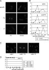
Recombination focus formation is normal in smc6 mutants. (A) Rad22Rad52 foci (live-cell imaging) in HU and upon release. rad22-GFP cells were arrested for 4 h (10 mM HU) at 30°C and released at 18°C. In smc6+, smc6-74, and smc6-X, the Rad22Rad52 signal was diffuse in the nucleus in HU, but cells with one or two nuclear foci became visible upon release. (B) Quantification of Rad22Rad52 foci and mitotic cells upon HU release. Foci formed with similar kinetics in smc6+, smc6-X, and smc6-74 cultures, peaking at ∼90 min after release, and disappeared prior to mitosis. (C and D) In contrast, smc6 cds1CHK2 double mutants, like cds1CHK2 null cells, showed multiple nuclear Rad22Rad52 foci during HU treatment (examples are shown in panel C) and these remained for the duration of the experiment after release (D).
FIG. 5.
FIG. 5.
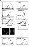
smc6 mutants have a checkpoint maintenance defect. Cells were arrested for 4 h (10 mM HU) at 30°C, and cell cycle progression was monitored (diamonds, mitotic index; open squares, septation index) following release at 30°C. (A) smc6+, smc6-X, and smc6-74 cultures all delayed mitosis for ∼90 min, indicating that the G2/M checkpoint is intact but smc6-X and smc6-74 cultures accumulate aberrant mitotic figures and “cut” cells (closed squares) (examples are shown in panel B). Under the same conditions, few “cut” cells were seen in rhp55-d, rhp55-d smc6-74, or rhp55-d smc6-X cultures. Thus, in smc6 mutants, recombination-dependent structures cause mitotic aberrations. These structures are not recognized by the G2/M checkpoint. (C) Overexpression of brc1 suppresses the accumulation of “cut” cells in smc6-74. Left, smc6-74 cells with an empty vector control (pREP41); right, smc6-74 cells with pREP41brc1. Symbols are as defined in panel A.
FIG. 6.
FIG. 6.
Smc6 is required in HU and upon release. smc6+, rad60-1, and smc6-T2 cells were incubated in HU (10 mM, 2.5 h) at the nonpermissive temperature for smc6-T2 and rad60-1 (36°C) and released into fresh media at the permissive temperature (22°C) (A), incubated in HU (10 mM, 4.5 h) at the permissive temperature (22°C) and released into fresh media at the nonpermissive temperature for smc6-T2 and rad60-1 (36°C) (B), or incubated in HU (10 mM, 4.5 h) at the permissive temperature and released at the same temperature (22°C) (C). Cell cycle progression following release (time zero) was monitored by septation index, and the percentages of aberrant mitotic or “cut” cells were scored. smc6+, rad60-1, and smc6-T2 entered mitosis with similar kinetics. rad60-1 accumulated aberrant mitotic cells only in regimen B (in which cells were released into the nonpermissive temperature) but not in regimen A (in which cells were HU blocked at the nonpermissive temperature and released into the permissive temperature), indicating that the Rad60 function is required only when the cells resume replication. In contrast, smc6-T2 accumulated aberrant cells in both regimen A and regimen B, suggesting that the Smc6 function is required both when replication is inhibited (Rad60 is not required) and when replication resumes upon release (a common requirement with Rad60). wt, wild type. (D) Viabilities observed after treatment with regimens A, B, and C or after incubation for 5 h at 36°C expressed as percentages of the smc6+ value.
FIG. 7.
FIG. 7.
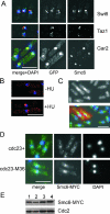
Smc6 localizes to the nucleolus and increases in intensity upon exposure to replication stress. (A). Indirect immunofluorescence with anti-Smc6 identifies foci that colocalize with Gar2-green fluorescent protein (GFP) (nucleolus) but not Swi6-GFP (heterochromatin) or Taz1-GFP (telomeres). Smc6 (red), GFP (green), and DNA visualized with DAPI (4′,6′-diamidino-2-phenylindole) (blue) are shown. Bar, 5 μm. (B). Smc6 foci are more intense in HU-blocked cells. Smc6 (red) and DNA visualized with DAPI (blue) are shown. (C) Asynchronous and HU-blocked cells under the same coverslip. HU-blocked cells, identified by Texas Red-conjugated lectin staining cell walls, have increased nuclear Smc6 (green). DNA visualized with DAPI (blue) is shown. (D) Increased nuclear Smc6 in the replication mutant cdc23-M36 (MCM10) at the nonpermissive temperature. Top panel, smc6-MYC cells at 35°C; bottom panel, smc6-MYC cdc23-M36 cells at 35°C. Smc6-MYC (green) and DNA visualized with DAPI (blue) are shown. (E) Total Smc6 protein levels do not change upon inhibition of replication. Column 1, smc6-MYC cells at 25°C; column 2, smc6-MYC cells at 35°C; column 3, smc6-MYC cdc23-M36 cells at 25°C; column 4, smc6-MYC cdc23-M36 cells at 35°C. Top, anti-MYC; bottom, anti-Cdc2 loading control.
FIG. 8.
FIG. 8.
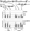
Smc6 is chromatin associated. Results for qPCR analysis of Smc6 chromatin IP are shown. Using either anti-Smc6 (smc6+ cells) or anti-MYC (smc6-MYC cells), Smc6 precipitates are enriched at all loci tested (see schematic). HU treatment modestly increased enrichment. Data shown are mean values for two or three independent experiments. Enrichment (n-fold) is calculated compared to beads only or untagged controls. Error bars indicate standard errors.
FIG. 9.
FIG. 9.
Model for the Smc5/6 complex function. Cds1Chk2 activity stabilizes stalled replication forks, and replication complexes remain associated. Forks collapse (replication complexes are disassociated), which can lead to the generation of a polar double-strand break. Restoration of the fork then occurs through break-induced replication (BIR) initiated by HR-dependent strand invasion of the intact template and subsequent HJ resolution (15). Rad22Rad52 and other recombination proteins (not shown) associate with the damaged chromatin at collapsed forks independently of Smc5/6, but the complex is required with Rad60 at a later stage for HR repair and/or restoration of replication (bottom right). Both smc6-74 and smc6-X are defective in this process (indicated by bars). Fork resetting without Rhp51Rad51-dependent HR occurs by multiple mechanisms, some of which require Smc5/6 (top right, shown in gray) or Brc1. In smc6-74 mutants, Brc1-dependent resetting of unstable forks can still occur inefficiently, whereas in smc6-X mutants, this process is nonfunctional (indicated by bar and ?X). Overexpression of brc1 could thus bypass the requirement for smc6-74 but not for smc6-X.

Similar articles

Cited by

References

    1. Akamatsu, Y., D. Dziadkowiec, M. Ikeguchi, H. Shinagawa, and H. Iwasaki. 2003. Two different Swi5-containing protein complexes are involved in mating-type switching and recombination repair in fission yeast. Proc. Natl. Acad. Sci. USA 100:15770-15775. - PMC - PubMed
    1. Andrews, E. A., J. Palecek, J. Sergeant, E. Taylor, A. R. Lehmann, and F. Z. Watts. 2005. Nse2, a component of the Smc5-6 complex, is a SUMO ligase required for the response to DNA damage. Mol. Cell. Biol. 25:185-196. - PMC - PubMed
    1. Bahler, J., J. Q. Wu, M. S. Longtine, N. G. Shah, A. McKenzie III, A. B. Steever, A. Wach, P. Philippsen, and J. R. Pringle. 1998. Heterologous modules for efficient and versatile PCR-based gene targeting in Schizosaccharomyces pombe. Yeast 14:943-951. - PubMed
    1. Betts Lindroos, H., L. Strom, T. Itoh, Y. Katou, K. Shirahige, and C. Sjogren. 2006. Chromosomal association of the Smc5/6 complex reveals that it functions in differently regulated pathways. Mol. Cell 22:755-767. - PubMed
    1. Boddy, M. N., P. Shanahan, W. H. McDonald, A. Lopez-Girona, E. Noguchi, I. J. Yates, and P. Russell. 2003. Replication checkpoint kinase Cds1 regulates recombinational repair protein Rad60. Mol. Cell. Biol. 23:5939-5946. - PMC - PubMed

Publication types

MeSH terms

LinkOut - more resources