. 2006 Oct;26(20):7529-38.
doi: 10.1128/MCB.00447-06. Epub 2006 Aug 5.

Phosphorylation of Chk1 by ATR is antagonized by a Chk1-regulated protein phosphatase 2A circuit

Affiliations

Phosphorylation of Chk1 by ATR is antagonized by a Chk1-regulated protein phosphatase 2A circuit

Van Leung-Pineda et al. Mol Cell Biol. 2006 Oct.

Abstract

In higher eukaryotic organisms, the checkpoint kinase 1 (Chk1) contributes essential functions to both cell cycle and checkpoint control. Chk1 executes these functions, in part, by targeting the Cdc25A protein phosphatase for ubiquitin-mediated proteolysis. In response to genotoxic stress, Chk1 is phosphorylated on serines 317 (S317) and 345 (S345) by the ataxia-telangiectasia-related (ATR) protein kinase. Phosphorylation of Chk1 on these C-terminal serine residues is used as an indicator of Chk1 activation in vivo. Here, we report that inhibition of Chk1 kinase activity paradoxically leads to the accumulation of S317- and S345-phosphorylated Chk1 in vivo and that ATR catalyzes Chk1 phosphorylation under these conditions. We demonstrate that Chk1 phosphorylation by ATR is antagonized by protein phosphatase 2A (PP2A). Importantly, dephosphorylation of Chk1 by PP2A is regulated, in part, by the kinase activity of Chk1. We propose that the ATR-Chk1-PP2A regulatory circuit functions to keep Chk1 in a low-activity state during an unperturbed cell division cycle but at the same time keeps Chk1 primed to respond rapidly in the event that cells encounter genotoxic stress.


Figures

FIG. 1.
FIG. 1.
Chk1 inhibitors induce Chk1 phosphorylation on S317 and S345 in vivo. (A) Asynchronously growing HeLa cells were treated with either vehicle (DMSO) or 1 μM Gö6976 for the indicated times. Western blotting was performed with the indicated antibodies. (B) HeLa cells, synchronized by a double-thymidine-block protocol, were released into S phase and treated with different concentrations of either UCN-01, SB-218078, or Gö6976 for 1.5 h. Total lysates were probed with antibodies specific for Cdc25A, phosphorylated Chk1 (pS345), total Chk1, and β-catenin. Controls included asynchronously growing cells (Asy), cells treated with the vehicle DMSO (V), and cells collected at 0 h before release (G1/S). The asterisk indicates phosphorylated Chk1. (C) HeLa cells synchronized by a double-thymidine-block protocol were released and harvested at the indicated times: 1.5 h (S phase), 3 h 45 min (G2 phase), and 10 h 50 min (G1 phase). In addition, nocadazole was added for 10 h 50 min after release to collect M-phase cells. DMSO, SB-218078, or Gö6976 (5 μM) was added 1.5 h prior to harvest of cells at each cell cycle stage. Total lysates were prepared and analyzed by Western blotting.
FIG. 2.
FIG. 2.
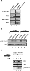
Gö6976-induced phosphorylation of Chk1 is mediated by ATR. (A) Asynchronously growing HeLa cells were treated with vehicle (DMSO), 50 μM wortmannin for 2 h, or 1 μM Gö6976 for 1 h. HeLa cells were also pretreated with wortmannin for 2 h and then treated with Gö6976 for 1 h. As a positive control for wortmannin, inhibition of Akt phosphorylation was analyzed. Western blotting was performed with the indicated antibodies. (B) Cells expressing functional ATM (ATM+) or cells lacking functional ATM (ATM−) were incubated with 1 μM Gö6976 for 1 h or 2 h. Western blotting was performed with the indicated antibodies. (C) HeLa cells were transfected with 100 nM ATR-specific siRNAs or scrambled siRNAs. After 48 h, cells were treated with DMSO or 1 μM Gö6976 for 1 h and then harvested for Western blotting with the indicated antibodies.
FIG. 3.
FIG. 3.
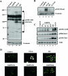
Gö6976 does not induce global phosphorylation of ATR targets or γH2AX foci. HeLa cells were untreated (control) or were incubated with 20 mM HU (2 h), 20 μM VP-16 (4 h), 1 μM Gö6976 (1 h), or 1 μM UCN-01 (2 h). Cell lysates were resolved by SDS-PAGE and subjected to Western blotting with antibodies that recognize phosphorylated substrates of ATM/ATR (A) or phosphorylated Brca1 (B). β-Catenin antibodies were used to verify equal loading. (C) HeLa cells were treated as in panels A and B, except that cells were incubated with Gö6976 for 1 h, 2 h, 3 h, and 6 h before harvest. Samples were analyzed by Western blotting for pS1981-ATM, pS966-Smc1, pS345-Chk1, and Chk1. (D) HeLa cells were treated as in panel C and analyzed by indirect immunofluorescence for γH2AX foci.
FIG. 4.
FIG. 4.
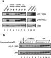
OA impairs Chk1 dephosphorylation in vivo and in vitro. (A) HeLa cells were untreated (lane 1) or were incubated with 20 mM HU for 2 h to generate phosphorylated Chk1 (lanes 2 to 8). The culture medium was then removed, and cells were incubated in complete medium containing DMSO, 1 μM Gö6976, or 1 μM OA. Chk1 phosphorylation was monitored at 45 min and 60 min after HU removal by Western blot analysis. Alternatively, HeLa cells were incubated with 1 μM Gö6976 or 1 μM OA for 1 h followed by Western blot analysis (lanes 10 and 11). (B) HeLa cells were incubated with 20 mM HU for 2 h to generate phosphorylated Chk1. Cells were washed, and lysates were prepared and either analyzed directly by SDS-PAGE and Western blotting (lane 1) or were incubated in vitro in the presence of DMSO (lane 2) or the indicated concentrations of Gö6976 (lanes 3 to 6) or OA (lanes 7 to 11) for 30 min at 30°C. Reaction products were resolved by SDS-PAGE, and endogenous Chk1 was monitored by Western blotting.
FIG. 5.
FIG. 5.
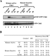
Kinase-inactive Chk1 exhibits higher basal phosphorylation and slower kinetics of dephosphorylation. HeLa cells were transiently transfected with kinase-active GFP-Chk1 or kinase-inactive GFP-Chk1. Twenty-four hours posttransfection, cells were left untreated (lanes 1 and 6) or were incubated with HU for 2 h. HU was removed, and cells were harvested immediately (lanes 2 and 7) or were cultured in complete medium for the indicated times. (A) A fraction of the cells were lysed and analyzed by Western blotting with the indicated antibodies. (B) The remaining cells were stained for DNA content, and GFP-positive cells were analyzed by flow cytometry. The percentages of cells in each phase of the cell cycle are indicated.
FIG. 6.
FIG. 6.
PP2A dephosphorylates Chk1 on S317 and S345. (A) HeLa cells were untreated (lane 1) or were incubated in the presence of HU for 2 h to induce Chk1 phosphorylation. Lysates derived from HU-treated cells were harvested immediately (lane 2) or were incubated in the presence of DMSO (lane 3), 1 μM Gö6976 (lane 4), 1 μM OA (lane 5), 0.5 μM fostriecin (lane 6), or 1 μM I-2 (lane 7) for 30 min at 30°C (lanes 3 to 7). Phosphorylation of Chk1 was assayed by immunoblotting. (B) Targeted knockdown of specific protein phosphatases was accomplished by transfecting HeLa cells with the indicated siRNAs. After 48 h, cells were either untreated (lanes 1 to 3) or were incubated with 1 μM Gö6976 for 1 h (lanes 4 to 6). Cell lysates were prepared and analyzed for phosphorylated Chk1, total Chk1, PP1, and PP2A by Western blotting using the Femto maximum sensitivity substrate (Pierce). Short (5 s) and long (15 s) exposures of the Western blot are shown. (C) HeLa cells were transfected with scrambled or PP2A-specific siRNAs. Lysates were prepared 48 h later and incubated in vitro with phosphorylated GFP-Chk1 for 30 min at 30°C in the presence or absence of OA. Western blotting was performed with the indicated antibodies. (D) Purified PP1 or PP2A was incubated with phosphorylated GFP-Chk1 immunoprecipitated from HU-treated cells for 30 min at 30°C in the presence or absence of 1 μM Gö6976 or 1 μM OA. Reaction products were resolved by SDS-PAGE and analyzed by Western blotting.
FIG. 7.
FIG. 7.
The ATR-Chkl-PP2A regulatory circuit. During an unperturbed cell division cycle, Chk1 is continually being phosphorylated on S317 and S345 by ATR. However, phosphorylated Chk1 does not accumulate to any significant extent because PP2A continually dephosphorylates Chk1 on S317 and S345. Importantly, the ability of PP2A to dephosphorylate Chk1 depends, in part, on Chk1 kinase activity as chemical inhibition of Chk1 promotes the accumulation of phosphorylated Chk1 in vivo. Thus, Chk1 promotes its own dephosphorylation during an unperturbed cell division cycle.

Similar articles

Cited by

References

    1. Adams, K. E., A. L. Medhurst, D. A. Dart, and N. D. Lakin. 2006. Recruitment of ATR to sites of ionising radiation-induced DNA damage requires ATM and components of the MRN protein complex. Oncogene 25:3894-3904. (First published 13 February 2006; doi:10.1038/sj.onc.1209426.) - DOI - PMC - PubMed
    1. Ali, A., J. Zhang, S. Bao, I. Liu, D. Otterness, N. M. Dean, R. T. Abraham, and X. F. Wang. 2004. Requirement of protein phosphatase 5 in DNA-damage-induced ATM activation. Genes Dev. 18:249-254. - PMC - PubMed
    1. al-Khodairy, F., E. Fotou, K. S. Sheldrick, D. J. Griffiths, A. R. Lehmann, and A. M. Carr. 1994. Identification and characterization of new elements involved in checkpoint and feedback controls in fission yeast. Mol. Biol. Cell 5:147-160. - PMC - PubMed
    1. Arena, S., S. Benvenuti, and A. Bardelli. 2005. Genetic analysis of the kinome and phosphatome in cancer. Cell Mol. Life Sci. 62:2092-2099. - PMC - PubMed
    1. Bakkenist, C. J., and M. B. Kastan. 2003. DNA damage activates ATM through intermolecular autophosphorylation and dimer dissociation. Nature 421:499-506. - PubMed

Publication types

MeSH terms

LinkOut - more resources