

doi: 10.1128/JVI.00995-06.
Human cytomegalovirus UL84 protein contains two nuclear export signals and shuttles between the nucleus and the cytoplasm
Affiliations
- PMID: 17005707
- PMCID: PMC1617278
- DOI: 10.1128/JVI.00995-06
Item in Clipboard
Human cytomegalovirus UL84 protein contains two nuclear export signals and shuttles between the nucleus and the cytoplasm
J Virol.
2006 Oct.
Abstract
Previous studies defined pUL84 of human cytomegalovirus as an essential regulatory protein with nuclear localization that was proposed to act during initiation of viral-DNA synthesis. Recently, we demonstrated that a complex domain of 282 amino acids within pUL84 functions as a nonconventional nuclear localization signal. Sequence inspection of this domain revealed the presence of motifs with homology to leucine-rich nuclear export signals. Here, we report the identification of two functional, autonomous nuclear export signals and show that pUL84 acts as a CRM-1-dependent nucleocytoplasmic shuttling protein. This suggests an unexpected cytoplasmic role for this essential viral regulatory protein.
Figures
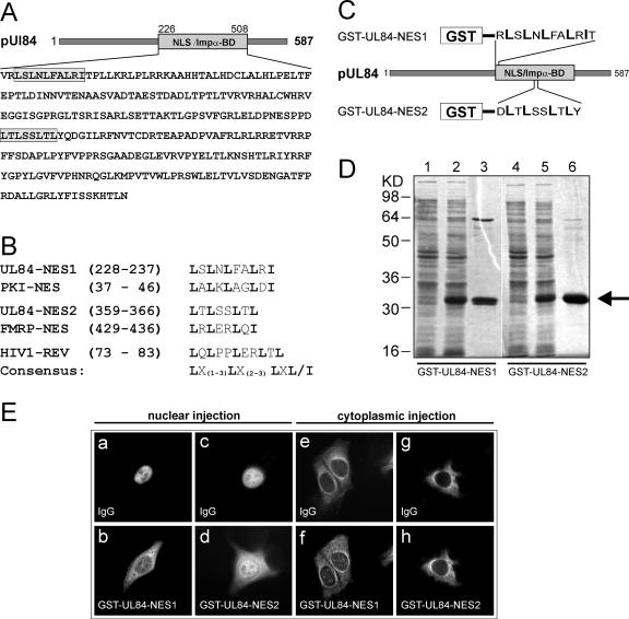
pUL69 contains two active leucine-rich NESs. (A) Schematic representation of the UL84 protein showing the NLS/importin alpha binding domain (20). Two putative leucine-rich NES motifs located within the nonconventional NLS/importin alpha binding domain of pUL84 are highlighted. (B) Alignment and comparison of the two putative pUL84 NES motifs with known leucine-rich NESs and with the consensus sequence for leucine-rich NESs. The indicated pUL84 motifs are compared with the NES of the protein kinase inhibitor α (PKI) (31) or the fragile X mental retardation protein (FMRP) (11). Additionally, the NES of the human immunodeficiency virus type 1 Rev protein (HIV1-REV) (8) and a derived consensus NES are listed. Conserved residues in the NESs are shown in boldface. The numbers refer to the positions of the amino acid sequences within each protein. (C) Schematic representation of the UL84 coding sequence showing the two putative NESs fused to the C terminus of GST to produce GST-UL84-NES1 and GST-UL84-NES2. (D) Procaryotic expression and purification of GST-UL84-NES1 and GST-UL84-NES2. Shown is a Coomassie blue-stained gel: extracts from E. coli cells grown without isopropyl-β-d -thiogalactopyranoside (IPTG) (lanes 1 and 4) and grown in the presence of IPTG (lanes 2 and 5) are shown; lanes 3 and 6, purified GST fusion proteins. (E) GST fusion proteins were microinjected into the nuclei (a to d) or the cytoplasm (e to h) of HeLa cells, together with rabbit IgG as a marker for the injection site. At 1 h after injection, the cells were fixed and immunostained for GST-UL84-NES1 or GST-UL84-NES2 and the coinjected IgG control.
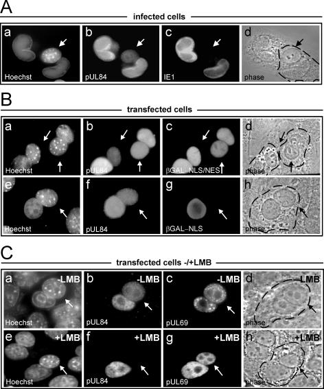
Nucleocytoplasmic shuttling of pUL84 in infected and transfected cells. (A) Heterokaryons were generated by fusion of HCMV-infected primary HFF and murine NIH 3T3 cells. Prior to and following heterokaryon formation, de novo protein synthesis was inhibited using cycloheximide. At 3.5 h after fusion, the cells were fixed, and a double-immunofluorescence analysis was performed with a polyclonal antiserum directed against pUL84 (b) and a monoclonal antibody against the IE1 protein (c). Staining with Hoechst 33258 (a) was used to differentiate between human and murine nuclei within the heterokaryon. Murine nuclei display a characteristic punctate pattern, whereas human nuclei are diffusely stained with the reagent; murine nuclei are indicated by arrows. Panel d shows the phase-contrast image of the heterokaryons; the cytoplasmic edge is highlighted by a broken line. (B) HeLa cells were cotransfected with expression plasmids for pUL84 and one of the internal control plasmids, β-Gal-NLS/NES or β-Gal-NLS, as indicated (a to d, UL84 and β-Gal-NLS/NES; e to h, UL84 and β-Gal-NLS). The transfected cells were subsequently analyzed in heterokaryon assays as described in the legend to panel A. Double-immunofluorescence analysis with polyclonal anti-pUL84 serum and a monoclonal antibody against β-Gal was performed in order to detect the expressed proteins. (C) pUL84 shuttles between the nucleus and the cytoplasm in a CRM1-dependent manner. Expression constructs for pUL84 and pUL69 were cotransfected into HeLa cells, and the transfected cells were subjected to heterokaryon assays. Three hours prior to fusion and throughout the experiment, the cells were incubated in the absence (−) (a to d) or presence (+) (e to h) of LMB. The indicated proteins were detected by double-label immunofluorescence using a polyclonal pUL84 antiserum and a monoclonal antibody directed against pUL69 (19).
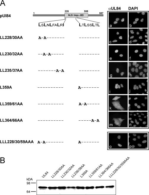
Subcellular localization of UL84 mutants carrying mutated nuclear export signals. (A) A series of pUL84 mutants carrying alanine replacement mutations either in NES1 or NES2 was generated, and the subcellular localization of the resulting mutants was analyzed via indirect immunofluorescence analysis. The mutants indicated on the left were transiently expressed in HeLa cells, which were subsequently fixed and immunostained (right) using an anti-pUL84 antiserum (αUL84); DAPI, DNA staining of the transfected HeLa cells. (B) Western blot analysis of expression levels of the indicated UL84 mutants in transfected HeLa cell cultures.

Nucleocytoplasmic shuttling activities of pUL84 mutants carrying alanine substitutions within NES1, NES2, or NES1 and NES2. (Left) Schematic representation of the respective pUL84 mutants and β-Gal-NLS/NES, which was used as an internal shuttling control in the interspecies heterokaryon analysis. (Right) Heterokaryon experiments (as described in the legend to Fig. 2) were performed to visualize the nuclear-export activities of the indicated proteins.
Similar articles
-
A novel transferable nuclear export signal mediates CRM1-independent nucleocytoplasmic shuttling of the human cytomegalovirus transactivator protein pUL69.EMBO J. 2001 Dec 17;20(24):7271-83. doi: 10.1093/emboj/20.24.7271. EMBO J. 2001. PMID: 11743003 Free PMC article.
-
A methionine-rich domain mediates CRM1-dependent nuclear export activity of Borna disease virus phosphoprotein.J Virol. 2006 Feb;80(3):1121-9. doi: 10.1128/JVI.80.3.1121-1129.2006. J Virol. 2006. PMID: 16414989 Free PMC article.
-
The redox state of SECIS binding protein 2 controls its localization and selenocysteine incorporation function.Mol Cell Biol. 2006 Jul;26(13):4895-910. doi: 10.1128/MCB.02284-05. Mol Cell Biol. 2006. PMID: 16782878 Free PMC article.
-
Leucine-rich nuclear-export signals: born to be weak.Trends Cell Biol. 2005 Mar;15(3):121-4. doi: 10.1016/j.tcb.2005.01.005. Trends Cell Biol. 2005. PMID: 15752974 Review.
-
Interactions of human cytomegalovirus proteins with the nuclear transport machinery.Curr Top Microbiol Immunol. 2008;325:167-85. doi: 10.1007/978-3-540-77349-8_10. Curr Top Microbiol Immunol. 2008. PMID: 18637506 Review.
Cited by
-
Viral Subversion of the Chromosome Region Maintenance 1 Export Pathway and Its Consequences for the Cell Host.Viruses. 2023 Nov 6;15(11):2218. doi: 10.3390/v15112218. Viruses. 2023. PMID: 38005895 Free PMC article. Review.
-
Human cytomegalovirus UL84 interacts with an RNA stem-loop sequence found within the RNA/DNA hybrid region of oriLyt.J Virol. 2007 Jul;81(13):7077-85. doi: 10.1128/JVI.00058-07. Epub 2007 Apr 25. J Virol. 2007. PMID: 17459920 Free PMC article.
-
The IE2 60-kilodalton and 40-kilodalton proteins are dispensable for human cytomegalovirus replication but are required for efficient delayed early and late gene expression and production of infectious virus.J Virol. 2007 Mar;81(6):2573-83. doi: 10.1128/JVI.02454-06. Epub 2007 Jan 3. J Virol. 2007. PMID: 17202222 Free PMC article.
-
Interaction of HCMV UL84 with C/EBPalpha transcription factor binding sites within oriLyt is essential for lytic DNA replication.Virology. 2009 Sep 15;392(1):16-23. doi: 10.1016/j.virol.2009.06.035. Epub 2009 Jul 23. Virology. 2009. PMID: 19631360 Free PMC article.
-
Dynamic and nucleolin-dependent localization of human cytomegalovirus UL84 to the periphery of viral replication compartments and nucleoli.J Virol. 2014 Oct;88(20):11738-47. doi: 10.1128/JVI.01889-14. Epub 2014 Jul 30. J Virol. 2014. PMID: 25078694 Free PMC article.
References
-
- Boehmer, P. E., M. S. Dodson, and I. R. Lehman. 1993. The herpes simplex virus type-1 origin binding protein. DNA helicase activity. J. Biol. Chem. 268:1220-1225. - PubMed
-
- Bruckner, R. C., J. J. Crute, M. S. Dodson, and I. R. Lehman. 1991. The herpes simplex virus 1 origin binding protein: a DNA helicase. J. Biol. Chem. 266:2669-2674. - PubMed
-
- Colletti, K. S., Y. Xu, I. Yamboliev, and G. S. Pari. 2005. Human cytomegalovirus UL84 is a phosphoprotein that exhibits UTPase activity and is a putative member of the DExD/H box family of proteins. J. Biol. Chem. 280:11955-11960. - PubMed
-
- Dodson, M. S., and I. R. Lehman. 1993. The herpes simplex virus type I origin binding protein. DNA-dependent nucleoside triphosphatase activity. J. Biol. Chem. 268:1213-1219. - PubMed
Publication types
MeSH terms
Substances
LinkOut - more resources
Full Text Sources
