. 2006 Sep 25;174(7):973-83.
doi: 10.1083/jcb.200605106.

Exomer: A coat complex for transport of select membrane proteins from the trans-Golgi network to the plasma membrane in yeast

Affiliations

Exomer: A coat complex for transport of select membrane proteins from the trans-Golgi network to the plasma membrane in yeast

Chao-Wen Wang et al. J Cell Biol. .

Abstract

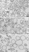
A yeast plasma membrane protein, Chs3p, transits to the mother-bud neck from a reservoir comprising the trans-Golgi network (TGN) and endosomal system. Two TGN/endosomal peripheral proteins, Chs5p and Chs6p, and three Chs6p paralogues form a complex that is required for the TGN to cell surface transport of Chs3p. The role of these peripheral proteins has not been clear, and we now provide evidence that they create a coat complex required for the capture of membrane proteins en route to the cell surface. Sec7p, a Golgi protein required for general membrane traffic and functioning as a nucleotide exchange factor for the guanosine triphosphate (GTP)-binding protein Arf1p, is required to recruit Chs5p to the TGN surface in vivo. Recombinant forms of Chs5p, Chs6p, and the Chs6p paralogues expressed in baculovirus form a complex of approximately 1 MD that binds synthetic liposomes in a reaction requiring acidic phospholipids, Arf1p, and the nonhydrolyzable GTPgammaS. The complex remains bound to liposomes centrifuged on a sucrose density gradient. Thin section electron microscopy reveals a spiky coat structure on liposomes incubated with the full complex, Arf1p, and GTPgammaS. We termed the novel coat exomer for its role in exocytosis from the TGN to the cell surface. Unlike other coats (e.g., coat protein complex I, II, and clathrin/adaptor protein complex), the exomer does not form buds or vesicles on liposomes.


Figures
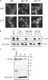
Figure 1.
Figure 1.
Chs5p is regulated by a Sec7p- and Arf1p-dependent machinery. (A) Chs5p-GFP localizes to Golgi membranes, and this association is Sec7p dependent. Wild-type (WT; CWY512), pik1-83 (CWY559), and sec7-4 (CWY612) cells that bear chromosomally tagged Chs5p-GFP were grown at 26°C to mid-log phase and either kept at 26°C or shifted to 37°C for 40 min for fluorescence microscopy. Arrowheads indicate exaggerated Golgi. (B) Chs5p interacts with Arf1p-PA. CWY506 cells harboring Arf1p-PA integrated at the chromosomal locus were harvested at mid-log phase as described in Materials and methods. A clear cell lysate (input) was distributed in aliquots and incubated with 0.5 mM GTP, GMP-PNP, or GTPγS for 10 min at 30°C. Arf1p–protein A was absorbed by IgG-coated Dynabeads at 4°C for 2 h. Beads were recovered using a magnet, and the unbound proteins were removed in the flow through (FT) followed by wash (W) steps. Protein remaining bound (B) to the beads was resuspended in buffer and analyzed by anti-Chs5p (also recognizes protein A [PA]) immunoblotting. (C) CWY506 was lysed as described in B followed by incubation with 0.5 mM GTPγS in the presence of buffer (−) or 2 μg/ml brefeldin A (BFA) at 30°C for 10 min. Samples were processed as in B except that only the bound (B) samples are shown.
Figure 2.
Figure 2.
Functional mapping of Chs5p. (A) Mapping Chs5p lipid interaction and Chs6p interaction regions. Full-length Chs5p (aa 1–671) contains two motifs: FN3 (aa 79–160) and BRCT (aa 160–260). The fragments positive for lipid interaction are shown by a plus sign based on GST fusion constructs purified from E. coli that showed the lipid interaction profile on PIP strips (Echenlon). These fragments were also cloned into the yeast two-hybrid construct pGBD to test interaction with pGAD-Chs6. Growth on an SD-Leu-Ura-His plate is shown by a plus sign. (B) Wild-type (WT; SEY6210) cells harboring GST, GST-Chs5 (aa 1–79), and GST-Chs5 (aa 401–671) cloned into pRS424 (2μ; tryptophan) were streaked on SD-Trp plates in addition to 50 (CF 50) or 100 μg/ml (CF 100) calcofluor. For each construct, two colonies were restreaked and examined on the plates.
Figure 3.
Figure 3.
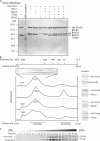
Purification and characterization of the Chs5p and Chs6p protein complexes. (A) Chs5p and all Chs6-like proteins were purified as complexes. Amplified baculovirus stocks were inoculated (+) or not inoculated (−) into the insect culture cell line Sf-9. Cultures were harvested after 4 d postinoculation, and proteins were purified on a Ni-NTA column (for binding to 6× His-tagged Chs5p). Purified proteins were examined by SDS-PAGE followed by Coomassie blue staining. MW, mol wt. (B) Protein complexes purified on Ni-NTA were size fractionated on a Superose 6 FPLC column. Elution during 6–18 ml is shown, and the localization of mol wt standards is indicated. Peak fractions of the complex (size as indicated) were analyzed by SDS-PAGE and Sypro red staining. As examined on gels, His-Chs5p and Chs6p-like proteins such as Bch1p, Bud7p, and Chs6p are found together in the ∼1-MD fractions. (C) The purified Chs5–Chs6[all] complex was analyzed by a 10–50% sucrose gradient. A total of 20 × 100-μl fractions were collected from the top, and proteins were analyzed by SDS-PAGE and Sypro red staining. Gels were visualized using a Typhoon imager.
Figure 4.
Figure 4.
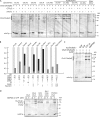
Recruitment of the Chs5–Chs6[all] complex by mArf1p. (A) Liposomes composed of various phospholipid formulations were tested for recruitment of the Chs5–Chs6[all] complex in the presence of GTPγS alone, mArf1p(Q71L) alone, or mArf1p(Q71L) and GTPγS. Liposomes were floated through a step sucrose gradient as described in Materials and methods. Liposome-bound proteins were analyzed by SDS-PAGE followed by Sypro red staining. Gels were visualized using a Typhoon imager. (B) Comparison of binding as shown in A. mArf1p recovery from different liposome formulations as shown in A was set at 100% (light gray bar), relative recoveries of the His-Chs5p amount (wt/wt) are compared (gray bar), and the amount of His-Chs5p floated in the absence of mArf1p was subtracted (black bar) from the gray bar. The y axis measures the relative binding index. (C) Standard recruitment assay. Major-minor liposomes were incubated with 1 μM mArf1p in the presence of buffer (−), 0.1 mM GDP, GTP, GTPγS, or GMP-PNP at 30°C for 1 h in a chelating condition used to trigger nucleotide exchange. 0.5 μM Chs5–Chs6[all] complex was tested for binding as described in Materials and methods. Floated proteins were examined by Sypro red staining. (D) The same mArf1p exchange conditions were performed as in C, but different proteins or protein complexes were tested for their association with mArf1p in the presence of 0.1 mM GDP (D) or GTPγS (T). PC, phosphatidylcholine; PS, phosphatidylserine; PI, phosphatidylinositol.
Figure 5.
Figure 5.
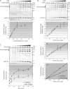
Recruitment of the Chs5–Chs6[all] complex via mArf1p in the presence of GTPγS is a saturable process. (A) Titration of the Chs5–Chs6[all] complex. 1 μM mArf1p incubated with GTPγS and various concentrations of the Chs5–Chs6[all] complex at room temperature for 15 min. Recruitment was determined by a step flotation gradient as described in Materials and methods. Sypro red staining of one experiment is shown, and quantitative results from several experiments are plotted below. (B) Titration of mArf1p with a fixed amount of the Chs5–Chs6[all] complex. Various amounts of mArf1p were incubated with GTPγS for 1 h at 30 min. 0.8 μM of a fixed concentration of the Chs5–Chs6[all] complex was then tested for binding at room temperature for 15 min. Recruitments were determined by a step flotation gradient as described in Materials and methods. Sypro red staining of one experiment is shown, and quantitative results from several experiments are plotted below. (i and ii) The membrane-associated His-Chs5p (i) and mArf1p (ii) are quantified. (iii) The membrane-associated His-Chs5p versus mArf1p (wt/wt) before the saturation concentration is quantified. (C) Recruitment time course experiment. 1 μM mArf1p and 0.5 μM Chs5–Chs6[all] complex were mixed at t = 0. Reactions were incubated at 30°C and stopped at the indicated times. Membrane association of His-Chs5p versus mArf1p (wt/wt) was determined by a step flotation gradient as described in Materials and methods. Sypro red staining of one experiment is shown, and quantitative results from several experiments are plotted below. Error bars represent SD.
Figure 6.
Figure 6.
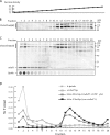
Enrichment of coated membrane on a sucrose density gradient. (A) Sucrose density index for the gradient centrifuged at 4°C for 16 h at 100,000 g in a TLS55 rotor. (B) Distribution of the Chs5–Chs6[all] complex (11–20) alone. (C) Cofractionation of lipids, mArf1p, and the Chs5p–Chs6[all] complex in the lighter fractions (1–10). (D) Quantification of the distribution in B and C.
Figure 7.
Figure 7.
Coating of the Chs5–Chs6[all] complex on liposomes. (A) Major-minor liposomes were incubated with mArf1p and GTPγS at 30°C for 1 h, and the Chs5–Chs6[all] complex was then added and incubated at room temperature for 15 min followed by fixation for thin section electron microscopy. An extensive coating on liposomes can be seen as spikes formed by patches. The inset is a higher magnification view of the boxed area. (B) Major-minor liposomes were incubated with mArf1p-GDP for 1 h at 30°C, and the Chs5–Chs6[all] complex was added at room temperature for 15 min. (C) Major-minor liposomes were incubated with mArf1p-GTPγS alone at 1 h at 30°C, and buffer was added at room temperature for 15 min.

Similar articles

Cited by

References

    1. Ang, A.L., T. Taguchi, S. Francis, H. Folsch, L.J. Murrells, M. Pypaert, G. Warren, and I. Mellman. 2004. Recycling endosomes can serve as intermediates during transport from the Golgi to the plasma membrane of MDCK cells. J. Cell Biol. 167:531–543. - PMC - PubMed
    1. Antonny, B., S. Beraud-Fufour, P. Chardin, and M. Chabre. 1997. N-terminal hydrophobic residues of the G-protein ADP-ribosylation factor-1 insert into membrane phospholipids upon GDP to GTP exchange. Biochemistry. 36:4675–4684. - PubMed
    1. Antonny, B., P. Gounon, R. Schekman, and L. Orci. 2003. Self-assembly of minimal COPII cages. EMBO Rep. 4:419–424. - PMC - PubMed
    1. Audhya, A., M. Foti, and S.D. Emr. 2000. Distinct roles for the yeast phosphatidylinositol 4-kinases, Stt4p and Pik1p, in secretion, cell growth, and organelle membrane dynamics. Mol. Biol. Cell. 11:2673–2689. - PMC - PubMed
    1. Barlowe, C., L. Orci, T. Yeung, M. Hosobuchi, S. Hamamoto, N. Salama, M.F. Rexach, M. Ravazzola, M. Amherdt, and R. Schekman. 1994. COPII: a membrane coat formed by Sec proteins that drive vesicle budding from the endoplasmic reticulum. Cell. 77:895–907. - PubMed

Publication types

MeSH terms