Comparative Study
. 2006 Sep 20;26(38):9750-60.
doi: 10.1523/JNEUROSCI.2745-06.2006.

A specific survival response in dopamine neurons at most risk in Parkinson's disease

Affiliations
Comparative Study

A specific survival response in dopamine neurons at most risk in Parkinson's disease

Sachiko Murase et al. J Neurosci. .

Abstract

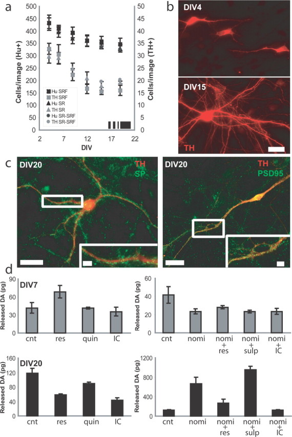
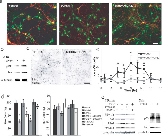
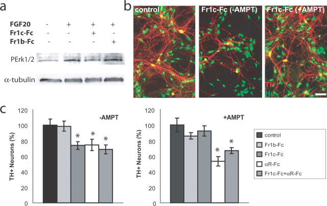
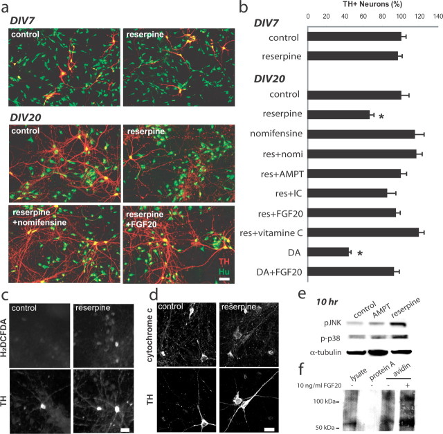
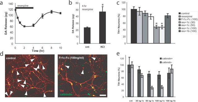
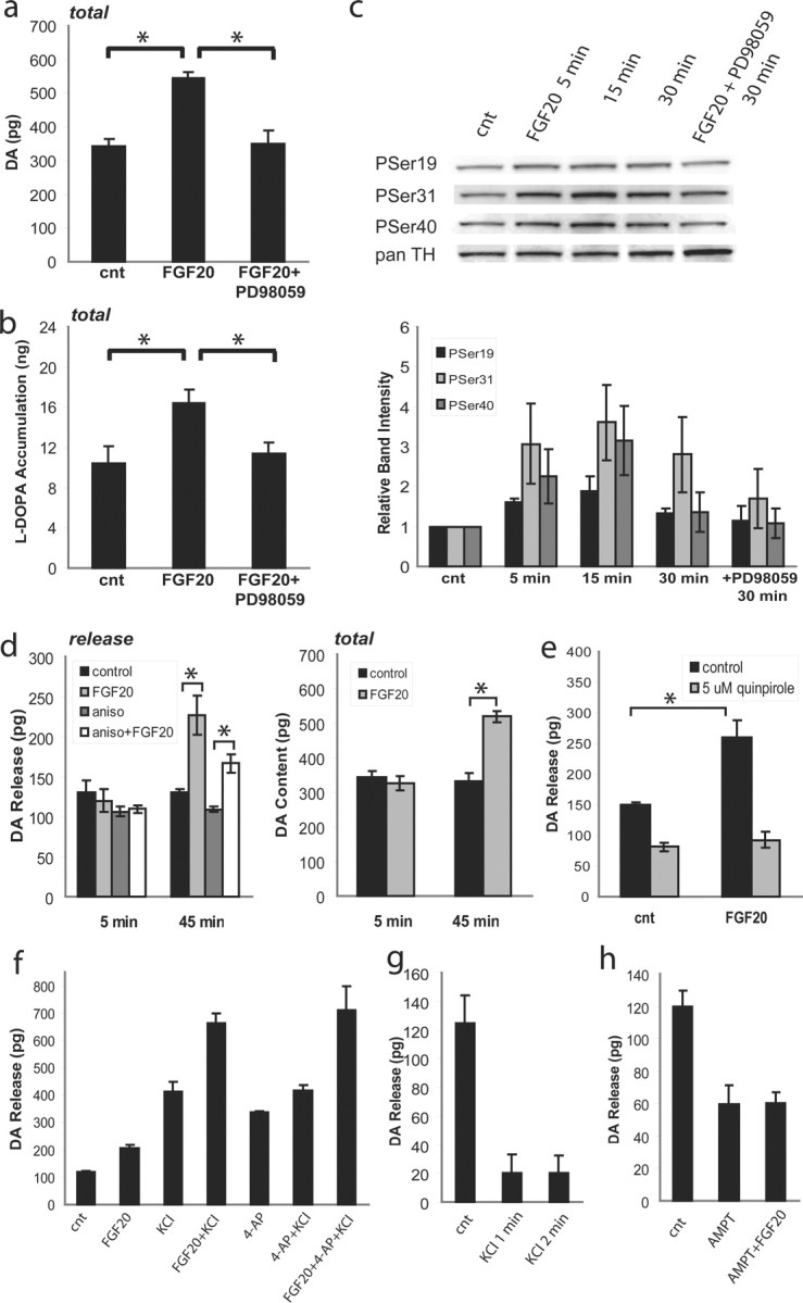
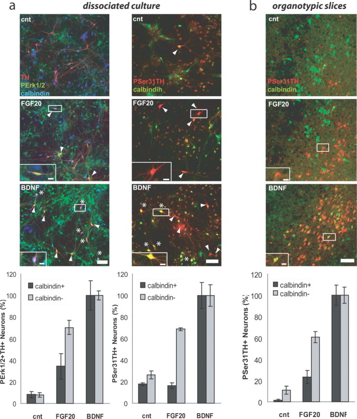
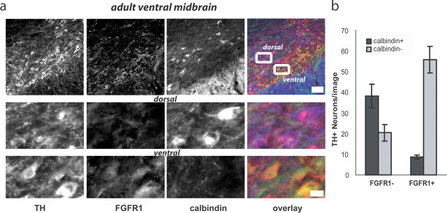
The specific expression of fibroblast growth factor 20 (FGF-20) in the adult substantia nigra and the association between FGF-20 mutations and Parkinson's disease provoked exploration of the function of this growth factor. We show by gain- and loss-of-function in vitro experiments that FGF-20 promotes survival and stimulates dopamine (DA) release in a calbindin-negative subset of cells that are preferentially lost in Parkinson's disease. FGF-20 selectively activates tyrosine hydroxylase in calbindin-negative neurons. In the adult substantia nigra, calbindin-negative neurons specifically express high levels of FGFR1 (FGF receptor 1). These data show that FGF signals to elevate DA levels and protect the specific midbrain neuron type at most risk in Parkinson's patients.


Figures

Figure 1.
Figure 1.
Development of DA neurons in dissociated ventral midbrain culture. a, Neurons including DA neurons go through natural cell death. Shown are numbers of cells/image from the cultures without serum (SRF; Neurobasal and B27) (Hu+, black square; TH+, gray square), from the cultures with serum (SR; modified MEM) (Hu+, black triangle; TH+, gray triangle), and the cultures switched from SR to SRF at DIV12 (Hu+, black circle; TH+, gray circle; n = 40). All of the experiments were done after DIV19 except for the long incubation with FGFR-Fc (DIV16). b, Extension of processes. DA neurons are stained with anti-TH antibody. Top, DIV4; bottom, DIV15. Scale bar, 20 μm. c, Formation of synaptic structure. By DIV20, processes of DA neurons (TH, red) are well colocalized with both pre- [synaptophysin (SP), green, left] and post- (PSD95, green, right) synaptic markers. Scale bars, 50 μm. Insets are magnified images (scale bars, 5 μm). d, DA release from young (top) and mature (bottom) DA neurons. cnt, Control; res, 1.5 μm reserpine; quin, 5 μm quinpirole; IC, inhibitor cocktail (1 μm TTX, 100 μm APV, 50 μm CNQX, 30 μm bicuculin); nomi, 5 μm nomifensine; sulp, 5 μm sulpiride. n = 4. Error bars indicate SE.
Figure 2.
Figure 2.
6-OHDA lesion rescued by FGF-20. a, Immunostaining with TH (red) and Hu (green). 6-OHDA treatment causes DA neuron-specific loss, which is rescued by FGF-20. Thirty millimolars 6-OHDA in PBS was freshly prepared and filtered immediately before application. Neurons were incubated with 30 μm 6-OHDA (and 10 ng/ml FGF-20) overnight. Scale bar, 100 μm. b, 6-OHDA induces stress response. Cells were incubated with 30 μm 6-OHDA for 6 h. c, Cleaved-caspase-3 immunostaining (left) when incubated for 9 h with 6-OHDA (and FGF-20). Scale bar, 50 μm. Shown are numbers of cleaved-caspase-3+ cells after incubating with 30 μm 6-OHDA (and 10 ng/ml FGF-20) for the indicated time (right) (n = 10). *p < 0.05. d, Effect of FGF-20 and contribution of MAPK and PI3 kinase. Neurons were incubated with 50 μm PD98059 or 25 μm LY294002 (n = 30). *p < 0.05. e, FGF-20 activates both MAPK and PI3K pathways in ventral midbrain culture, and protects mitochondria. α-Tubulin was blotted as a loading control. To minimize the contribution of astrocytes, the glia-free cultures were used. Error bars indicate SE.
Figure 3.
Figure 3.
Role of endogenous activation of FGFR1c. a, FGFR1c-Fc selectively inhibits PErk1/2 activation by FGF-20. Fc-chimeric proteins were preincubated with FGF-20 at 37°C for 2 h before applied to cultured neurons. Final concentrations of FGF-20 and Fc-chimeric protein were 10 ng/ml and 200 ng/ml, respectively. b, Cell death of DA neurons induced by FGFR1c-Fc. Neurons were incubated for 4 d with 100 ng/ml FGFR1c-Fc in the presence or absence of 500 μm AMPT. Half of the medium was changed everyday. Immunostaining with TH (red) and Hu (green) is shown. Scale bar, 100 μm. c, Effect of Fc-chimeric proteins. Incubation with 100 ng/ml Fc-chimeric proteins with or without 500 μm AMPT for 4 d (n = 40). *p < 0.05. Error bars indicate SE.
Figure 4.
Figure 4.
DA-induced toxicity rescued by FGF-20. a, Inhibition of VMAT2 caused loss of DA neurons in mature but not young cultures. Neurons were incubated overnight with 1.5 μm reserpine (and 10 ng/ml FGF-20). Shown are representative images stained with TH (red) and Hu (green) of control, 1.5 μm reserpine, 1.5 μm reserpine plus 5 μm nomifensine, and 1.5 μm reserpine plus 10 ng/ml FGF-20 overnight-treated neurons. Scale bar, 100 μm. b, Neurons were incubated overnight with 1.5 μm reserpine, 5 μm nomifensine, 500 μm AMPT (preincubated with AMPT alone for 10 h), inhibitor cocktail (IC; 1 μm TTX, 100 μm APV, 50 μm CNQX, 30 μm bicuculin), 200 μm vitamin C and 10 ng/ml FGF-20. For DA treatments, cells were exposed to 2 mm dopamine (and 10 ng/ml FGF-20) for 30 min, then incubated overnight with neuron medium (and 10 ng/ml FGF-20; n = 30). *p < 0.05. c, Reserpine induces H2O2 production in DA neurons. Cells were incubated with 1.5 μm reserpine for 6 h before carboxy-H2DCFDA staining. Scale bar, 20 μm. d, Reserpine changes cytochrome c distribution in DA neurons. Cells were incubated with 1.5 μm reserpine for 10 h. Scale bar, 20 μm. e, Reserpine induces stress response in ventral midbrain culture. Cells were incubated with 1.5 μm reserpine or 500 μm AMPT for 10 h. f, Surface labeling of DAT. Cells were treated with 10 ng/ml FGF-20 for 1 h before biotinylation. Error bars indicate SE.
Figure 5.
Figure 5.
Short-term treatment with reserpine and FGFR1c-Fc causes loss of DA neurons. a, Effect of reserpine on DA release. The cultured neurons were incubated with 1.5 μm reserpine for 4 h (bar). DA release decreased rapidly after the application of 1.5 μm reserpine, and recovered after the removal of reserpine (n = 4). b, DA release is stimulated by high K+ after 4 h of incubation with reserpine (n = 4; p = 0.026). c, FGFR1c-Fc but not FGFR1b-Fc promotes DA neuron cell death induced by cytosolic DA in a dose-dependent manner. Neurons were incubated for 4 h with 1.5 μm reserpine and the indicated concentrations of FGFR1c-Fc, then incubated with the medium overnight (n = 30). *p < 0.05. d, Immunostaining of TH (red) and calbindin (green) is shown. Number of calbindin DA neurons is decreased by 4 h treatment with1.5 μm reserpine and 100 ng/ml FGFR1c-Fc (right), as compared with control (left). Scale bar, 100 μm. Arrowheads, Calbindin DA neurons. e, Calbindin DA neurons are sensitive to the treatment with reserpine and FGFR1c-Fc. No effect was observed with 100 ng/ml FGFR1b-Fc. Data were normalized with the total number of DA neurons in the control (n = 30). Error bars indicate SE.
Figure 6.
Figure 6.
FGF-20 increases DA level. a, Total DA level is increased by FGF-20. Neurons were incubated with 10 ng/ml of FGF-20 and 50 μm PD98059 for 45 min (n = 4). *p < 0.05. b, l-DOPA accumulation induced by FGF-20 in the presence of 100 μm NSD-1015 (n = 4). *p < 0.05. c, Phosphorylation of Ser residues in TH by FGF-20. Shown are Western blot probed with anti-phospho Ser-19, Ser-31, and Ser-40 antibodies, and band intensities of phospho-Ser-19, Ser-31, and Ser-40. n = 3. d, DA release (left) and total DA content (right) are both increased by FGF-20. Neurons were incubated with or without 10 ng/ml of FGF-20 for the indicated time before DA release measurements. To inhibit protein synthesis, cells were preincubated with 10 μm anisomycin for 30 min before FGF-20 application. Note that the increase was not acute (5 min; n = 4). *p < 0.05. e, Effect of D2 autoreceptor agonist, quinpirole. Preincubation with 10 ng/ml FGF-20 for 45 min resulted in a significant increase in DA release, but no difference when release is measured in 5 μm quinpirole (n = 4), *p < 0.05. f, Evoked DA release by 50 mm KCl was also increased by FGF-20. Samples were incubated with 10 ng/ml FGF-20 for 45 min before the DA release measurements. 10 mm 4-AP was applied (n = 11, 6, 7, 5, 3, 3, and 3, respectively). g, Effect of pre-exposure to high K+ on DA release. DA release was compared in 50 mm KCl-MKB. Only 1 min of pre-exposure to 50 mm KCl was enough to decrease DA release to 16% (n = 3). h, Effect of TH inhibition on the increased high K+ stimulated DA release by FGF-20. The culture was incubated with 500 μm AMPT for 20 min before the application of 10 ng/ml of FGF-20. DA release was compared in 50 mm KCl-MKB, 45 min after the application of FGF-20 (control, AMPT, AMPT plus FGF-20; n = 5, 8, and 9, respectively). Error bars indicate SE.
Figure 7.
Figure 7.
FGF-20 acts on calbindin DA neurons. a, Dissociated cultured neurons were stimulated by neurotrophins (n = 30). Left, Immunostaining with TH (red), phospho-Erk1/2 (green), and calbindin (blue). Right, Immunostaining with phospho-Ser-31 TH (red) and calbindin (green). Samples were incubated with 10 ng/ml FGF-20 (or BDNF) for 10 min. *Calbindin+; arrowheads, calbindin. b, Organotypic midbrain slices were stimulated by neurotrophins (n = 10). Immunostaining with phospho-Ser31 TH (red) and calbindin (green) is shown. Samples were incubated with 10 ng/ml FGF-20 (or BDNF) for 45 min. Scale bars, 50 μm. Insets, magnified images. Scale bars, 10 μm. Relative numbers of neurons are shown at the bottom. Calbindin DA neurons were selectively activated by FGF-20. Error bars indicate SE.
Figure 8.
Figure 8.
FGFR1 is highly expressed in calbindin DA neurons. a, Adult midbrain slice was immunostained with TH (red), FGFR1 (green), and calbindin (blue). Scale bar, 50 μm. Magnified images of dorsal region with calbindin+ DA neurons and ventral region with calbindin DA neurons were shown in bottom panels. Scale bar, 20 μm. b, Numbers of neurons are shown (n = 10). Error bars indicate SE.

Similar articles

Cited by

References

    1. Abeliovich A, Schmitz Y, Farinas I, Choi-Lundberg D, Ho WH, Castillo PE, Shinsky N, Verdugo JM, Armanini M, Ryan A, Hynes M, Phillips H, Sulzer D, Rosenthal A. Mice lacking alpha-synuclein display functional deficits in the nigrostriatal dopamine system. Neuron. 2000;25:239–252. - PubMed
    1. Acsadi G, Anguelov RA, Yang H, Toth G, Thomas R, Jani A, Wang Y, Ianakova E, Mohammad S, Lewis RA, Shy ME. Increased survival and function of SOD1 mice after glial cell-derived neurotrophic factor gene therapy. Hum Gene Ther. 2002;13:1047–1059. - PubMed
    1. Adams JD, Jr, Chang ML, Klaidman L. Parkinson's disease-redox mechanisms. Curr Med Chem. 2001;8:809–814. - PubMed
    1. Backman C, Rose GM, Hoffer BJ, Henry MA, Bartus RT, Friden P, Granholm AC. Systemic administration of a nerve growth factor conjugate reverses age-related cognitive dysfunction and prevents cholinergic neuron atrophy. J Neurosci. 1996;16:5437–5442. - PMC - PubMed
    1. Baquet ZC, Gorski JA, Jones KR. Early striatal dendrite deficits followed by neuron loss with advanced age in the absence of anterograde cortical brain-derived neurotrophic factor. J Neurosci. 2004;24:4250–4258. - PMC - PubMed

Publication types

MeSH terms