. 2006 Sep 25;174(7):1087-96.
doi: 10.1083/jcb.200605022. Epub 2006 Sep 18.

p120 catenin is essential for mesenchymal cadherin-mediated regulation of cell motility and invasiveness

Affiliations

p120 catenin is essential for mesenchymal cadherin-mediated regulation of cell motility and invasiveness

Masahiro Yanagisawa et al. J Cell Biol. .

Abstract

During epithelial tumor progression, the loss of E-cadherin expression and inappropriate expression of mesenchymal cadherins coincide with increased invasiveness. Reexpression experiments have established E-cadherin as an invasion suppressor. However, the mechanism by which E-cadherin suppresses invasiveness and the role of mesenchymal cadherins are poorly understood. We show that both p120 catenin and mesenchymal cadherins are required for the invasiveness of E-cadherin-deficient cells. p120 binding promotes the up-regulation of mesenchymal cadherins and the activation of Rac1, which are essential for cell migration and invasiveness. p120 also promotes invasiveness by inhibiting RhoA activity, independently of cadherin association. Furthermore, association of endogenous p120 with E-cadherin is required for E-cadherin-mediated suppression of invasiveness and is accompanied by a reduction in mesenchymal cadherin levels. The data indicate that p120 acts as a rheostat, promoting a sessile cellular phenotype when associated with E-cadherin or a motile phenotype when associated with mesenchymal cadherins.


Figures
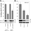
Figure 1.
Figure 1.
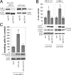
Endogenous p120 promotes invasiveness. (A) p120 depletion blocks invasiveness. E-cadherin–deficient MDA-231 and UMRC3 cells were infected with control pRS retrovirus or with a retrovirus expressing anti-human p120-specific shRNA. Polyclonal cell populations were generated, and invasion assays were performed in vitro using 20 ng/ml HGF as a chemoattractant for MDA-231 cells or 5% FBS for the UMRC3 cells. After a 20-h incubation, cells on the underside of a Matrigel-coated transwell membrane were counted under a 20× objective (n = 6). (B) Endogenous p120 levels are directly correlated to cell invasion. MDA-231 cells were infected with control retrovirus or retrovirus expressing p120-specific shRNA. Individual clones of shRNA-expressing cells were selected and tested for levels of endogenous p120 and invasiveness in vitro, toward HGF (n = 6). (bottom) Lysates from all cell lines were subjected to SDS-PAGE and Western blotted for expression of endogenous p120 (using mAb pp120) and actin. The two p120 bands correspond to p120 isoforms 1 (top) and 3 (bottom). Error bars indicate SEM. *, P < 0.05; **, P < 0.01; +, P < 0.05 as compared to pRS-infected (control) UMRC3 cells.
Figure 2.
Figure 2.
A cadherin-uncoupled p120 mutant does not promote motility and invasiveness. (A) Ectopic expression of murine p120 rescues the migration of p120-depleted cells. p120-depleted MDA-231 cells (clone 3; Fig. 1 B) were infected with control neo retrovirus or retrovirus expressing murine p120. After G418 selection, stable polyclonal cell lines were used in a scratch-wound assay to test their migration in response to HGF. Images shown are 0 and 12 h after HGF addition. The line denotes the cell front at time 0. The number of individual cells that crossed the line after 12 h was counted, and results were expressed as a percentage of migrating cells compared with control (MDA-231-pRS). n = 9. (B) The increased migration of p120-expressing cells is not due to reduced cell–cell adhesion. Cells were suspended as hanging drops and allowed to aggregate overnight. The strength of cell adhesion was assessed after passing the cell aggregates 10 times through a standard 200-μl pipette tip. Note that p120-expressing cells are more resistant to dissaggregation than cells depleted of endogenous p120. (C) MDA-231 cells with knocked down expression of endogenous p120 (clone 3) were infected with retroviruses expressing neo resistance alone or together with full-length murine p120 or a murine p120 mutant (mp120-A1), which is unable to bind E-cadherin. The invasiveness of p120-reexpressing cells was tested in vitro toward HGF (n = 6). (bottom) Lysates from all cell lines were subjected to SDS-PAGE and Western blotted for expression of p120 (using mAb pp120) and actin. Error bars indicate SEM. **, P < 0.01.
Figure 3.
Figure 3.
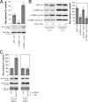
Endogenous mesenchymal cadherins promote cell invasiveness. (A) Expression of the E-cadherin juxtamembrane domain inhibits cadherin 11 levels and blocks invasiveness. MDA-231 cells were infected with control retrovirus (zeo) or retrovirus expressing a small, p120-binding, myc-tagged fragment of the E-cadherin cytoplasmic tail (ΔCB) that cannot associate with β-catenin. Polyclonal stable cell lines were subjected to invasion assays, toward a gradient of HGF (n = 6). (bottom) Lysates from both cell lines were subjected to SDS-PAGE and Western blotted for expression of cadherin 11, ΔCB (using the myc tag–specific mAb 9E10), and actin. (B) p120 regulates the levels of endogenous mesenchymal cadherins. Polyclonal populations of p120-depleted MDA-231 or UMRC3 cells were examined for their expression of cadherin 11 or N-cadherin, respectively. Note that upon p120 depletion (top), levels of endogenous mesenchymal cadherins (middle and graphs) are reduced (n = 3). (C) Endogenous mesenchymal cadherins promote cell invasiveness. MDA-231 and UMRC3 cells were transiently transfected by electroporation with either control siRNA or siRNA specific for human cadherin 11 or N-cadherin, respectively. Control experiments verified maximal knock down of cadherin expression 3 d after transfection. 2 d after transfection, cells were serum starved overnight and plated in Matrigel-coated transwells. Invasiveness was determined 24 h later in response to a gradient of HGF (MDA-231) or serum (UMRC3). n = 6. Error bars indicate SEM. *, P < 0.05; **, P < 0.01; ***, P < 0.001.
Figure 4.
Figure 4.
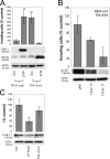
p120 binding regulates the levels of endogenous mesenchymal cadherins. (A) The p120-A1–deletion mutant does not bind mesenchymal cadherins. Lysates of p120-depleted MDA-231 cells (clone 3) reexpressing murine p120 (mp120) or a murine p120 mutant lacking the first armadillo domain (mp120-A1) were immunoprecipitated with the murine p120-specific mAb 8D11. Precipitates were then subjected to SDS-PAGE and Western blotted for the presence of cadherin 11 or p120. As shown, cadherin 11 coimmunoprecipitates with wt p120 but not with mp120-A1. (right) Expression of cadherin 11 in total cell lysates of both cell lines. (B) The p120-A1–deletion mutant does not regulate the levels of endogenous mesenchymal cadherins. p120-depleted MDA-231 cells (clone 3) stably expressing control neo, mp120, or mp120-A1 were lysed and subjected to SDS-PAGE and Western blotted for expression of p120, cadherin 11, or actin as a loading control (left). (right) Similar Western blots of p120-depleted UMRC3 cells blotted for expression of N-cadherin. In both cases, reexpression of mp120, but not mp120-A1, increases mesenchymal cadherin levels. (C) p120 binding is required for the invasion-promoting effects of N-cadherin. UMRC3 cells were transiently transfected with human N-cadherin–specific siRNA together with control YFP, murine N-cadherin–YFP, or N-cad-AAA-YFP, a murine N-cadherin mutant unable to bind p120 (N-cad-AAA). In contrast to wt N-cadherin, N-cad-AAA failed to promote invasiveness. Error bars indicate SEM. *, P < 0.05; **, P < 0.01.
Figure 5.
Figure 5.
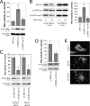
p120-mediated Rac activation requires mesenchymal cadherin binding. (A) Endogenous p120 regulates basal Rac1 activity. Levels of total and GTP-bound Rac1 were determined in serum-starved control MDA-231 cells (pRS-neo), p120-depleted cells (p120KD-neo), or cells reexpressing p120 (p120KD-mp120). Data are also expressed as a percentage of Rac1 activity, determined by densitometric analysis of immunoblots compared with the pRS-neo controls (n = 6). (B) p120 regulates HGF-induced Rac1 activity. Levels of total and GTP-bound Rac1 were determined in the same serum-starved cell lines as above, as well as in p120KD cells expressing mp120-A1, after incubation for 15 min with HGF. Both the p120-depleted cells and cells expressing mp120-A1 failed to induce Rac1 in response to HGF. Data are also expressed as a percentage of Rac activity at time 0 (control). n = 3. (C) Cadherin 11 is required for p120-dependent Rac1 induction. p120KD MDA-231 cells expressing either neo control or mp120 were transiently transfected using electroporation with control or cadherin 11 siRNA. Cells were then serum starved overnight, and the activity of Rac1 was determined using pull-down assays after a 15-min incubation with HGF. Data was also expressed as a percentage of Rac1 activity compared with the neo controls (n = 6). Error bars indicate SEM. *, P < 0.05; **, P < 0.01.
Figure 6.
Figure 6.
p120 inhibits RhoA independently of mesenchymal cadherin expression. (A) Endogenous p120 regulates basal RhoA activity. Levels of total and GTP-bound RhoA were determined in serum-starved control MDA-231 cells (pRS-neo), p120-depleted cells (p120KD-neo), or cells reexpressing p120 (p120KD-mp120). Densitometric analysis of immunoblots was used to express the data as a percentage of RhoA activity, as compared with the pRS-neo controls (n = 6). (B) p120 regulates HGF-induced effects on RhoA activity. RhoA activity was determined in the same cell lines as explained above after incubation for 20 min with HGF. Data are also expressed as a percentage of Rho activity at time 0 (n = 3). (C) Cadherin 11 is dispensable for p120-dependent RhoA inhibition. p120KD MDA-231 cells expressing neo control or mp120 were transiently transfected with control or cadherin 11 siRNA. Cells were then serum starved overnight, and the activity of RhoA was determined after a 20-min incubation with HGF. Treatment with cadherin 11 siRNA failed to block the p120-mediated inhibition of RhoA under these conditions (n = 3). (D) The cadherin-uncoupled p120-A1 mutant can inhibit RhoA activity. p120-depleted MDA-231 cells expressing neo control or mp120-A1 were subjected to Rho pull-down assays to determine levels of active RhoA after 20 min of HGF treatment. Data are also expressed as a percentage of RhoA activity (n = 6). (E) mp120-A1 can induce “branching” in NIH3T3 cells. NIH3T3 cells were transiently transfected with mp120, mp120-A1, or pcDNA control. 24 h later, cells were fixed in methanol and stained for p120 with mAb 12F4. Photographs show the characteristic branching phenotype of p120-overexpressing cells. Error bars indicate SEM. *, P < 0.05; **, P < 0.01.
Figure 7.
Figure 7.
Restoration of Rho GTPase signaling rescues the invasiveness of p120-depleted cells. (A) Inhibition of Rac1 and activation of RhoA activities block MDA-231 cell invasiveness. MDA-231 cells were transiently transfected by electroporation with a control pcDNA construct or with a myc-tagged pcDNA construct expressing either a dominant-negative N17-Rac1 mutant (DN-Rac1) or a constitutively active V14-RhoA mutant (CA-RhoA). 2 d after transfection, cells were plated in transwell filters, and invasion assays were performed a day later using HGF as a chemoattractant (n = 6). The expression of DN-Rac1 or CA-RhoA was monitored in cell lysates using the myc tag–specific mAb 9E10. (B) Activation of Rac1 and inhibition of Rho signaling promote invasiveness in p120-depleted cells. p120-depleted MDA-231 cells were infected either with retrovirus expressing control neo or with constitutively active Rac1. Stable cell lines were subjected to invasion assays in either control media or media containing a Rho kinase inhibitor (H-1152; 1.6 nM). n = 3. Error bars indicate SEM. *, P < 0.05; **, P < 0.01.
Figure 8.
Figure 8.
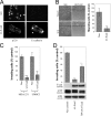
p120-uncoupled E-cadherin is less able to suppress invasiveness. (A) Ectopic E-cadherin recruits p120 to cell junctions. MDA-231 cells were infected with retroviruses expressing neomycin resistance alone or in combination with wt E-cadherin. More than 95% of G418-selected cells expressed wt E-cadherin. Wt E-cadherin accumulated at areas of cell–cell contact (mAb HECD-1). Endogenous p120 was primarily cytoplasmic in control cells and was recruited to cell–cell junctions in cells expressing wt E-cadherin (detected with polyclonal antibody F1αSH). (B) E-cadherin expression suppresses cell migration. After overnight serum starvation, confluent monolayers of control neo cells or MDA-231 cells expressing wt E-cadherin were scratched with a pipette tip, and cell migration into the wound was monitored over time in the presence of HGF. The number of individual cells that crossed the original cell front after 12 h was counted, and results were expressed as a percentage of migrating cells compared with neo control (n = 6). (C) E-cadherin suppresses cell invasiveness. The invasiveness of E-cadherin–expressing MDA-231 or UMRC3 cells was tested in vitro using HGF (MDA-231) or serum (UMRC3) as the chemoattractant (n = 6). (D) p120-uncoupled E-cadherin is less able to suppress invasiveness. Polyclonal populations of MDA-231 cells expressing neo control, wt E-cadherin, or a p120-uncoupled E-cadherin mutant (764-AAA) were subjected to invasion assays in response to HGF (n = 6). Error bars indicate SEM. **, P < 0.01; ***, P < 0.001; ++, P < 0.01, as compared with cells expressing wt E-cadherin (ANOVA).
Figure 9.
Figure 9.
p120 binding to E-cadherin suppresses invasiveness and reduces mesenchymal cadherin levels. (A) E-cadherin–free p120 promotes invasiveness. MDA-MB-231 cells stably expressing wt E-cadherin or a p120-uncoupled E-cadherin mutant (764-AAA) were infected with retroviruses expressing zeomycin resistance alone (zeo) or together with murine p120 or ΔCB. ΔCB is a myc-tagged truncation mutant of the E-cadherin cytoplasmic domain, which binds p120 but not β-catenin. Invasion assays using HGF as chemoattractant were performed as described earlier (n = 6). (bottom) Lysates from all four cell lines were subjected to SDS-PAGE and Western blotted for expression of exogenous p120 (mAb 8D11 recognizes murine but not endogenous human p120), ΔCB (9E10), and actin as a loading control. (B) Endogenous p120 promotes the invasiveness of p120-uncoupled E-cadherin in a dose-dependent manner. MDA-231 cells expressing the p120-uncoupled 764-AAA E-cadherin mutant were infected with control pRS retrovirus or viruses expressing p120-specific shRNA. Individual clones of shRNA-expressing cells were selected and examined for levels of endogenous p120 and in vitro invasiveness toward HGF (n = 6). Note that levels of endogenous p120 in the clonal cell lines correlate directly to their invasiveness in vitro. (C) p120 recruitment to E-cadherin inhibits cadherin 11 levels. Lysates of MDA-231 cells stably expressing control neo, wt E-cadherin, or the p120-uncoupled 764-AAA E-cadherin mutant were subjected to SDS-PAGE and Western blotted for cadherin 11 levels or actin as a loading control. (top) Cadherin 11 levels are also expressed as a percentage of neo control (n = 3). Error bars indicate SEM. *, P < 0.05; **, P < 0.01.

Similar articles

Cited by

References

    1. Anastasiadis, P.Z., and A.B. Reynolds. 2001. Regulation of Rho GTPases by p120-catenin. Curr. Opin. Cell Biol. 13:604–610. - PubMed
    1. Anastasiadis, P.Z., S.Y. Moon, M.A. Thoreson, D.J. Mariner, H.C. Crawford, Y. Zheng, and A.B. Reynolds. 2000. Inhibition of RhoA by p120 catenin. Nat. Cell Biol. 2:637–644. - PubMed
    1. Bellovin, D.I., R.C. Bates, A. Muzikansky, D.L. Rimm, and A.M. Mercurio. 2005. Altered localization of p120 catenin during epithelial to mesenchymal transition of colon carcinoma is prognostic for aggressive disease. Cancer Res. 65:10938–10945. - PubMed
    1. Berx, G., and F. Van Roy. 2001. The E-cadherin/catenin complex: an important gatekeeper in breast cancer tumorigenesis and malignant progression. Breast Cancer Res. 3:289–293. - PMC - PubMed
    1. Braga, V.M. 2002. Cell-cell adhesion and signalling. Curr. Opin. Cell Biol. 14:546–556. - PubMed

Publication types