

Persistent expression of autoantibodies in SLE patients in remission
- PMID: 16966430
- PMCID: PMC2118096
- DOI: 10.1084/jem.20061446
Persistent expression of autoantibodies in SLE patients in remission
Abstract
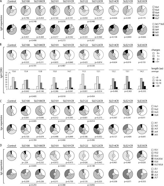
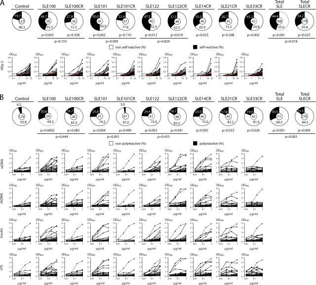
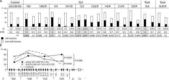
A majority of the antibodies expressed by nascent B cells in healthy humans are self-reactive, but most of these antibodies are removed from the repertoire during B cell development. In contrast, untreated systemic lupus erythematosus (SLE) patients fail to remove many of the self-reactive and polyreactive antibodies from the naive repertoire. Here, we report that SLE patients in clinical remission continue to produce elevated numbers of self-reactive and polyreactive antibodies in the mature naive B cell compartment, but the number of B cells expressing these antibodies is lower than in patients with active disease. Our finding that abnormal levels of self-reactive mature naive B cells persist in the majority of patients in clinical remission suggests that early checkpoint abnormalities are an integral feature of SLE.
Figures
Similar articles
-
Expression of B-cell activating factor of the tumour necrosis factor family (BAFF) in T cells in active systemic lupus erythematosus: the role of BAFF in T cell-dependent B cell pathogenic autoantibody production.Rheumatology (Oxford). 2007 Jul;46(7):1083-6. doi: 10.1093/rheumatology/kem097. Epub 2007 May 11. Rheumatology (Oxford). 2007. PMID: 17500077
-
Defective B cell tolerance checkpoints in systemic lupus erythematosus.J Exp Med. 2005 Mar 7;201(5):703-11. doi: 10.1084/jem.20042251. Epub 2005 Feb 28. J Exp Med. 2005. PMID: 15738055 Free PMC article.
-
Role of pathogenic auto-antibody production by Toll-like receptor 9 of B cells in active systemic lupus erythematosus.Rheumatology (Oxford). 2008 Feb;47(2):145-9. doi: 10.1093/rheumatology/kem327. Epub 2007 Dec 26. Rheumatology (Oxford). 2008. PMID: 18160420
-
B cell loss leading to remission in severe systemic lupus erythematosus.J Rheumatol. 2003 Feb;30(2):412-4. J Rheumatol. 2003. PMID: 12563705 Review.
-
Peripheral B cell abnormalities and disease activity in systemic lupus erythematosus.Lupus. 2008 Dec;17(12):1064-9. doi: 10.1177/0961203308095138. Lupus. 2008. PMID: 19029273 Review.
Cited by
-
B cell receptor light chain repertoires show signs of selection with differences between groups of healthy individuals and SLE patients.Mol Immunol. 2012 Jul;51(3-4):273-82. doi: 10.1016/j.molimm.2012.03.028. Epub 2012 Apr 18. Mol Immunol. 2012. PMID: 22516082 Free PMC article.
-
Decreased somatic hypermutation induces an impaired peripheral B cell tolerance checkpoint.J Clin Invest. 2016 Nov 1;126(11):4289-4302. doi: 10.1172/JCI84645. Epub 2016 Oct 4. J Clin Invest. 2016. PMID: 27701145 Free PMC article.
-
Reduced receptor editing in lupus-prone MRL/lpr mice.J Exp Med. 2007 Nov 26;204(12):2853-64. doi: 10.1084/jem.20071268. Epub 2007 Oct 29. J Exp Med. 2007. PMID: 17967905 Free PMC article.
-
A fully synthetic human Fab antibody library based on fixed VH/VL framework pairings with favorable biophysical properties.MAbs. 2013 May-Jun;5(3):445-70. doi: 10.4161/mabs.24218. Epub 2013 Apr 9. MAbs. 2013. PMID: 23571156 Free PMC article.
-
Serum from patients with SLE instructs monocytes to promote IgG and IgA plasmablast differentiation.J Exp Med. 2012 Jul 2;209(7):1335-48. doi: 10.1084/jem.20111644. Epub 2012 Jun 11. J Exp Med. 2012. PMID: 22689824 Free PMC article.
References
-
- Wardemann, H., S. Yurasov, A. Schaefer, J.W. Young, E. Meffre, and M.C. Nussenzweig. 2003. Predominant autoantibody production by early human B cell precursors. Science. 301:1374–1377. - PubMed
-
- Tan, E.M. 1989. Antinuclear antibodies: diagnostic markers for autoimmune diseases and probes for cell biology. Adv. Immunol. 44:93–151. - PubMed
-
- Davidson, A., and B. Diamond. 2001. Autoimmune diseases. N. Engl. J. Med. 345:340–350. - PubMed
-
- Stichweh, D., E. Arce, and V. Pascual. 2004. Update on pediatric systemic lupus erythematosus. Curr. Opin. Rheumatol. 16:577–587. - PubMed
Publication types
MeSH terms
Substances
LinkOut - more resources
Full Text Sources
Other Literature Sources
Medical
