Comparative Study
. 2006 Sep 7:6:42.
doi: 10.1186/1471-213X-6-42.

Cytomegalovirus-induced embryopathology: mouse submandibular salivary gland epithelial-mesenchymal ontogeny as a model

Affiliations
Comparative Study

Cytomegalovirus-induced embryopathology: mouse submandibular salivary gland epithelial-mesenchymal ontogeny as a model

Michael Melnick et al. BMC Dev Biol. .

Abstract

Background: Human studies suggest, and mouse models clearly demonstrate, that cytomegalovirus (CMV) is dysmorphic to early organ and tissue development. CMV has a particular tropism for embryonic salivary gland and other head mesenchyme. CMV has evolved to co-opt cell signaling networks so to optimize replication and survival, to the detriment of infected tissues. It has been postulated that mesenchymal infection is the critical step in disrupting organogenesis. If so, organogenesis dependent on epithelial-mesenchymal interactions would be particularly vulnerable. In this study, we chose to model the vulnerability by investigating the cell and molecular pathogenesis of CMV infected mouse embryonic submandibular salivary glands (SMGs).

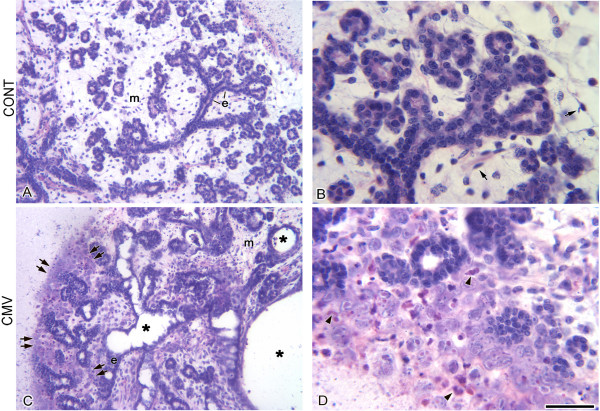
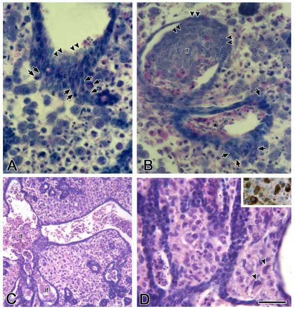
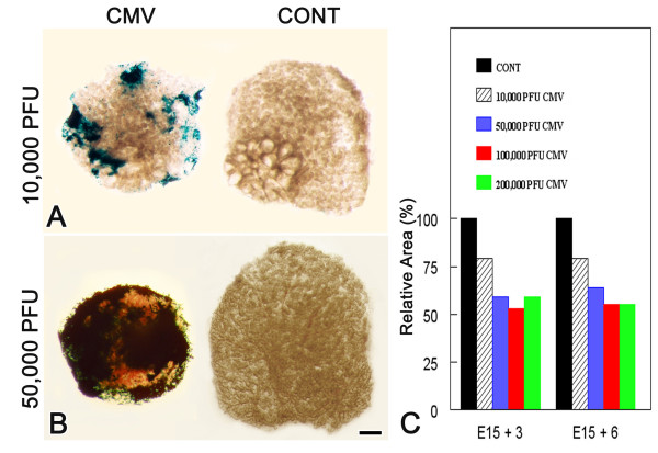
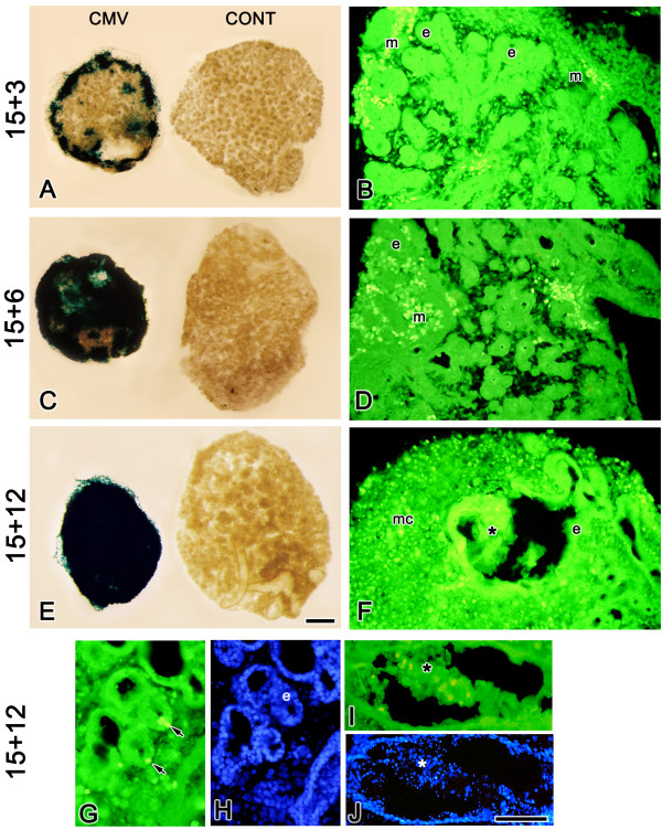
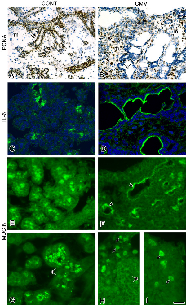
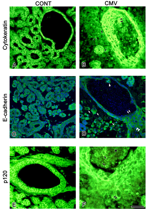
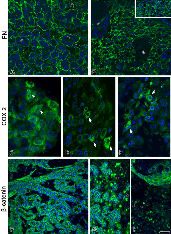
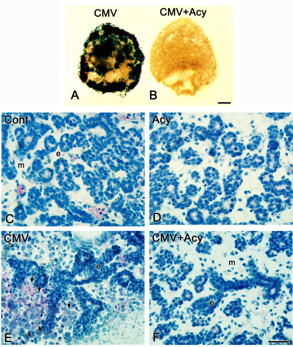
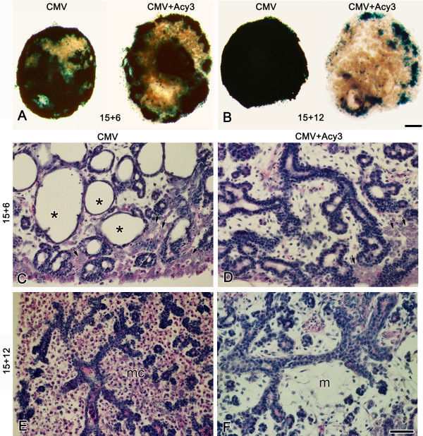
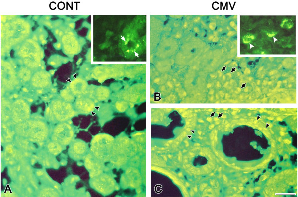
Results: We infected E15 SMG explants with mouse CMV (mCMV). Active infection for up to 12 days in vitro results in a remarkable cell and molecular pathology characterized by atypical ductal epithelial hyperplasia, apparent epitheliomesenchymal transformation, oncocytic-like stromal metaplasia, beta-catenin nuclear localization, and upregulation of Nfkb2, Relb, Il6, Stat3, and Cox2. Rescue with an antiviral nucleoside analogue indicates that mCMV replication is necessary to initiate and maintain SMG dysmorphogenesis.

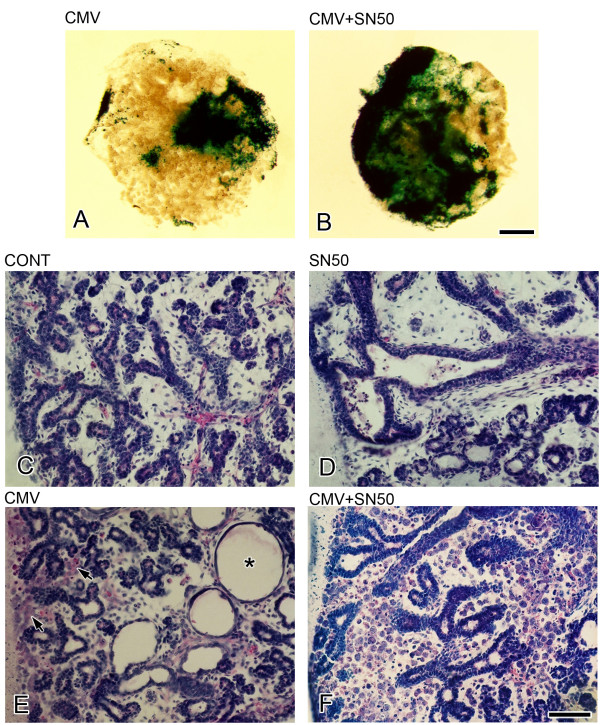
Conclusion: mCMV infection of embryonic mouse explants results in dysplasia, metaplasia, and, possibly, anaplasia. The molecular pathogenesis appears to center around the activation of canonical and, perhaps more importantly, noncanonical NFkappaB. Further, COX-2 and IL-6 are important downstream effectors of embryopathology. At the cellular level, there appears to be a consequential interplay between the transformed SMG cells and the surrounding extracellular matrix, resulting in the nuclear translocation of beta-catenin. From these studies, a tentative framework has emerged within which additional studies may be planned and performed.


Figures

Figure 1
Figure 1
Histopathology of mCMV-infected E15 + 6 SMGs. A, B. E15 + 6 SMGs have achieved the Terminal Bud Stage, consisting of ductal and terminal bud epithelia (e) which surround distinct lumina. The epithelial component is embedded in loosely-packed mesenchyme (m) sparsely populated by fibroblasts (arrows). C, D. E15 + 6 SMGs infected with 100,000 PFU mCMV exhibit a marked decrease in branching epithelia and greatly dilated lumina (*); clusters of large, basophilic, pleiomorphic cells, often with inclusion bodies (arrowheads), are seen in peripherally-localized mesenchyme (double arrows); centrally-localized cells retain their fibroblastic morphology (m). Bar: A, C: 100 μm; B, D: 20 μm.
Figure 2
Figure 2
Morphology of progressive mCMV infection in SMGs. A, B. In E15 + 10 mCMV-infected SMGs, the stromal cells are composed of 2 distinct cell types: large basophilic round cells and smaller eosinophilic cells. Basophilic cells can be seen emigrating from the epithelium into the stroma (arrows) and invaginating into the lumina (double arrowheads). Note the presence of pycnotic cells in ductal lumina (*). C-D. E15 + 12 mCMV infected SMGs exhibit a further decrease in branching epithelia and the large, dilated lumina are partially filled with eosinophilic lumina-filling cells (lf). The stroma is entirely composed of large polygonal cells with darkly staining nuclei, eosinophilic cytoplasm, and the frequent presence of inclusion bodies (arrowheads). Insert: Giant polygonal cells exhibit an increase in ATP synthetase protein (brown color), a marker of mitochondrial activity. Bar: A, B, D: 20 μm; C: 100 μm.
Figure 3
Figure 3
Dose-dependent effect of mCMV-infection on embryonic SMG development. E15+6 SMGs infected with 10,000 PFU mCMV (A) exhibit modest β-gal staining (viral presence) in the periphery, whereas SMGs infected with 50,000 PFU mCMV (B) exhibit staining almost completely throughout the gland. No β-gal staining is seen in control (CONT) SMGs. Bar:50 μm C. Relative to controls, there are significant (P < 0.01) declines in the size of mCMV-infected SMGs (as measured by area); this decline is dose dependent (P < 0.01). Comparisons of control and infected SMGs were based on matched pairs (right v. left in the same embryo). Samples sizes for each bar ranged from 6 to 29 matched pairs. True differences were determined by matched-pairs t-test. See text for details.
Figure 4
Figure 4
Time-dependent increase in mCMV infection of cultured SMGs. A, C, E. β-gal (mCMV) staining of SMGs demonstrates a marked increase in viral infection with increased days in culture. B, D, F, G, H. Immunolocalization of viral IE1 protein. In E15 + 3 (B) and E15 + 6 (D) mCMV-infected SMGs, IE1 protein is localized to mesenchymal cells (m) and not epithelial cells (e). F, G, I. In E15 + 12 mCMV-infected SMGs, IE1 protein is seen in metaplastic stromal cells (mc), in a few epithelial cells (arrows) and in lumen-filling cells (*). H. J. DAPI staining of E15 + 12 mCMV-infected SMGs shown in G, I. Bar: A: 62 μm; B-F: 50 μm; G, H-J: 25 μm.
Figure 5
Figure 5
mCMV infection, cell proliferation, IL-6 expression, and mucin expression. A, B. Cell proliferation. Cell proliferation was determined by the distribution of PCNA (brown color). In control E15 + 6 SMGs (A), PCNA positive nuclei are primarily seen in branching epithelia (double arrows) and rarely in mesenchyme (m). With mCMV infection (B), PCNA-positive nuclei are primarily seen in mesenchymal cells (arrowheads) and, to a lesser degree, in epithelial cells (e). C, D. Immunolocalization of IL-6. A substantial increase in immunodetectable IL-6 is seen in mCMV-infected SMGs ductal epithelia (D) compared to controls (C). E-I. Immunolocalization of mucin protein. In E15 + 6 (E) and E15 + 12 (G) SMGs, mucin is localized to the cytoplasm of terminal bud epithelia (e). In mCMV-infected E15 + 6 SMGs (F), there is an increase in mucin localized to epithelial apical surfaces surrounding dilated lumina (arrowheads). By day 12 (H, I), mCMV-infected SMGs are characterized by a notable decline in epithelial-localized mucin; however, mucin is found in a subpopulation of metaplastic stromal cells (arrows). Sections C, D were counterstained with DAPI. Bar: A, B: 23 μm; C-F: 20 μm; G-I: 16 μm.
Figure 6
Figure 6
Cell characterization: cytokeratin, E-cadherin, p120. A, B. In control SMGs (A), cytokeratin is immunodetected in branching epithelia and not in mesenchyme; with mCMV infection (B), cytokeratin is detected in the abnormal epithelia, in lumen-filling cells (double black arrowheads), and in the cytoplasm of stromal cells (black arrows) adjacent to pseudostratified epithelia. C, D. In control SMGs (C), E-cadherin is localized solely to epithelial cell membranes. In mCMV infected SMGs (D), a decrease in E-cadherin immunostain is seen in epithelial cells facing the lumina (double white arrows) and in pseudostratified epithelia facing the stroma (double white arrowheads); E-cadherin is also localized to membranes of lumina-filling cells (white arrowhead) and to the cytoplasm of some metaplastic stromal cells (white arrow) adjacent to abnormal epithelia. E, F. p120 localization. In control SMGs (E), p120 is detected adjacent to epithelial plasma membranes and is absent from mesenchyme. With mCMV infection (F), p120 is seen in all epithelia, as well as in cells emigrating from ductal epithelia to stroma (double black arrow) and lumen-filling cells (black arrows). C, D were counterstained with DAPI. * lumen-filling cells. Bar: A, B, E, F: 20 μm; C, D: 27 μm.
Figure 7
Figure 7
Cell characterization: fibronectin, α5β1 integrin, COX-2, and β-catenin. A, B. In control E15 + 12 SMGs (A), fibronectin (FN) is primarily localized to epithelial (e) basement membranes (arrows). In mCMV-infected SMGs (B), FN surrounds individual stromal cells and there is a marked decrease of FN in epithelial basement membranes. α5 integrin (B, insert) and β1 integrin (data not shown) are similarly localized. C-E. COX-2 is localized in E15 + 3 (C) mCMV-infected SMG fibroblasts (white arrowheads) and in metaplastic stromal cells (white arrows) of E15 + 6 (D) and E 15 + 12 (E) mCMV-infected SMGs. F-H. In control E15 + 12 SMGs (F), β-catenin is immunodetected in the cytoplasm adjacent to epithelial, but not mesenchymal, plasma membranes. With mCMV infection (G, H), β-catenin is seen in intact epithelia, as well as in nuclei of metaplastic stromal cells (arrowheads). All sections were counterstained with DAPI. Bar: 20 μm.
Figure 8
Figure 8
Acyclovir treatment inhibits mCMV replication and rescues the mCMV-induced abnormal phenotype. β-gal (mCMV) staining is seen throughout E15 + 6 SMGs infected with 50,000 PFU mCMV (A) but is absent from SMGs cultured with mCMV + acyclovir (CMV + Acy) (B). C-F. Histological analysis of control (C), acyclovir-treated (Acy) (D), mCMV-infected (E), and mCMV-infected explants treated with acyclovir (CMV + Acy) (F) SMGs. The epithelial (e) and mesenchymal (m) cellular morphology in CMV + Acy glands (F) is similar to that seen in control (C) and acyclovir-treated (D) SMGs. Note that acyclovir treatment of mCMV-infected SMGs (F) maintained the fibroblastic appearance of the mesenchyme (m); typically, mCMV-infected glands (E) exhibit clusters of large, basophilic abnormal cells (arrows) in the periphery. Bar: A, B: 50 μm; C-F: 20 μm.
Figure 9
Figure 9
Acyclovir rescues E15 SMGs infected for 3 days with mCMV and cultured for an additional 3 days (E15 + 6) (A, C, D) or 9 days (E15 + 12) (B, C, F) in the presence (CMV + Acy3) or absence (CMV) of acyclovir treatment. On day 6 (A), there is a decrease in β-gal (mCMV) staining in CMV + Acy3 SMGs compared to mCMV SMGs; by day 12 (B), acyclovir treatment results in sparse β-gal staining. At E15+6, with mCMV infection alone (C), abnormal epithelia surround dilated lumina (*) and clusters of atypical basophilic cells are seen in the periphery (arrows); in contrast, acyclovir treatment (D) partially restores the epithelial phenotype to that seen in control SMGs, with fewer atypical mesenchymal cells (arrows). At E15 + 12, acyclovir treatment (F) results in the near normal appearance of epithelia and mesenchyme (m); this markedly differs from the histopathology seen in E15 + 12 CMV-infected SMGs (E). mc-metaplastic stromal cells. Bar: A, B: 50 μm; C-F: 30 μm.
Figure 10
Figure 10
mCMV infection and SN50 inhibition of canonical NFκB nuclear translocation. With mCMV+SN50 treatment of E15 SMGs (B), there is a notable increase in β-gal (mCMV) staining compared to mCMV infection alone (A). Control E15 + 6 SMGs (C) are at the Terminal Bud Stage with epithelia surrounding distinct lumina; E15 + 6 SN50-treated SMGs (D) are characterized by a modest decrease in epithelia and larger, somewhat dilated lumina. The glandular morphology of CMV + SN50 (F) differs from that seen with SN50 (D) or mCMV infection (E) alone. mCMV-infected E15 + 6 SMGs (E) exhibit peripherally-localized clusters of large, atypical, basophilic cells (arrows) and greatly dilated lumen (*); by contrast, the stroma of the CMV + SN50 E15 + 6 SMGs is almost entirely composed of polygonal cells with darkly staining nuclei and often eosinophilic cytoplasm, the metaplastic cell type typical of E15 + 12 mCMV-infected SMGs (compare to Fig. 9E). Bar: A, B: 50 μm; C-F: 30 μm.
Figure 11
Figure 11
Immunolocalization of RelB and NFκB2 in mCMV-infected E15 + 6 SMGs. A-C. RelB localization. Insert: NFκB2 localization. In control SMGs, RelB (A) (black arrowheads) and NFκB2 (insert, white arrows) are seen in epithelia surrounding forming lumina and not in the stroma. With CMV infection (B, C), RelB is seen in the nuclei of stromal cells (black arrows), many of which exhibit inclusion bodies. RelB is also seen in the nuclei of ductal epithelia (black arrowheads) surrounding enlarged lumina (C). In CMV-infected glands, NFκB2 is seen in the cytoplasm of large stroma cells (insert, white arrowheads) and epithelia surrounding lumina (data not shown). Bar: 50 μm.
Figure 12
Figure 12
Working model of CMV-induced salivary gland dysmorphology. → Single or multistep stimulatory modification. formula image 
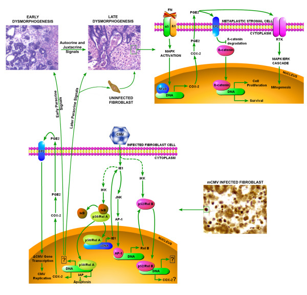
single or multistep stimulatory modification of unknown mechanism. ⊣ single or multistep inhibitory modification. RKT: Receptor Tyrosine Kinase. DNA: host DNA. vDNA: viral DNA.

Similar articles

Cited by

References

    1. Farber S. Wolbach SB Intranuclear and cytoplasmic inclusions ("protozoan-like bodies") in the salivary glands and other organs of infants. Am J Pathol. 1932;8:123–135. - PMC - PubMed
    1. Smith MG Propagation in tissue cultures of a cytopathogenic virus from human salivary gland virus (SGV) disease. Proc Soc Exp Biol Med. 1956;92:424–430. - PubMed
    1. Weller TH The cytomegaloviruses: Ubiquitous agents with protean clinical manifestations. I. N Engl J Med. 1971;285:203–214. - PubMed
    1. Weller TH The cytomegaloviruses: Ubiquitous agents with protean clinical manifestations. II. N Engl J Med. 1971;285:267–274. - PubMed
    1. Davison AJ . Comparative betaherpesvirus genome and virion structure. In: Arvin AM, Mocarski ES, Moore P, Whitley R, Yamanishi K, Gampadelli-Fiume G and Roizman B, editor. Human Herpesviruses: Biology, Therapy, and Immunoprophylaxis. Cambridge, Cambridge Press; 2006. - PubMed

Publication types