PD-1 is a regulator of virus-specific CD8+ T cell survival in HIV infection
- PMID: 16954372
- PMCID: PMC2118095
- DOI: 10.1084/jem.20061496
PD-1 is a regulator of virus-specific CD8+ T cell survival in HIV infection
Abstract
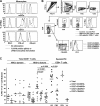
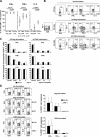
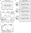
Here, we report on the expression of programmed death (PD)-1 on human virus-specific CD8(+) T cells and the effect of manipulating signaling through PD-1 on the survival, proliferation, and cytokine function of these cells. PD-1 expression was found to be low on naive CD8(+) T cells and increased on memory CD8(+) T cells according to antigen specificity. Memory CD8(+) T cells specific for poorly controlled chronic persistent virus (HIV) more frequently expressed PD-1 than memory CD8(+) T cells specific for well-controlled persistent virus (cytomegalovirus) or acute (vaccinia) viruses. PD-1 expression was independent of maturational markers on memory CD8(+) T cells and was not directly associated with an inability to produce cytokines. Importantly, the level of PD-1 surface expression was the primary determinant of apoptosis sensitivity of virus-specific CD8(+) T cells. Manipulation of PD-1 led to changes in the ability of the cells to survive and expand, which, over several days, affected the number of cells expressing cytokines. Therefore, PD-1 is a major regulator of apoptosis that can impact the frequency of antiviral T cells in chronic infections such as HIV, and could be manipulated to improve HIV-specific CD8(+) T cell numbers, but possibly not all functions in vivo.
Figures


Similar articles
-
HCV-specific T cells in HCV/HIV co-infection show elevated frequencies of dual Tim-3/PD-1 expression that correlate with liver disease progression.Eur J Immunol. 2010 Sep;40(9):2493-505. doi: 10.1002/eji.201040340. Eur J Immunol. 2010. PMID: 20623550
-
Combined Env- and Gag-specific T cell responses in relation to programmed death-1 receptor and CD4 T cell loss rates in human immunodeficiency virus-1 infection.Clin Exp Immunol. 2010 Aug;161(2):315-23. doi: 10.1111/j.1365-2249.2010.04179.x. Epub 2010 May 10. Clin Exp Immunol. 2010. PMID: 20491784 Free PMC article.
-
Differential association of programmed death-1 and CD57 with ex vivo survival of CD8+ T cells in HIV infection.J Immunol. 2009 Jul 15;183(2):1120-32. doi: 10.4049/jimmunol.0900182. Epub 2009 Jun 29. J Immunol. 2009. PMID: 19564339 Free PMC article.
-
Revisiting immune exhaustion during HIV infection.Curr HIV/AIDS Rep. 2011 Mar;8(1):4-11. doi: 10.1007/s11904-010-0066-0. Curr HIV/AIDS Rep. 2011. PMID: 21188556 Free PMC article. Review.
-
Costimulatory molecule programmed death-1 in the cytotoxic response during chronic hepatitis C.World J Gastroenterol. 2009 Nov 7;15(41):5129-40. doi: 10.3748/wjg.15.5129. World J Gastroenterol. 2009. PMID: 19891011 Free PMC article. Review.
Cited by
-
Molecular signatures of T-cell inhibition in HIV-1 infection.Retrovirology. 2013 Mar 20;10:31. doi: 10.1186/1742-4690-10-31. Retrovirology. 2013. PMID: 23514593 Free PMC article. Review.
-
CD4:CD8 lymphocyte ratio as a quantitative measure of immunologic health in HIV-1 infection: findings from an African cohort with prospective data.Front Microbiol. 2015 Jul 1;6:670. doi: 10.3389/fmicb.2015.00670. eCollection 2015. Front Microbiol. 2015. PMID: 26191056 Free PMC article.
-
Interleukin 15 upregulates the expression of PD-1 and TIM-3 on CD4+ and CD8+ T cells.Am J Clin Exp Immunol. 2020 Jun 15;9(3):10-21. eCollection 2020. Am J Clin Exp Immunol. 2020. PMID: 32704430 Free PMC article.
-
Decrease of T-cells exhaustion markers programmed cell death-1 and T-cell immunoglobulin and mucin domain-containing protein 3 and plasma IL-10 levels after successful treatment of chronic hepatitis C.Sci Rep. 2020 Sep 29;10(1):16060. doi: 10.1038/s41598-020-73137-6. Sci Rep. 2020. PMID: 32994477 Free PMC article.
-
A participant-derived xenograft model of HIV enables long-term evaluation of autologous immunotherapies.J Exp Med. 2021 Jul 5;218(7):e20201908. doi: 10.1084/jem.20201908. Epub 2021 May 14. J Exp Med. 2021. PMID: 33988715 Free PMC article.
References
-
- Gougeon, M.L. 2003. Apoptosis as an HIV strategy to escape immune attack. Nat. Rev. Immunol. 3:392–404. - PubMed
-
- Johnson, W.E., and R.C. Desrosiers. 2002. Viral persistance: HIV's strategies of immune system evasion. Annu. Rev. Med. 53:499–518. - PubMed
-
- van Kooyk, Y., and T.B. Geijtenbeek. 2003. DC-SIGN: escape mechanism for pathogens. Nat. Rev. Immunol. 3:697–709. - PubMed
Publication types
MeSH terms
Substances
Grants and funding
LinkOut - more resources
Full Text Sources
Other Literature Sources
Medical
Research Materials

