Regulation of peripheral inflammation by spinal p38 MAP kinase in rats
- PMID: 16953659
- PMCID: PMC1560929
- DOI: 10.1371/journal.pmed.0030338
Regulation of peripheral inflammation by spinal p38 MAP kinase in rats
Abstract
Background: Somatic afferent input to the spinal cord from a peripheral inflammatory site can modulate the peripheral response. However, the intracellular signaling mechanisms in the spinal cord that regulate this linkage have not been defined. Previous studies suggest spinal cord p38 mitogen-activated protein (MAP) kinase and cytokines participate in nociceptive behavior. We therefore determined whether these pathways also regulate peripheral inflammation in rat adjuvant arthritis, which is a model of rheumatoid arthritis.
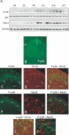
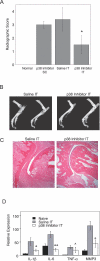
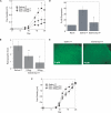
Methods and findings: Selective blockade of spinal cord p38 MAP kinase by administering the p38 inhibitor SB203580 via intrathecal (IT) catheters in rats with adjuvant arthritis markedly suppressed paw swelling, inhibited synovial inflammation, and decreased radiographic evidence of joint destruction. The same dose of SB203580 delivered systemically had no effect, indicating that the effect was mediated by local concentrations in the neural compartment. Evaluation of articular gene expression by quantitative real-time PCR showed that spinal p38 inhibition markedly decreased synovial interleukin-1 and -6 and matrix metalloproteinase (MMP3) gene expression. Activation of p38 required tumor necrosis factor alpha (TNFalpha) in the nervous system because IT etanercept (a TNF inhibitor) given during adjuvant arthritis blocked spinal p38 phosphorylation and reduced clinical signs of adjuvant arthritis.
Conclusions: These data suggest that peripheral inflammation is sensed by the central nervous system (CNS), which subsequently activates stress-induced kinases in the spinal cord via a TNFalpha-dependent mechanism. Intracellular p38 MAP kinase signaling processes this information and profoundly modulates somatic inflammatory responses. Characterization of this mechanism could have clinical and basic research implications by supporting development of new treatments for arthritis and clarifying how the CNS regulates peripheral immune responses.
Conflict of interest statement
Figures


Comment in
-
Spinal delivery of p38: TNF-alpha inhibitors.PLoS Med. 2006 Nov;3(11):e511. doi: 10.1371/journal.pmed.0030511. PLoS Med. 2006. PMID: 17132064 Free PMC article. No abstract available.
Similar articles
-
Inhibition of spinal p38 MAPK prevents articular neutrophil infiltration in experimental arthritis via sympathetic activation.Fundam Clin Pharmacol. 2018 Apr;32(2):155-162. doi: 10.1111/fcp.12338. Epub 2017 Dec 22. Fundam Clin Pharmacol. 2018. PMID: 29206314 Free PMC article.
-
Predominant activation of MAP kinases and pro-destructive/pro-inflammatory features by TNF alpha in early-passage synovial fibroblasts via TNF receptor-1: failure of p38 inhibition to suppress matrix metalloproteinase-1 in rheumatoid arthritis.Ann Rheum Dis. 2007 Aug;66(8):1043-51. doi: 10.1136/ard.2006.062521. Epub 2007 Jan 12. Ann Rheum Dis. 2007. PMID: 17223661 Free PMC article.
-
Activation of p38 MAPK is a key step in tumor necrosis factor-mediated inflammatory bone destruction.Arthritis Rheum. 2006 Feb;54(2):463-72. doi: 10.1002/art.21626. Arthritis Rheum. 2006. PMID: 16447221
-
Does gamma-aminobutyric acid (GABA) influence the development of chronic inflammation in rheumatoid arthritis?J Neuroinflammation. 2008 Jan 3;5:1. doi: 10.1186/1742-2094-5-1. J Neuroinflammation. 2008. PMID: 18171484 Free PMC article. Review.
-
[p38 MAP Kinase inhibitor].Nihon Rinsho Meneki Gakkai Kaishi. 2007 Oct;30(5):390-7. doi: 10.2177/jsci.30.390. Nihon Rinsho Meneki Gakkai Kaishi. 2007. PMID: 17984579 Review. Japanese.
Cited by
-
p38(MAPK): stress responses from molecular mechanisms to therapeutics.Trends Mol Med. 2009 Aug;15(8):369-79. doi: 10.1016/j.molmed.2009.06.005. Epub 2009 Aug 6. Trends Mol Med. 2009. PMID: 19665431 Free PMC article. Review.
-
The as-yet unfulfilled promise of p38 MAPK inhibitors.Nat Rev Rheumatol. 2009 Sep;5(9):475-7. doi: 10.1038/nrrheum.2009.171. Nat Rev Rheumatol. 2009. PMID: 19710669 No abstract available.
-
Regulation of peripheral inflammation by the central nervous system.Curr Rheumatol Rep. 2010 Oct;12(5):370-8. doi: 10.1007/s11926-010-0124-z. Curr Rheumatol Rep. 2010. PMID: 20676807 Free PMC article.
-
Anti-hyperalgesic effects of photobiomodulation therapy (904 nm) on streptozotocin-induced diabetic neuropathy imply MAPK pathway and calcium dynamics modulation.Sci Rep. 2022 Oct 6;12(1):16730. doi: 10.1038/s41598-022-19947-2. Sci Rep. 2022. PMID: 36202956 Free PMC article.
-
Go-sha-jinki-Gan Alleviates Inflammation in Neurological Disorders via p38-TNF Signaling in the Central Nervous System.Neurotherapeutics. 2021 Jan;18(1):460-473. doi: 10.1007/s13311-020-00948-w. Epub 2020 Oct 20. Neurotherapeutics. 2021. PMID: 33083995 Free PMC article.
References
-
- Sluka KA, Westlund KN. Centrally administered non-NMDA but not NMDA receptor antagonists block peripheral knee joint inflammation. Pain. 1993;55:217–225. - PubMed
-
- Boyle DL, Moore J, Yang L, Sorkin LS, Firestein GS. Spinal adenosine receptor activation inhibits inflammation and joint destruction in rat adjuvant-induced arthritis. Arthritis Rheum. 2002;46:3076–3082. - PubMed
-
- Svensson CI, Marsala M, Westerlund A, Calcutt NA, Campana WM, et al. Activation of p38 mitogen-activated protein kinase in spinal microglia is a critical link in inflammation-induced spinal pain processing. J Neurochem. 2003;86:1534–1544. - PubMed
-
- Yaksh TL, Rudy RA. Chronic catheterization of the spinal subarachnoid space. Physiol Behav. 1976;17:1031–1036. - PubMed
Publication types
MeSH terms
Substances
Grants and funding
LinkOut - more resources
Full Text Sources
Other Literature Sources
Medical
Miscellaneous

