. 2006 Nov;17(11):4748-59.
doi: 10.1091/mbc.e06-03-0183. Epub 2006 Aug 30.

Dual role of the cysteine-string domain in membrane binding and palmitoylation-dependent sorting of the molecular chaperone cysteine-string protein

Affiliations

Dual role of the cysteine-string domain in membrane binding and palmitoylation-dependent sorting of the molecular chaperone cysteine-string protein

Jennifer Greaves et al. Mol Biol Cell. 2006 Nov.

Abstract

S-palmitoylation occurs on intracellular membranes and, therefore, membrane anchoring of proteins must precede palmitate transfer. However, a number of palmitoylated proteins lack any obvious membrane targeting motifs and it is unclear how this class of proteins become membrane associated before palmitoylation. Cysteine-string protein (CSP), which is extensively palmitoylated on a "string" of 14 cysteine residues, is an example of such a protein. In this study, we have investigated the mechanisms that govern initial membrane targeting, palmitoylation, and membrane trafficking of CSP. We identified a hydrophobic 31 amino acid domain, which includes the cysteine-string, as a membrane-targeting motif that associates predominantly with endoplasmic reticulum (ER) membranes. Cysteine residues in this domain are not merely sites for the addition of palmitate groups, but play an essential role in membrane recognition before palmitoylation. Membrane association of the cysteine-string domain is not sufficient to trigger palmitoylation, which requires additional downstream residues that may regulate the membrane orientation of the cysteine-string domain. CSP palmitoylation-deficient mutants remain "trapped" in the ER, suggesting that palmitoylation may regulate ER exit and correct intracellular sorting of CSP. These results reveal a dual function of the cysteine-string domain: initial membrane binding and palmitoylation-dependent sorting.


Figures
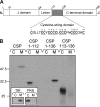
Figure 1.
Figure 1.
The cysteine-string domain of CSP is not sufficient for membrane association in vivo. (A) Schematic diagram of the domains present and their location in mammalian CSP1. The amino acids in the cysteine-string domain are shown, with the cysteine residues underlined and numbered consecutively. The amino acid numbers corresponding to domain boundaries are indicated. (B) PC12 cells were transfected with wild-type EGFP-CSP, EGFP-CSP(1-112), EGFP-CSP(1-136), or EGFP-CSP(113-136). Forty-eight hours after transfection, the cells were separated into cytosolic (C) and membrane (M) fractions. Expression of the EGFP-CSP constructs in each fraction was detected by immunoblotting with an anti-GFP mAb. The inset shows the distribution of control proteins, the transferrin receptor (TfR, membrane protein), and protein kinase B (PKB, cytosolic protein) in cytosol and membrane fractions. Position of molecular-weight standards are indicated.
Figure 2.
Figure 2.
Analysis of the minimum membrane-binding domain of CSP. PC12 cells transfected with EGFP-CSP truncation mutants were separated into cytosolic (C) and membrane (M) fractions, and the distribution of the mutants was determined by immunoblotting with an anti-GFP antibody. (A) Distribution of CSP truncation mutants containing the linker domain and cysteine-string domain only (residues 84-136), residues 93-136, residues 103-136, and the cysteine-string domain alone (residues 113-136). (B) Distribution of CSP truncation mutants containing residues 106-136, 107-136, and 108-136 of CSP. (C) Alignment of the primary sequence of residues 106-136 of CSP1 from various species. Conserved residues are shaded, cysteine residues are shown in bold and numbering of amino acids is indicated at the top.
Figure 3.
Figure 3.
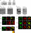
CSP mutants that lack the C-terminal domain are inefficiently palmitoylated and colocalize with ER marker proteins. (A) PC12 cells expressing EGFP-CSP, EGFP-CSP(1-136), and EGFP-CSP(106-136) were fractionated into cytosolic (C) and membrane (M) fractions. Isolated membranes were further incubated in 1 M Tris, pH 7 (T) or 1 M hydroxylamine, pH 7 (HA), for 20 h at room temperature. Membranes were recovered by centrifugation and analyzed by immunoblotting with an anti-GFP mAb. Note that HA treatment promotes an ∼8-kDa band-shift in EGFP-CSP as a result of depalmitoylation. In contrast, EGFP-CSP(1-136) and EGFP-CSP(106-136) exhibit no detectable band-shift, suggesting these constructs are inefficiently palmitoylated. Position of molecular weight standards are indicated. (B) PC12 cells transfected with CSP or CSP(1-136) were incubated for 4 h in media containing 1 mCi/ml [3H]palmitic acid. GFP-labeled proteins were isolated by magnetic separation of microbeads coupled to GFP antibody, and the recovered proteins were resolved by SDS-PAGE and transferred to duplicate nitrocellulose membranes. Left panel, nitrocellulose membrane immunoblotted with anti-GFP. Right panel, nitrocellulose membrane processed for fluorographic detection of [3H]palmitic acid. The arrowheads in the panels denote position of palmitoylated forms of CSP, whereas the asterisks indicated nonpalmitoylated CSPs. (C) As in B, except with increased gel loading of the CSP(1-136) mutant and longer 3H exposure time. Note that when gel loading of immunoprecipitated CSP(1-136) is increased, a faint higher molecular weight species is detected (arrowhead). The 3H label is only incorporated into this minor higher molecular weight band, whereas the major pool of CSP(1-136) (asterisk) is completely devoid of the 3H signal. The faint lower molecular weight band labeled with 3H in WT CSP is a degraded form of palmitoylated CSP and migrates faster than nonpalmitoylated CSP. (D) PC12 cells expressing EGFP-CSP(1-136) or EGFP-CSP(106-136) were fixed, permeabilized, and incubated with a polyclonal anti-CSP antibody to label endogenous CSP. The CSP staining was detected with a rhodamine-conjugated anti-rabbit IgG (red) and compared with the GFP signal. Scale bar, 10 μm. (E) PC12 cells transfected with EGFP-CSP(1-136) were fixed, permeablized, and stained with a polyclonal anti-calreticulin antibody. Calreticulin staining was detected with a rhodamine-conjugated anti-rabbit IgG (red) and compared with the GFP signal. Scale bar, 10 μm. The highlighted region of the cells shown in this image has been enlarged and is shown in the bottom panel. (F) PC12 cells were cotransfected with either EGFP-CSP, EGFP-CSP(1-136), or EGFP-CSP(106-136) and dsRed-ER. Colocalization of the transfected proteins was examined by confocal imaging. Scale bar, 10 μm.
Figure 4.
Figure 4.
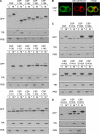
Residues downstream of the cysteine-string domain are required for efficient palmitoylation and correct intracellular sorting of CSP. (A) Cytosol (C) and membrane (M) distribution of EGFP-CSP C-terminal truncation mutants. Top, in contrast to CSP(1-136), CSP(1-146), CSP(1-156), and CSP(1-166) are all efficiently palmitoylated, as indicated by the band-shift detected for membrane-bound CSP compared with cytosolic CSP. Middle and bottom panels, the intensity of palmitoylation increases with the addition of successive C-terminal amino acid residues. (B) PC12 cells transfected with EGFP-CSP(1-146) were costained for calreticulin (red) and analyzed by confocal imaging. Note that the CSP(1-146) staining does not overlap with calreticulin, but displays PM staining similar to that of wild-type EGFP-CSP, shown in Figure 3F. (C) Cytosolic (C) and membrane (M) distribution of EGFP-CSP and the following point mutants: K137A, P138A, K139A, A140L, P141A, E142A, and G143A. Note that the K137A mutation decreases membrane association and palmitoylation of CSP. (D) Fractionation analysis of the following mutations introduced into EGFP-CSP: K137A, K137E, and K137R. As a control, the distribution of transferrin receptor (TfR) and protein kinase B (PKB) in the recovered cytosolic and membrane fractions is also shown.
Figure 5.
Figure 5.
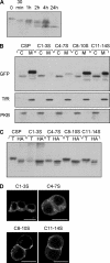
Membrane binding and subcellular localization of cysteine-to-serine mutants. (A) A membrane fraction was prepared from PC12 cells and incubated in 1 M Tris (pH 7) for 20 h (0), or 1 M hydroxylamine (pH 7) for various time points (30 min and 1, 2, 4, or 24 h). The treated membranes were recovered by centrifugation and analyzed by immunoblotting with an anti-CSP polyclonal antibody. Note that a number of immunoreactive bands were detected, consistent with multiple palmitoylation of CSP. (B) Cytosolic (C) and membrane (M) fractions prepared from cells expressing wild-type EGFP-CSP, C1-3S, C4-7S, C8-10S, or C11-14S. Samples were probed with a GFP antibody. As a control, the distribution of transferrin receptor (TfR) and protein kinase B (PKB) in the recovered cytosolic and membrane fractions is also shown. (C) Membrane fractions of PC12 cells expressing C1-3S, C4-7S, C8-10S, or C11-14S were incubated in 1 M Tris (pH 7) or 1 M hydroxylamine (pH 7) for 20 h at room temperature. Membranes were recovered and immunoblotted with anti-GFP antibody. (D) Immunolocalization of the cysteine-string domain serine mutants (C1-3S, C4-7S, C8-10S, and C11-14S) in transfected PC12 cells. Scale bar, 10 μm.
Figure 6.
Figure 6.
Analysis of membrane binding, palmitoylation, and subcellular localization of C1-3A, C4-7A, and C4-7L mutants. (A) PC12 cells transfected with C1-3S and C1-3A (left panel) or C4-7S, C4-7A, and C4-7L (right panel) were fractionated into cytosolic (C) and membrane (M) fractions. Protein distribution was examined by immunoblotting with a GFP antibody. As a control, the distribution of transferrin receptor (TfR) and protein kinase B (PKB) in the recovered cytosolic and membrane fractions is also shown. (B) PC12 cells transfected with wild-type CSP, CSP(1-136), C4-7L, or C1-3A mutants were incubated for 4 h in media containing 1 mCi/ml [3H]palmitic acid. GFP-labeled proteins were isolated by magnetic separation of microbeads coupled to GFP antibody, and the recovered proteins were resolved by SDS-PAGE and transferred to duplicate nitrocellulose membranes. Left, nitrocellulose membrane immunoblotted with anti-GFP. Right, nitrocellulose membrane processed for fluorographic detection of [3H]palmitic acid. The arrowheads in the panels denote position of palmitoylated forms of CSP, whereas the asterisks indicated nonpalmitoylated CSPs. The faint lower molecular weight band labeled with 3H in WT CSP and C1-3A samples is a degraded form of palmitoylated CSP, detected upon longer exposure of immunoblots, and migrates faster than nonpalmitoylated CSP. (C) Distribution of the C1-3A and C4-7L mutants (green) in PC12 cells was compared with calreticulin (red). Note that cells expressing C4-7L mutant were permeabilized with digitonin before fixation to allow leakage of cytosolic proteins. Scale bar, 10 μm.
Figure 7.
Figure 7.
Analysis of membrane binding of EGFP-CSP, CSP(1-136), and CSP(C4-7L). (A) Membranes were prepared from PC12 cells transfected with EGFP-CSP, CSP(1-136), and CSP(C4-7L). The membranes were incubated in either HES buffer, 1 M NaCl, 0.1 M sodium carbonate, pH 11.5 (Na2CO3), or 1% Triton X-100 for 30 min at 4°C. Supernatant (S) and pelleted membrane (P) fractions were separated by centrifugation and probed with antibodies against GFP, munc18-1, and alpha-SNAP. (B) Membranes prepared from cells expressing EGFP-CSP and C4-7L were incubated in the presence and absence of proteinase K and 0.2% Triton X-100 for 30 min on ice (as indicated). The samples were resolved by SDS-PAGE and transferred to nitrocellulose for immunoblotting analysis using antibodies recognizing GFP, CSP, or calreticulin. The band highlighted by the arrowhead in the center panel represents endogenous CSP.

Similar articles

Cited by

References

    1. Apolloni A., Prior I. A., Lindsay M., Parton R. G., Hancock J. F. H-ras but not K-ras traffics to the plasma membrane through the exocytic pathway. Mol. Cell. Biol. 2000;20:2475–2487. - PMC - PubMed
    1. Bartels D. J., Mitchell D. A., Dong X., Deschenes R. J. Erf2, a novel gene product that affects the localisation and palmitoylation of Ras2 in Saccharomyces cerevisiae. Mol. Biol. Cell. 1999;19:6775–6787. - PMC - PubMed
    1. Boal F., Zhang H., Tessier C., Scotti P., Lang J. The variable C-terminus of cysteine string proteins modulates exocytosis and protein-protein interactions. Biochemistry. 2004;43:16212–16223. - PubMed
    1. Braun J. E., Scheller R. H. Cysteine string protein, a DnaJ family member, is present on diverse secretory vesicles. Neuropharmacology. 1995;34:1361–1369. - PubMed
    1. Brown H., et al. Cysteine string protein (CSP) is an insulin secretory granule-associated protein regulating beta-cell exocytosis. EMBO J. 1998;17:5048–5058. - PMC - PubMed

Publication types

MeSH terms

LinkOut - more resources