. 2006 Nov;26(21):8159-72.
doi: 10.1128/MCB.00680-06. Epub 2006 Aug 28.

Specific recognition of ZNF217 and other zinc finger proteins at a surface groove of C-terminal binding proteins

Affiliations

Specific recognition of ZNF217 and other zinc finger proteins at a surface groove of C-terminal binding proteins

Kate G R Quinlan et al. Mol Cell Biol. 2006 Nov.

Abstract

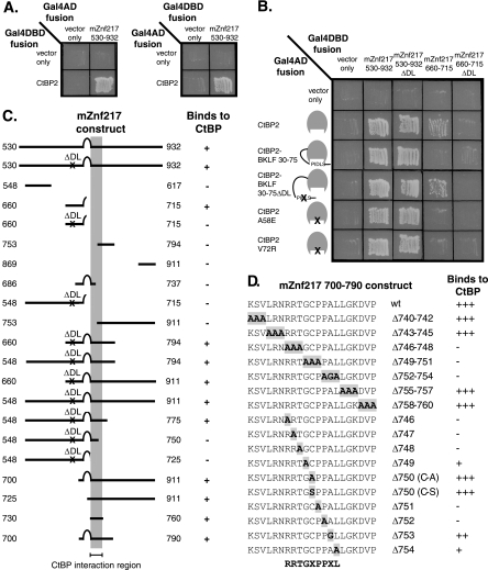
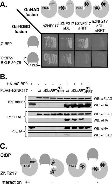
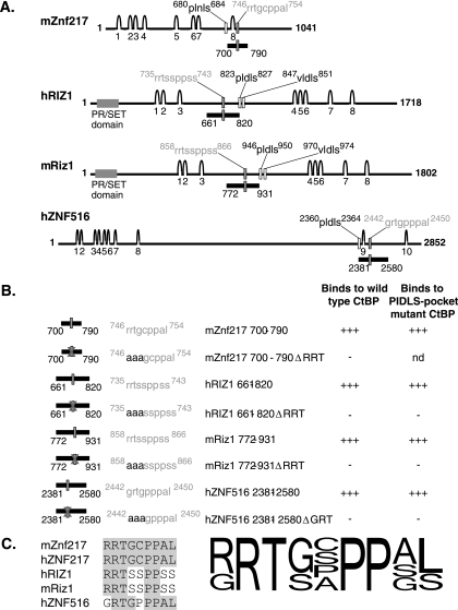
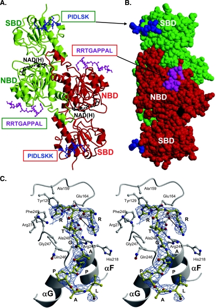
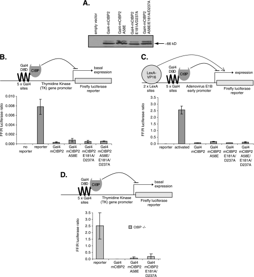
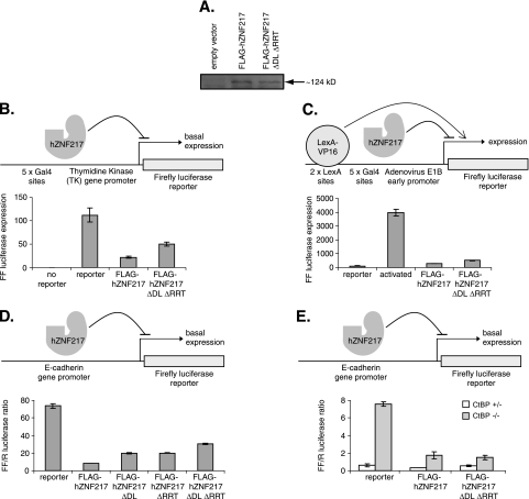
Numerous transcription factors recruit C-terminal binding protein (CtBP) corepressors. We show that the large zinc finger protein ZNF217 contacts CtBP. ZNF217 is encoded by an oncogene frequently amplified in tumors. ZNF217 contains a typical Pro-X-Asp-Leu-Ser (PXDLS) motif that binds in CtBP's PXDLS-binding cleft. However, ZNF217 also contains a second motif, Arg-Arg-Thr (RRT), that binds a separate surface on CtBP. The crystal structure of CtBP bound to an RRTGAPPAL peptide shows that it contacts a surface crevice distinct from the PXDLS binding cleft. Interestingly, both PXDLS and RRT motifs are also found in other zinc finger proteins, such as RIZ. Finally, we show that ZNF217 represses several promoters, including one from a known CtBP target gene, and mutations preventing ZNF217's contact with CtBP reduce repression. These results identify a new CtBP interaction motif and establish ZNF217 as a transcriptional repressor protein that functions, at least in part, by associating with CtBP.


Figures

FIG. 1.
FIG. 1.
The human and murine ZNF217 protein sequences show significant homology. The sequences of full-length human ZNF217 (NM_006526) and murine Znf217 (NM_001033299) proteins are shown. The zinc finger regions (1 to 8) are underlined with solid gray lines. The conserved PXDLS motifs and RRT motifs are underlined with gray dashed lines. Residues 530 (lysine) and 932 (glycine) of mZnf217, the first and last amino acids of the yeast two-hybrid screen isolate, are indicated with asterisks. Identical and similar residues are boxed in black and gray, respectively. Dashes indicate gaps introduced to maintain alignment.
FIG. 2.
FIG. 2.
Znf217 interacts with CtBP2, and the non-PXDLS interaction site was mapped to the motif RRTGXPPXL. A. Yeast two-hybrid assays were performed to examine the interactions between mZnf217 530-932 and CtBP2. These assays were performed with each of the two test proteins fused to the C terminus of either Gal4AD or Gal4DBD. Growth on -His-Leu-Trp plates (pictured) indicates that the two test proteins interact. B. The PXDLS motifs in Znf217 530-932 and Znf217 660-715 were mutated, and both wild-type and mutant proteins were tested for binding to wild-type CtBP and CtBP with mutations in the PXDLS cleft. Interaction with only wild-type CtBP (and the BKLF 30-75 ΔDL control protein) indicates binding which is dependent on a PXDLS motif. Interaction with both wild-type and mutant CtBPs indicates binding which is not dependent on a PXDLS motif. C. Deletion mapping was performed to determine the minimal portion of murine Znf217 capable of interacting with CtBP in a PXDLS motif-independent manner. The Gal4DBD-Znf217 proteins are depicted schematically, and results of yeast two-hybrid assays with these proteins and Gal4AD-CtBP are shown as either plus (for growth of yeast) or minus. A ΔDL mutation (NL-AS in the PLNLS motif) was introduced into many of the mZnf217 proteins so that only non-PXDLS binding was being examined, and this is indicated. The minimal region of mZnf217 required for interaction with CtBP, amino acids 730 to 760, is indicated by a gray column. D. Both single and triple mutations were introduced into amino acids 740 to 760 of Gal4DBD-Znf217 700-790. The mutations in each of the constructs are highlighted within the sequence of Znf217 amino acids 740 to 760. The results of yeast two-hybrid assays with these mutant Gal4DBD-mZnf217 proteins and Gal4AD-CtBP are shown as either pluses (for relative growth of yeast) or minus. The consensus motif suggested, RRTGXPPXL, is shown below.
FIG. 3.
FIG. 3.
Mutation of the PXDLS and RRT motifs of ZNF217 reduce the ability to bind to CtBP, and the double mutant has a severe reduction in binding. A. Gal4AD-fused wild-type hZNF217 or hZNF217 with mutations in the PLNLS motif (ΔDL), RRTGCPPAL motif (ΔRRT), or both motifs (ΔDL ΔRRT) was examined for its ability to interact with Gal4DBD-fused wild-type or cleft-filled (CtBP2-BKLF 30-75) CtBP in yeast two-hybrid assays. B. Fusion of FLAG and wild-type hZNF217 or hZNF217 with mutations in the PLNLS motif (ΔDL), RRTGCPPAL motif (ΔRRT), or both motifs (ΔDL ΔRRT) was examined for its ability to interact with HA-CtBP2 in coimmunoprecipitation experiments. COS-1 cells were transfected with the expression vectors indicated, and whole-cell extracts were immunoprecipitated (IP) separately with both the anti-FLAG (αFLAG) and anti-HA (αHA) antibodies. Expression of each of the FLAG-fused and HA-fused proteins is shown in the top two panels (10% input). FLAG-ZNF217 immunoprecipitated by the anti-FLAG antibody and the resulting coimmunoprecipitated HA-CtBP2 is shown in the middle two panels (IP: αFLAG). HA-CtBP2 immunoprecipitated by the anti-HA antibody and the resulting coimmunoprecipitated FLAG-hZNF217 is shown in the bottom two panels (IP: αHA). C. A summary diagram combining the results of interaction studies between CtBP and wild-type or mutant ZNF217.
FIG. 4.
FIG. 4.
RRT motifs are also found in RIZ and ZNF516 and are capable of mediating binding to CtBP. A. mZnf217, hRIZ1, mRiz1, and hZNF516 are large zinc finger proteins which possess both PXDLS motifs and putative RRT motifs. The features of each protein are shown. The predicted zinc fingers are shown as arches and numbered, the consensus PXDLS motifs are indicated by hollow rectangles with the motifs outlined above in black, and the putative RRT motifs are indicated by gray filled rectangles with the motifs outlined above in gray. The PR/SET domains in the RIZ proteins are indicated by wide gray rectangles. The portion of each protein containing the putative RRT motif which was tested for interaction with CtBP is indicated below each sequence as a black bar with the numbers of the flanking amino acids indicated. B. Segments of ZNF516 and both murine and human RIZ1 with and without mutations in the putative RRT motifs fused to Gal4DBD were tested for their ability to bind to wild-type and cleft mutant CtBP fused to Gal4AD in yeast two-hybrid assays. nd, not determined. C. An alignment is shown of the RRT motifs that have been shown to mediate binding to CtBP. Amino acids within the sequences which are identical to the amino acids in the mZnf217 RRT motif are boxed in gray. The consensus combines information obtained from validated natural RRT motifs and also from mutagenesis studies. The height of each amino acid at each position is representative of the relative frequency in the naturally occurring RRT motif proteins and the tolerance for various amino acids as determined by mutational analysis.
FIG. 5.
FIG. 5.
X-ray crystal structure of the RRTGAPPAL peptide bound to t-CtBP1-S. A. Ribbon diagram of the t-CtBP1-S dimer. The protein subunits composing the dimer are shown in green and red. The substrate- and nucleotide-binding domains of each subunit are labeled as SBD and NBD, respectively. The bound NAD(H) and RRTGAPPAL peptide molecules are shown in ball-and-stick representations (black and magenta, respectively). The PXDLS binding site is reported from the crystal structure of the complex formed by t-CtBP1-S and the PIDLSKK peptide, shown in blue (PDB entry code 1HL3) (prepared with MOLSCRIPT [21] and Raster3D [27]). B. CPK representation of the t-CtBP1-S dimer. In this space-filling representation, the molecular complex displayed in panel A has been rotated by about 90° around the vertical axis. In this view the location of the PXDLS and RRTGAPPAL binding sites belonging to different subunits that fall on the same face of the dimeric assembly are clearly depicted. C. Consensus peptide binding site. Stereo view of the consensus RRTGAPPAL peptide (yellow) bound to the t-CtBP1-S nucleotide-binding domain. Salt bridges (black lines) between R1 and D220 and between R2 and E164 are highlighted. The 2Fo-Fc electron density map at 2.85-Å resolution is shown as a blue grid.
FIG. 6.
FIG. 6.
Mutagenesis confirms the RRT contact residues of CtBP. A. Gal4AD-hZNF217 was examined for its ability to interact with the Gal4DBD-CtBP2 wild type or with a PXDLS motif binding cleft (A58E) mutant, the newly identified RRT motif binding cleft (E181A D237A) mutant, or with CtBP2 with mutations in both clefts in yeast two-hybrid assays. Interactions between the Gal4DBD CtBP2 mutants and with Gal4AD wild-type CtBP were also examined as a positive control for the expression and folding of the CtBP2 mutants in yeast. B. The ability of FLAG-hZNF217 to interact with YFP-CtBP2 wild type or with a PXDLS motif binding cleft (A58E) or RRT motif binding cleft (E181A D237A) mutant or with a construct with mutations in both clefts in coimmunoprecipitation experiments. COS-1 cells were transfected with the expression vectors indicated above each lane, and whole-cell extracts of those cells were immunoprecipitated (IP) with anti-FLAg (αFLAG) antibody. Expression of each of the FLAG-fused or YFP-fused proteins is shown in the top two panels (5% input). FLAG-ZNF217 immunoprecipitated by the anti-FLAG antibody and the resulting coimmunopreciptated YFP-CtBP2 are shown in the bottom two panels (IP: αFLAG). C. A summary diagram combining the results of interaction studies between ZNF217 and wild-type or mutant CtBP. αYFP, anti-YFP antibody; WB, Western blot.
FIG. 7.
FIG. 7.
CtBP repression activity does not depend on its ability to bind ZNF217. A. Western blotting was performed to examine the expression level of Gal4DBD-fused CtBP2 wild type or A58E, E181A D237A, or A58E E181A D237A mutant in transiently transfected COS-1 cells. B and C. Gal4DBD-CtBP2 constructs were tested for their abilities to repress (B) basal firefly luciferase reporter gene expression from the TK promoter or (C) LexA-VP16 activated firefly luciferase reporter gene expression from the E1B promoter in COS-1 cells following transient transfection (n = 4; ± standard deviation; representative experiment). D. Gal4DBD-CtBP2 constructs were tested for their abilities to repress basal firefly luciferase reporter gene expression from the TK promoter in CtBP−/− cells following transient transfection (n = 2; ± standard deviation; representative experiment).
FIG. 8.
FIG. 8.
ZNF217 represses gene transcription, and this effect is partially dependent on its ability to bind to CtBP. A. A Western blot was performed to examine the expression levels of the FLAG-fused hZNF217 wild type and an ΔDL ΔRRT mutant in transiently transfected COS-1 cells. B to E. FLAG-hZNF217 constructs were tested for their ability to repress (B) firefly luciferase reporter gene expression from the TK promoter in COS-1 cells (n = 4; ± standard deviation; representative experiment), (C) LexA-VP16 activated firefly luciferase reporter gene expression from the E1B promoter in COS-1 cells (n = 4; ± standard deviation; representative experiment), (D) basal firefly luciferase reporter gene expression from the E-cadherin promoter in HEK293 cells (n = 2; range of values; representative experiment), or (E) basal firefly luciferase reporter gene expression from the E-cadherin promoter in CtBP+/− and CtBP−/− cells (n = 2; range of values; representative experiment), following transient transfection.

Similar articles

Cited by

References

    1. Alpatov, R., G. C. Munguba, P. Caton, J. H. Joo, Y. Shi, Y. Shi, M. E. Hunt, and S. P. Sugrue. 2004. Nuclear speckle-associated protein Pnn/DRS binds to the transcriptional corepressor CtBP and relieves CtBP-mediated repression of the E-cadherin gene. Mol. Cell. Biol. 24:10223-10235. - PMC - PubMed
    1. Berman, H. M., J. Westbrook, Z. Feng, G. Gilliland, T. N. Bhat, H. Weissig, I. N. Shindyalov, and P. E. Bourne. 2000. The Protein Data Bank. Nucleic Acids Res. 28:235-242. - PMC - PubMed
    1. Chinnadurai, G. 2002. CtBP, an unconventional transcriptional corepressor in development and oncogenesis. Mol. Cell 9:213-224. - PubMed
    1. Collaborative Computational Project, no. 4. 1994. The CCP4 suite. Programs for protein crystallography. Acta Crystallogr. D Biol. Crystallogr. 50:760-763. - PubMed
    1. Collins, C., J. M. Rommens, D. Kowbel, T. Godfrey, M. Tanner, S. I. Hwang, D. Polikoff, G. Nonet, J. Cochran, K. Myambo, K. E. Jay, J. Froula, T. Cloutier, W. L. Kuo, P. Yaswen, S. Dairkee, J. Giovanola, G. B. Hutchinson, J. Isola, O. P. Kallioniemi, M. Palazzolo, C. Martin, C. Ericsson, D. Pinkel, D. Albertson, W. B. Li, and J. W. Gray. 1998. Positional cloning of ZNF217 and NABC1: genes amplified at 20q13.2 and overexpressed in breast carcinoma. Proc. Natl. Acad. Sci. USA 95:8703-8708. - PMC - PubMed

Publication types

MeSH terms