. 2006 Sep 1;20(17):2383-96.
doi: 10.1101/gad.1448206. Epub 2006 Aug 18.

Brd4 links chromatin targeting to HPV transcriptional silencing

Affiliations

Brd4 links chromatin targeting to HPV transcriptional silencing

Shwu-Yuan Wu et al. Genes Dev. .

Abstract

The E2 protein encoded by human papillomaviruses (HPVs) inhibits expression of the viral E6 oncoprotein, which, in turn, regulates p53 target gene transcription. To identify cellular proteins involved in E2-mediated transcriptional repression, we isolated an E2 complex from human cells conditionally expressing HPV-11 E2. Surprisingly, the double bromodomain-containing protein Brd4, which is implicated in cell cycle control and viral genome segregation, was found associated with E2 and conferred on E2 the ability to inhibit AP-1-dependent HPV chromatin transcription in an E2-binding site-specific manner as illustrated by in vitro reconstituted chromatin transcription experiments. Knockdown of Brd4 in human cells alleviates E2-mediated repression of HPV transcription. The E2-interacting domain at the extreme C terminus and the chromatin targeting activity of a bromodomain-containing region are both essential for the corepressor activity of Brd4. Interestingly, E2-Brd4 blocks the recruitment of TFIID and RNA polymerase II to the HPV E6 promoter region without inhibiting acetylation of nucleosomal histones H3 and H4, indicating an acetylation-dependent role of Brd4 in the recruitment of E2 for transcriptional silencing of HPV gene activity. Our finding that Brd4 is a component of the virus-assembled transcriptional silencing complex uncovers a novel function of Brd4 as a cellular cofactor modulating viral gene expression.


Figures

Figure 1.
Figure 1.
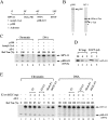
Identification of Brd4 as a component of a cellular complex assembled by HPV-11 E2, but not by dsE2C. (A) Inducible expression of hexahistidine/FLAG-tagged E2 and dsE2C in 293-derived cell lines. Western blotting was performed with anti-hexahistidine antibodies using cell lysates, harvested in the presence (+) or absence (−) of tetracycline (Tc). (B) Purification scheme for E2–P.5 and dsE2C–P.5 complexes. Detection of hexahistidine/FLAG-tagged E2 and dsE2C in nuclear extract (NE) and different P11 fractions, prepared from 293-E2 and 293-dsE2C cell lines, respectively, was conducted by Western blotting with anti-FLAG M2 monoclonal antibody. (C) Brd4 is a component of the E2–P.5 complex. The presence of Brd4 in E2–P.5, dsE2C–P.5, and a control sample purified in parallel from parental 293 cells (Mock-P.5) was analyzed by silver staining (lanes 13) and by Westernblotting with anti-mouse Brd4 antibodies (lanes 47, top panel). The existence of tagged E2 and dsE2C in the purified complexes was confirmed by Western blotting with anti-hexahistidine antibodies (lanes 57, bottom two rows). The P.5 fraction derived from 293-E2 cells was loaded as input (InP) control.
Figure 2.
Figure 2.
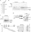
Mapping of promoter-proximal nucleosome positioning on the HPV E6 promoter in vitro and in vivo. (A) G-less cassette templates used for chromatin assembly and transcriptional analysis. (B) Purified ACF, core histones (C.H.), and hNAP-1 used for chromatin assembly. (C) Outline of in vitro chromatin assembly for MNase digestion, nucleosome mapping, and transcriptional analysis. (D) Formation of regularly spaced nucleosomes on in vitro reconstituted HPV-11 chromatin analyzed by MNase digestion at different incubation times. A 123-bp DNA ladder was used as a size marker (M). (E) Mapping the upstream boundary of a promoter-proximal nucleosome assembled on in vitro reconstituted HPV-11 chromatin. Increasing MNase digestion times were used to cleave HPV-11 chromatin or the same mock-assembled (i.e., protein-free) DNA. A size marker (T, C, G, and A lanes) was generated from a DNA sequencing reaction using the same primer for nucleosome mapping. Transcription factor-binding sites in the E6 promoter-proximal region are indicated on the right, with +1 specifying the transcription start site and the oval shape (drawn on the left) depicting the nucleosome-embedded region. (F) Nucleosome positioning on HPV-18 chromatin isolated from HeLa cells (in vivo) and on in vitro reconstituted HPV-18 chromatin. LM-PCR was conducted with primer A or primer B, in the presence (+) or absence (−) of T4 DNA kinase, for in vivo mapping of nucleosome boundaries, whereas primer extension (with primer A) was used for in vitro analysis. Numbers in parentheses represent the lengths of DNA fragments extending from the 5′ end of the primer to the downstream (lane 3) or upstream (lanes 1,5,6) boundary of the E6 promoter-proximal nucleosome (indicated by an asterisk). The number preceding each parenthesis additionally includes the length of the linker DNA (23 nt) used for LM-PCR. A DNA size marker, generated from a sequencing reaction, is shown on the left.
Figure 3.
Figure 3.
E2–P.5 inhibits AP-1-dependent HPV transcription specifically from chromatin, but not from DNA template. (A) Outline of transcription assay with HPV-11 chromatin or DNA template. (B) Purified p300 and AP-1 used for transcription assays. Asterisk indicates degradation products derived from hexahistidine-tagged c-Fos. (C) p300 and acetyl-CoA are critical for AP-1-mediated activation from HPV chromatin. In vitro transcription was performed in HeLa nuclear extracts (NE) using HPV-11 chromatin or DNA template, in the presence (+) or absence (−) of p300, acetyl-CoA, and AP-1 as indicated. The pMLΔ53 control DNA template was added at the same time with nucleoside triphosphates (NTPs), as outlined in A. (D) Normalization of recombinant E2 and the E2–P.5 complex by Western blotting. (E) E2–P.5, but not recombinant E2 or dsE2C–P.5 and Mock-P.5, significantly represses transcription from HPV chromatin. In vitro transcription was performed as described in C in the presence of p300 and acetyl-CoA, with (+) or without (−) AP-1, E2, E2–P.5, and dsE2C–P.5 added at the beginning of the reaction with HPV chromatin or DNA template.
Figure 4.
Figure 4.
The #4 promoter-proximal E2-binding site is critical for transcriptional silencing by the E2–P.5 complex. (A) HPV-11 G-less cassette templates used for transcriptional analysis. Protein-binding sites are indicated on the map with mutated E2-binding sites marked by X. (B) Formation of regularly spaced nucleosomes on in vitro reconstituted HPV-11 chromatin templates analyzed by MNase digestion. A 123-bp DNA ladder was used as a size marker (M). (C,D) The #4 E2-binding site is critical for transcriptional silencing by the E2–P.5 complex. In vitro transcription was performed with HPV chromatin templates as indicated, in the presence of p300 and acetyl-CoA, with (+) or without (−) AP-1 and recombinant E2 or an equivalent amount of the E2–P.5 complex. Transcription from each chromatin template was repeated at least three times, and only a representative set of data from each panel is shown.
Figure 5.
Figure 5.
Recombinant Brd4 is necessary and sufficient for E2-mediated silencing of HPV chromatin transcription. (A) Purified recombinant Brd4 and the in vivo reconstituted recombinant Brd4–E2 complex. Proteins purified from insect Sf9 cells were resolved on a gel and visualized by Coomassie blue staining. Asterisk indicates degradation products derived from FLAG-tagged Brd4. (B) Brd4 and E2, reconstituted in vivo (Brd4/E2) or individually combined in vitro, strongly inhibit HPV transcription from chromatin. In vitro transcription was performed as described in Figure 3E, except that recombinant Brd4 and recombinant E2 were used as indicated. (C) The E2–Brd4 complex does not inhibit Gal4-VP16-mediated chromatin transcription. In vitro transcription was performed as described in B, except that Gal4-VP16 and a chromatin template containing five Gal4-binding sites (pG5MLT) were used in the experiment. (D) The N-terminal domain of E2 is necessary for interaction with Brd4. A GST pull-down assay was performed by incubating recombinant FLAG-tagged Brd4 (f:Brd4) with immobilized GST-E2(FL), GST-E2(1–200), or GST-dsE2C as indicated. Bound f:Brd4 was then detected by Western blotting with anti-FLAG M2 monoclonal antibody. (E) The extreme C terminus and a region containing the second bromodo-main of Brd4 are involved in E2 interaction. A GST pull-down assay was performed by incubating individually purified FLAG-tagged Brd4 deletion domains (middle panel, Coomassie-stained gel) with immobilized GST or GST-E2(FL). Bound f:Brd4 domains were then detected by Western blotting with anti-FLAG M2 monoclonal antibody. Amino acid residues spanning BDI and BDII, an ET domain, and the CTD of Brd4 are depicted on the left.
Figure 6.
Figure 6.
Knockdown of Brd4 significantly alleviates E2-mediated repression of HPV transcription in cultured human cells. (A) Brd4 enhances repression of HPV E6 promoter activity mediated by E2, but not dsE2C, in transfected human C-33A cells. Transfection in C-33A cells was conducted by cotransfecting an HPV-11 URR-E6 promoter-driven reporter (pGL7072-161) with increasing amounts of an E2 (pRSE2-11) or dsE2C (pRS-CM4) expression plasmid, in the absence (−) or presence (+) of an f:Brd4 expression plasmid. Luciferase activity was measured 48 h after transfection. Relative activity represents the ratio of luciferase activity measured in the presence over the absence of E2. Detection of protein expression was performed as described in Materials and Methods. The open arrow points to the putative position of E2. (B) Knockdown of Brd4 alleviates E2-mediated repression in a transient transfection assay. C-33A cells were transfected with pGL7072-161, an HA-tagged HPV-11 E2 expression plasmid (pcDNA3-HA:11E2, 200 ng), and a retroviral vector expressing #1 or #2 Brd4 shRNA. Cell lysates were harvested 24 h later for Western blotting and luciferase analyses. (C) Restoration of E2-mediated repression of HPV-11 E6 promoter activity by transfecting a wild-type Brd4 expression plasmid into a C-33A-derived cell line constitutively expressing #1 Brd4 shRNA. The pGL7072-161 reporter plasmid was cotransfected with pcDNA3-HA:11E2 (400 ng), in the absence (lanes 1,2) or presence (lanes 3,4) of an f:Brd4 expression plasmid into #1-13 cells that express #1 Brd4 shRNA or into vector-harboring V-1 cells. Western blotting and luciferase assays were then carried out as described in B. (D) Schematic of the protein domains in mouse Brd4 proteins. (E) The CTD and a bromodomain-containing region are both necessary for the corepressor activity of Brd4. Reporter gene assay was carried out in #1-13 cells as described in C, except that different Brd4 deletion constructs were used for comparison with the full-length (FL) Brd4 clone. Detection of the expressed proteins by coimmunoprecipitation and Western blotting was conducted as described in Materials and Methods. Antibodies against the C-terminal region (residues 1199–1362) or the N-terminal domain (residues 149–284) of human Brd4 were used for Western blotting.
Figure 7.
Figure 7.
Brd4 recruits E2, which in turn prevents TFIID and pol II association with the HPV E6 promoter region. (A) E2 inhibits the expression of the E6 transcript. RT–PCR was performed using primer pairs annealing to the coding region of E2, E6, and β-actin, respectively. (B) Recruitment of E2 prevents TFIID and pol II association with the E6 promoter region. ChIP assays were conducted with chromatin samples isolated from HeLa and HeLa-E2 cells using different anti-protein antibodies or no antibody (mock) as indicated. PCR products, amplified by a primer pair annealing to the HPV-18 E6 promoter-proximal region (see C), were resolved on an 8% polyacrylamide gel and visualized after ethidium-bromide staining. (C) Locations of HPV-18-specific primer pairs used for ChIP assays. (D) Treatment of HeLa-E2 cells with sodium butyrate enhances Brd4-mediated recruitment of E2 to the endogenous HPV-18 E6 promoter and the −0.5-kb region that contains an E2-binding site. ChIP assays were performed with primer pairs outlined in C. (E) Brd4 enhances E2 recruitment to the E6 promoter region. ChIP assay was performed with anti-FLAG M2 or anti-E2 antibodies with chromatin samples isolated from C-33A cells transfected with pGL7072-161, with (+) or without (−) E2, dsE2C, and f:Brd4 expression plasmids as indicated. PCR products, amplified by a primer pair annealing to the HPV-11 E6 promoter-proximal region (see the top drawing), were resolved on an 8% polyacrylamide gel and visualized after ethidium-bromide staining. (F) The CTD and a bromodomain-containing region are both necessary for Brd4-mediated recruitment of E2 to the endogenous HPV-18 E6 promoter in stable HeLa-derived Brd4-knockdown cells (HeLa-shBrd4) cotransfected with HA:E2 and various Brd4 expression plasmids. The ChIP assay was performed with a primer pair that amplifies the HPV-18 E6 promoter region (see C).

Similar articles

Cited by

References

    1. Abbate E.A., Berger J.M., Botchan M.R. The X-ray structure of the papillomavirus helicase in complex with its molecular matchmaker E2. Genes&Dev. 2004;18:1981–1996. - PMC - PubMed
    1. Abroi A., Ilves I., Kivi S., Ustav M. Analysis of chromatin attachment and partitioning functions of bovine papillomavirus type 1 E2 protein. J. Virol. 2004;78:2100–2113. - PMC - PubMed
    1. Bellanger S., Demeret C., Goyat S., Thierry F. Stability of the human papillomavirus type 18 E2 protein is regulated by a proteasome degradation pathway through its amino-terminal transactivation domain. J. Virol. 2001;75:7244–7251. - PMC - PubMed
    1. Bernard B.A., Bailly C., Lenoir M.-C., Darmon M., Thierry F., Yaniv M. The human papillomavirus type 18 (HPV18)E2 gene product is a repressor of the HPV18 regulatory region in human keratinocytes. J. Virol. 1989;63:4317–4324. - PMC - PubMed
    1. Blachon S., Bellanger S., Demeret C., Thierry F. Nucleo-cytoplasmic shuttling of high risk human papillomavirus E2 proteins induces apoptosis. J. Biol. Chem. 2005;280:36088–36098. - PubMed

Publication types

MeSH terms

LinkOut - more resources