. 2006 Oct;17(10):4513-25.
doi: 10.1091/mbc.e06-05-0390. Epub 2006 Aug 16.

Clathrin-dependent association of CVAK104 with endosomes and the trans-Golgi network

Affiliations

Clathrin-dependent association of CVAK104 with endosomes and the trans-Golgi network

Michael Düwel et al. Mol Biol Cell. 2006 Oct.

Abstract

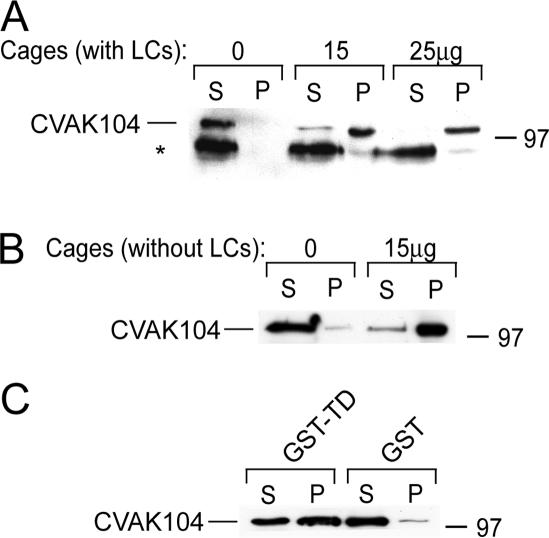
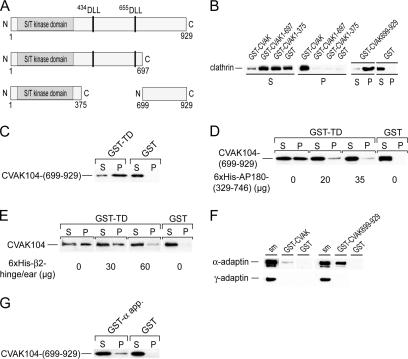
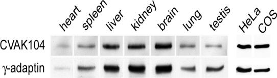
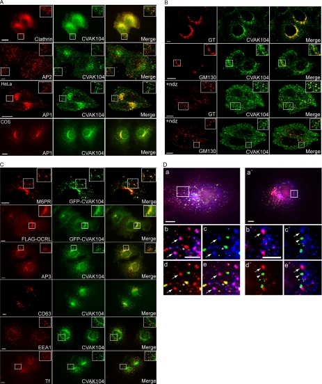
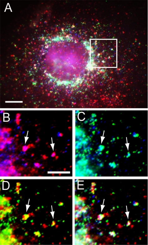
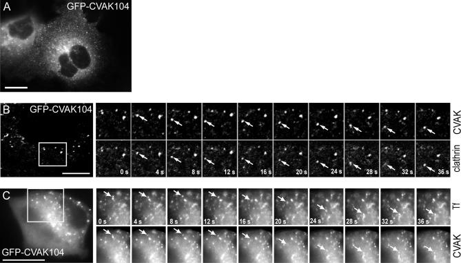
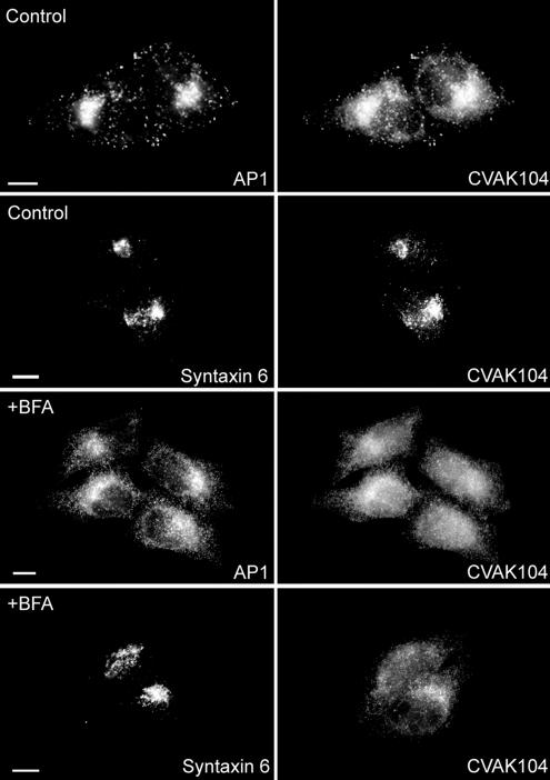
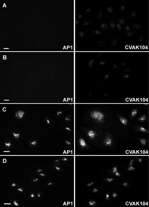
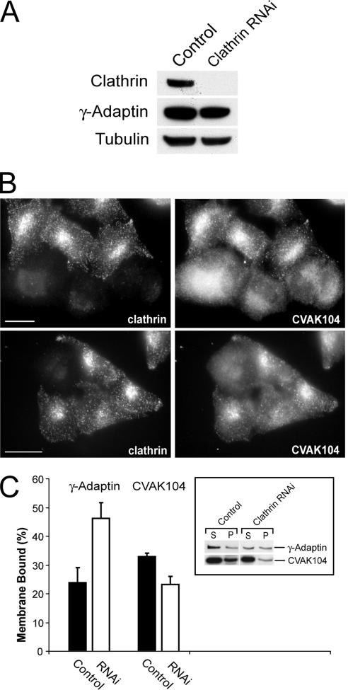
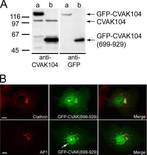
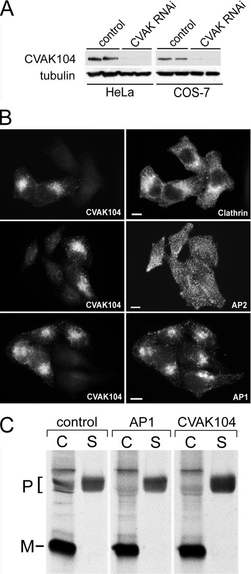
CVAK104 is a novel coated vesicle-associated protein with a serine/threonine kinase homology domain that was recently shown to phosphorylate the beta2-subunit of the adaptor protein (AP) complex AP2 in vitro. Here, we demonstrate that a C-terminal segment of CVAK104 interacts with the N-terminal domain of clathrin and with the alpha-appendage of AP2. CVAK104 localizes predominantly to the perinuclear region of HeLa and COS-7 cells, but it is also present on peripheral vesicular structures that are accessible to endocytosed transferrin. The distribution of CVAK104 overlaps extensively with that of AP1, AP3, the mannose 6-phosphate receptor, and clathrin but not at all with its putative phosphorylation target AP2. RNA interference-mediated clathrin knockdown reduced the membrane association of CVAK104. Recruitment of CVAK104 to perinuclear membranes of permeabilized cells is enhanced by guanosine 5'-O-(3-thio)triphosphate, and brefeldin A redistributes CVAK104 in cells. Both observations suggest a direct or indirect requirement for GTP-binding proteins in the membrane association of CVAK104. Live-cell imaging showed colocalization of green fluorescent protein-CVAK104 with endocytosed transferrin and with red fluorescent protein-clathrin on rapidly moving endosomes. Like AP1-depleted COS-7 cells, CVAK104-depleted cells missort the lysosomal hydrolase cathepsin D. Together, our data suggest a function for CVAK104 in clathrin-dependent pathways between the trans-Golgi network and the endosomal system.


Figures

Figure 1.
Figure 1.
Interaction of CVAK104 with clathrin. (A and B) Purified recombinant CVAK104 was incubated with the indicated amounts of clathrin cages with (A) or without light chains (B). Samples were ultracentrifuged and the resulting supernatants (S) and pellets (P) were analyzed by SDS-PAGE and Western blotting with a CVAK104 antibody. Note that an occasionally visible degradation product of CVAK104 fails to bind to the clathrin cages (asterisk in A). (C) Pull down of recombinant CVAK104 by recombinant clathrin TD fused to GST and immobilized to glutathione-Sepharose. Immobilized GST served as a control. S and P fractions were analyzed by SDS-PAGE and Western blotting. The results demonstrate that CVAK104 interacts directly with the TD of clathrin. In the cage binding assays (A and B), the loadings are directly comparable. In C, the pellet fractions were twofold concentrated compared with the supernatants.
Figure 2.
Figure 2.
Clathrin and AP2 binding domains of CVAK104. (A) Schematic view of the recombinantly expressed (GST-fusion) fragments of CVAK104 used in this work. Putative clathrin interaction motifs are indicated. (B) Incubation of rat brain cytosol with immobilized GST-CVAK104-fusion proteins. Note, that only full-length and the C-terminal segment 699-929 of CVAK104 pulled clathrin out of the cytosol. Pellet fractions are threefold concentrated compared with the supernatants. (C) Direct association of the immobilized GST-TD with recombinant CVAK104-(699-929). For this pull-down experiment, the GST moiety of CVAK104-(699-929) was removed by cleavage with PreScission protease. The pellet fractions are twofold concentrated over the supernatants. (D) Competition between CVAK104-(699-929) and His6-AP180-(329-746) for GST-TD. A constant amount of CVAK104-(699-929) was added to immobilized GST-TD together with increasing concentrations of His6-AP180-(329-746). Note that the recombinant AP180 fragment displaces the CVAK104 fragment. Loadings are directly comparable. (E) Competition between cytosolic CVAK104 and His6-β2-hinge/ear-(592-951) for the TD. A constant amount of rat brain cytosol was added to immobilized GST-TD together with increasing concentrations of His6-β2-hinge/ear-(592-951). Note, that the recombinant His6-β2 fragment displaces the CVAK104 fragment. The pellet fractions are threefold concentrated compared with the supernatants. (F) Association of CVAK104 with AP2. Fractions that were highly enriched in AP1 and AP2 from pig brain were incubated with immobilized GST-CVAK104 or its C-terminal fragment 699-929. Note that only AP2 and not AP1 associated with the C-terminal segment of CVAK104. One-tenth of starting material (sm) used in the binding reaction was run in parallel. The monoclonal antibodies AP.6 and 100/3 that specifically recognize α- and γ-adaptin, respectively, were used for Western blot analysis. (G) Direct association of the immobilized GST-α-appendage domain of AP2 with recombinant CVAK104-(699-929). Loadings are directly comparable.
Figure 3.
Figure 3.
Tissue distribution of CVAK104. Comparable amounts of lysed mouse organs and lysates of cultured cells were analyzed by SDS-PAGE and Western blotting with antibodies to CVAK104 and AP1.
Figure 4.
Figure 4.
Subcellular localization of CVAK104 in HeLa and COS-7 cells. (A) Endogenous CVAK104 localization in HeLa cells, determined by indirect immunofluorescence, is compared with that of clathrin heavy chain (HC), the α-adaptin subunit of AP2 (HeLa), and the γ-adaptin subunit of AP1 (HeLa and COS-7). Rows 2 and 3 are confocal images. (B) Confocal images of HeLa cells double labeled for CVAK104 and the trans-Golgi marker GT and the cis-Golgi marker GM130, respectively. The disruption of the Golgi stacks with nocodazole (+ndz) confirmed the close spatial relationship between GT and CVAK104, whereas GM130-positive vesicles did not contain CVAK104. (C) Spatial relationship of CVAK104 with the mannose 6-phosphate receptor (MPR), OCRL, AP3, CD63 (lamp II), EEA1, and transferrin (Tf) in COS-7 cells. Cells were allowed to take up prebound Texas Red-labeled Tf for 2 min. GFP-CVAK104–expressing cells were used for the staining of the cation-independent M6PR. To obtain information on the spatial relationship between OCRL and CVAK104, COS-7 cells were transfected with FLAG-tagged OCRL and GFP-CVAK104. The first panel in C (GFP-CVAK104 and MPR) shows confocal images. Insets show magnified views of the boxed regions. Bars, 10 μm. (D) Relationship between CVAK104, EEA1, and transferrin in COS-7 cells. Prebound transferrin was internalized for 2 (a–e) and 5 min (a′–e′), respectively. (a and a′) Overlay of CVAK104 (blue), transferrin (red),and EEA1 (green). (b–e and b′–e′) Enlarged regions of the cell boxed in a and a′, respectively. (b and b′) Overlay of CVAK104 (blue) and transferrin (red). (c and c′) Overlay of CVAK104 (blue) and EEA1 (green). (d and d′) Overlay of transferrin (red) and EEA1 (green). (e and e′) Overlay of CVAK104 (blue), transferrin (red), and EEA1 (green). Arrows indicate structures that are positive for transferrin and CVAK104 but not for EEA1. The asterisk marks a vesicular structure that contains transferrin and EEA1 but no CVAK104. The arrowheads point at EEA1-positive profiles that lack transferrin. Bars, 10 μm (a and a′) and 5 μm (b and b′). Note that after 2 min of internalization, a substantial amount of transferrin has passed EEA1-positive endosomes and entered CVAK104-positive structures. After 5 min, only very little transferrin is left in the EEA1 compartment (Supplemental Figure S1).
Figure 5.
Figure 5.
Transit of transferrin through CVAK104 and AP1-positive endosomes. Cell surface-bound Texas Red-labeled transferrin was chased for 2 min at 37°C into COS-7 cells. (A) Overlay of CVAK104 (blue), transferrin (red), and AP1 (green). (B–E) Enlarged regions of the cell boxed in A. (B) Overlay of CVAK104 (blue) and transferrin (red). (C) Overlay of CVAK104 (blue) and AP1 (green). (D) Overlay of transferrin (red) and AP1 (green). (E) Overlay of CVAK104 (blue), transferrin (red) and AP1 (green). Arrows indicate vesicular structures, which are positive for transferrin, CVAK104, and for AP1. Bars, 10 μm (A) and 5 μm (B–E).
Figure 6.
Figure 6.
Live fluorescence microscopy of GFP-CVAK104. (A) Frame of a video (see Supplemental Movie 6A.mov) showing COS-7 cells transfected with GFP-CVAK104. (B and C) COS-7 cells were transfected with GFP-CVAK104 and either cotransfected with RFP-tagged clathrin light chains (B) or incubated with Texas Red-labeled transferrin (C) and then analyzed by live cell imaging (see Supplemental Movies 6B.mov and 6C.mov). Frames were acquired every 4 s. The series of frames are taken from the movies and show an enlarged region of the cells. The arrows indicate moving structures positive for both GFP-CVAK104 and either RFP-CLC or transferrin. The frames of the movie illustrated in B are confocal images. Bars, 10 μm.
Figure 7.
Figure 7.
Effect of BFA on CVAK104 localization. HeLa cells were incubated with BFA-containing media (+BFA) or with medium alone (control) for 4 min at 37°C, fixed, and double stained for either CVAK104 and AP1 or for CVAK104 and the TGN-marker Syntaxin 6. Both AP1 and CVAK104 disperse in response to BFA, whereas the localization of Syntaxin 6 remains unaffected. Bars, 10 μm.
Figure 8.
Figure 8.
Recruitment of CVAK104 and AP1 by permeabilized cells. PtK cells were permeabilized by treatment with 40 μg/ml digitonin for 5 min on ice. The cells were then incubated at 30°C for 30 min with rat brain cytosol either alone (A) or in the presence of an ATP-regenerating system (B), GTPγS (C), or in the presence of both an ATP-regenerating system and GTPγS (D). Cells were fixed and immunostained for AP1 and CVAK104. Recruitment of CVAK104 only occurs in the presence of GTPγS, indicating that it requires a GTP binding protein. Bars, 10 μm.
Figure 9.
Figure 9.
Requirement of clathrin for the subcellular localization of CVAK104. (A) Western blot analysis of lysates from clathrin siRNA- and mock-transfected HeLa cells, respectively. Blots are stained for clathrin heavy chain, the γ-adaptin subunit of AP1, and for tubulin. (B) Distribution of CVAK104 in clathrin-depleted HeLa cells. To view knockdown and control cells within the same field, control and knockdown cells were trypsinized, mixed, and replated 24 h before immunostaining with antibodies to CVAK104 and clathrin HC. In clathrin-depleted cells, CVAK104 shows a more diffuse staining. Bars, 10 μm. (C) Membrane associations of AP1 and CVAK104 upon clathrin depletion. Cells were scraped off the tissue culture plates in lyses buffer and fractionated by ultracentrifugation into membrane and cytosol fraction. Both fractions were analyzed by SDS-PAGE and Western blotting. The membrane-bound fraction of CVAK104 is reduced in clathrin knockdown cells.
Figure 10.
Figure 10.
Subcellular localization of GFP-CVAK104-(699-929) in COS-7 cells. (A) Western blot analysis of lysates from COS-7 cells either expressing GFP-CVAK104 full-length (a) or GFP-CVAK104-(699-929) (b). Blots are stained for CVAK104 and for GFP. (B) GFP-CVAK104-(699-929)–expressing cells were stained for clathrin HC and AP1, respectively. A blob-like structure positive for GFP-CVAK104-(699-929) that does not colocalize with AP1 is marked by an arrow. Note, that the transiently expressed GFP-CVAK104-(699-929)-fusion protein is also localized to the nucleus (denoted by asterisks). Bars, 10 μm.
Figure 11.
Figure 11.
CVAK104 depletion by RNAi. (A) Western blot analysis of lysates from HeLa and COS-7 cells either mock transfected or transfected with a CVAK104-specific siRNA. Tubulin staining served as loading control. (B) Forty-eight hours after transfection, HeLa cells transfected with CVAK104-specific siRNAs and mock-transfected cells were trypsinized, mixed, and grown for another 24 h on coverslips. This made it possible to view knockdown and control cells within the same field. The cells were double stained for CVAK104 and either clathrin heavy chain, the α-adaptin subunit of AP2, or the γ-adaptin subunit of AP1. Bars, 10 μm. (C) COS-7 cells transfected with GFP (control), γ-adaptin (AP1), or CVAK104 siRNA were pulsed for 2 h with 35S-labeled methionine followed by a 4-h chase with unlabeled methionine. Cell-associated cathepsin D was immunoprecipitated from cell lysates (C) and secreted cathepsin D from culture media (S). Samples were analyzed by SDS-PAGE and autoradiography. The positions of the precursor (P) and mature (M) forms of the enzyme are indicated. AP1 and CVAK104 knockdown cells exhibit decreased levels of cell-associated and increased levels of secreted cathepsin D precursor compared with the levels in control cells.

Similar articles

Cited by

References

    1. Ahle S., Mann A., Eichelsbacher U., Ungewickell E. Structural relationships between clathrin assembly proteins from the Golgi and the plasma membrane. EMBO J. 1988;7:919–929. - PMC - PubMed
    1. Bar-Zvi D., Branton D. Clathrin-coated vesicles contain two protein kinase activities. Phosphorylation of clathrin beta-light chain by casein kinase II. J. Biol. Chem. 1986;261:9614–9621. - PubMed
    1. Behnia R., Munro S. Organelle identity and the signposts for membrane traffic. Nature. 2005;438:597–604. - PubMed
    1. Benesch S., Polo S., Lai F. P., Anderson K. I., Stradal T. E., Wehland J., Rottner K. N-WASP deficiency impairs EGF internalization and actin assembly at clathrin-coated pits. J. Cell Sci. 2005;118:3103–3115. - PubMed
    1. Berger E. G., Aegerter E., Mandel T., Hauri H. P. Monoclonal antibodies to soluble, human milk galactosyltransferase (lactose synthase A protein) Carbohydr. Res. 1986;149:23–33. - PubMed

Publication types

LinkOut - more resources