. 2006 Sep;80(17):8639-52.
doi: 10.1128/JVI.00560-06.

Highly conserved regions within the spike proteins of human coronaviruses 229E and NL63 determine recognition of their respective cellular receptors

Affiliations

Highly conserved regions within the spike proteins of human coronaviruses 229E and NL63 determine recognition of their respective cellular receptors

Heike Hofmann et al. J Virol. 2006 Sep.

Abstract

We have recently demonstrated that the severe acute respiratory syndrome coronavirus (SARS-CoV) receptor angiotensin converting enzyme 2 (ACE2) also mediates cellular entry of the newly discovered human coronavirus (hCoV) NL63. Here, we show that expression of DC-SIGN augments NL63 spike (S)-protein-driven infection of susceptible cells, while only expression of ACE2 but not DC-SIGN is sufficient for entry into nonpermissive cells, indicating that ACE2 fulfills the criteria of a bona fide hCoV-NL63 receptor. As for SARS-CoV, murine ACE2 is used less efficiently by NL63-S for entry than human ACE2. In contrast, several amino acid exchanges in human ACE2 which diminish SARS-S-driven entry do not interfere with NL63-S-mediated infection, suggesting that SARS-S and NL63-S might engage human ACE2 differentially. Moreover, we observed that NL63-S-driven entry was less dependent on a low-pH environment and activity of endosomal proteases compared to infection mediated by SARS-S, further suggesting differences in hCoV-NL63 and SARS-CoV cellular entry. NL63-S does not exhibit significant homology to SARS-S but is highly related to the S-protein of hCoV-229E, which enters target cells by engaging CD13. Employing mutagenic analyses, we found that the N-terminal unique domain in NL63-S, which is absent in 229E-S, does not confer binding to ACE2. In contrast, the highly homologous C-terminal parts of the NL63-S1 and 229E-S1 subunits in conjunction with distinct amino acids in the central regions of these proteins confer recognition of ACE2 and CD13, respectively. Therefore, despite the high homology of these sequences, they likely form sufficiently distinct surfaces, thus determining receptor specificity.


Figures
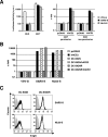
FIG. 1.
FIG. 1.
Expression of hACE2 but not DC-SIGN/DC-SIGNR renders otherwise-nonpermissive cells susceptible to NL63-S-dependent infection. (A) Expression of hACE2 renders otherwise-nonpermissive BHK cells susceptible to NL63-S-driven infection. Permissive 293T and nonpermissive BHK cells were transfected either with pcDNA3 or hACE2 in parallel and infected with infectivity-normalized pseudotypes bearing SARS-S or NL63-S (right panel). Normalization of input virus on 293T cells is shown in the left panel. Luciferase activities were determined 72 h postinfection, and fold activation was calculated based on the infectivity of the respective pseudotypes on target cells transfected with pcDNA3, which was set as 1. Error bars indicate standard deviations (SD). The results of a representative experiment carried out in quadruplicate is shown, and comparable results were obtained in an additional experiment using an independent virus preparation. (B) BHK cells were transfected with pcDNA3, hACE2, or the lectins DC-SIGN and DC-SIGNR or were cotransfected with hACE2 and DC-SIGN or DC-SIGNR. The amount of transfected DNA was kept constant by adding pcDNA3 plasmid. The cells were infected with pseudotypes bearing either VSV-G or the S-proteins normalized for comparable infectivity on Huh-7 cells in quadruplicate, and luciferase activities were determined 72 h postinfection. Fold activation is based on the infectivity on pcDNA3-transfected cells, which was set as 1. A representative experiment is shown and reflects the results of two independent experiments. Error bars indicate SD. (C) Soluble IgG protein (filled histograms) or IgG fusions with either NL63-S or SARS-S were incubated with B-THP cells stably expressing the lectins DC-SIGN and DC-SIGNR as indicated. Bound proteins were detected by FACS using a Cy5-coupled secondary antibody. An independent experiment yielded comparable results.
FIG. 2.
FIG. 2.
Analysis of SARS-S and NL63-S engagement of ACE2 variants expressed on nonpermissive BHK cells. (A) BHK cells expressing either mACE2, hACE2, or the indicated hACE2 mutants were stained with a goat-anti-ACE2 polyclonal antiserum and a Cy5-coupled secondary antibody. Filled histograms indicate staining of cells transfected with a control vector, while black lines indicate staining of ACE2-expressing cells. (B) BHK cells were transfected with ACE2 variants as for panel A and overinfected in quadruplicate with pseudotypes bearing VSV-G, SARS-S, or NL63-S. The pseudotypes had previously been normalized for comparable infectivity on Huh-7 cells. Luciferase activities were determined 72 h postinfection. Fold activation is based on the reporter gene activity in cells expressing pcDNA3. The data were confirmed in three additional experiments with independent pseudoparticle preparations. Error bars indicate standard deviations (SD). (C) BHK cells transfected with ACE2 variants as for panel A in combination with plasmid pGal5-luc were used as target cells for fusion with 293T effector cells carrying the S-proteins of SARS-CoV or hCoV-NL63 together with plasmid pGAL4-VP16 as indicated. Cells transfected with pcDNA3/pGAL4-VP16 served as a control. Fold activation was calculated based on the fusion activity (approximately 100 relative light units) of pcDNA3/pGal5-luc-transfected target with pcDNA3/pGAL4-VP16-transfected effector cells, which was set as 1. Each experiment was performed in triplicate. Error bars indicate SD. An independent experiment yielded similar results.
FIG. 3.
FIG. 3.
NL63-S exhibits a reduced dependence on low pH and protease cleavage compared to SARS-S. (A) Huh-7 cells were preincubated with the indicated concentrations of bafilomycin A1 followed by infection with pseudotypes carrying the envelope proteins of MLV, VSV, hCoV-NL63, and SARS-CoV. An experiment performed in quadruplicate is shown, which is representative for a total of three experiments performed with two independent pseudotype preparations. Error bars indicate standard deviations (SD). (B) 293T cells were preincubated with E64c followed by infection with pseudotypes carrying the glycoproteins of VSV, SARS-CoV, and hCoV-NL63 as indicated. Each experiment was performed in quadruplicate; similar results were obtained in an independent experiment. Error bars indicate SD.
FIG. 4.
FIG. 4.
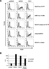
The unique domain in NL63-S is dispensable for binding to hACE2. (A) 293T cells transfected with either pcDNA3, hACE2, or CD13 were incubated with comparable amounts (as judged by Western blot analyses) of soluble IgG (filled histograms) or IgG fusions of NL63-S wild type (aa 16 to 741; panel 1), 229E-S wild type (aa 15 to 560; panel 2), the NL63-S unique domain (aa 16 to 178; panel 3) alone or fused to 229E-S (panel 4) and NL63-S lacking the unique domain (panel 5; black histograms). Bound proteins were detected by a Cy5-coupled anti-human secondary antibody, and binding was analyzed by FACS. Similar results were obtained in an independent experiment with a different batch of soluble IgG fusion proteins. (B) 293T cells expressing either pcDNA3, SARS-S, NL63-S, or an NL63-S variant lacking the unique domain (NL63-SΔ unique) were fused to cells expressing either pcDNA3 (gray) or hACE2 (black) as described in the legend for Fig. 2C. Fold fusion activity was calculated based on the activity of pcDNA3-transfected cells, which was set as 1. A representative experiment performed in triplicate is shown, and four independent experiments yielded comparable results. Error bars indicate standard deviations.
FIG. 5.
FIG. 5.
Schematic representation of NL63-S deletion variants and their interaction with hACE2. (A) The indicated NL63-S deletion mutants (amino acid positions are indicated relative to wild-type NL63-S) were expressed as IgG fusion proteins and analyzed for hACE2 interaction by FACS, as described in the legend to Fig. 4. The data are representative for two independent experiments performed with separate preparations of soluble S-proteins. +, hACE2 binding observed; −, no hACE2 interaction; n.e., respective mutants were not expressed in sufficient amounts. (B) The indicated NL63-S deletion mutants were expressed in 293T cells, concentrated from the supernatant, separated by 6% (left and middle panel) or 10% sodium dodecyl sulfate-polyacrylamide gel electrophoresis (right panel), and normalized for comparable protein amounts by Western blotting prior to the FACS-binding assays.
FIG. 6.
FIG. 6.
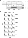
Binding of S-protein chimeras to hACE2 and CD13. (A) Overview depicting chimeras between NL63-S (dark gray) and 229E-S (light gray) and their interaction with hACE2 and CD13. The percent amino acid homology between the respective subdomains in both proteins is shown; the underlying amino acid residues are indicated. The data are representative of two independent experiments. +, hACE2 binding observed; −, no hACE2 interaction. (B) Representative experiment demonstrating binding of a panel of S-protein chimeras (depicted in panel A) to hACE2 and CD13. 293T cells were transfected with either pcDNA3, hACE2, or CD13, incubated with comparable amounts of soluble IgG (filled histograms) or the indicated IgG fusions (black histograms) (for nomenclature see panel A), and stained with a Cy5-coupled secondary antibody.
FIG. 7.
FIG. 7.
Identification of domains involved in the receptor interaction of NL63-S and 229E-S. (A) Schematic representation of NL63-S and 229E-S wild type and all analyzed chimeras and their ability to bind hACE2 or CD13, respectively. The deletion mutant 229E-S-h containing only the C-terminal CD13 interaction domain as determined by Bonavia et al. and Breslin et al. (6, 7) was also included. All chimeras were expressed as IgG fusion proteins and analyzed for receptor binding as described in the legend of Fig. 6. Each chimera was tested at least two times in independent experiments. +, interaction with hACE2 or CD13; −, no binding. The domains within NL63-S and 229E-S required for interaction with their receptors are shown by boxes. (B) The indicated chimeras fused to IgG were normalized for comparable protein amounts by Western blotting prior to the FACS-binding experiments.

Similar articles

Cited by

References

    1. Arden, K. E., M. D. Nissen, T. P. Sloots, and I. M. Mackay. 2005. New human coronavirus, HCoV-NL63, associated with severe lower respiratory tract disease in Australia. J. Med. Virol. 75:455-462. - PMC - PubMed
    1. Babcock, G. J., D. J. Esshaki, W. D. Thomas, Jr., and D. M. Ambrosino. 2004. Amino acids 270 to 510 of the severe acute respiratory syndrome coronavirus spike protein are required for interaction with receptor. J. Virol. 78:4552-4560. - PMC - PubMed
    1. Baer, G. S., D. H. Ebert, C. J. Chung, A. H. Erickson, and T. S. Dermody. 1999. Mutant cells selected during persistent reovirus infection do not express mature cathepsin L and do not support reovirus disassembly. J. Virol. 73:9532-9543. - PMC - PubMed
    1. Bastien, N., K. Anderson, L. Hart, P. Van Caeseele, K. Brandt, D. Milley, T. Hatchette, E. C. Weiss, and Y. Li. 2005. Human coronavirus NL63 infection in Canada. J. Infect. Dis. 191:503-506. - PMC - PubMed
    1. Birkmann, A., K. Mahr, A. Ensser, S. Yaguboglu, F. Titgemeyer, B. Fleckenstein, and F. Neipel. 2001. Cell surface heparan sulfate is a receptor for human herpesvirus 8 and interacts with envelope glycoprotein K8.1. J. Virol. 75:11583-11593. - PMC - PubMed

Publication types

MeSH terms

LinkOut - more resources