. 2006 Nov 1;91(9):3519-28.
doi: 10.1529/biophysj.106.080937. Epub 2006 Aug 11.

Kinetics of the formation and dissociation of actin filament branches mediated by Arp2/3 complex

Affiliations

Kinetics of the formation and dissociation of actin filament branches mediated by Arp2/3 complex

Rachel E Mahaffy et al. Biophys J. .

Abstract

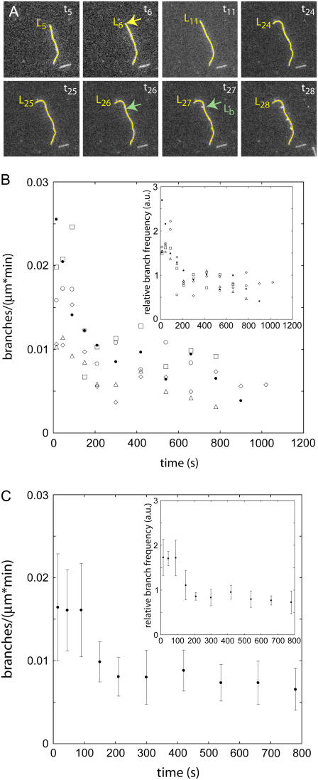
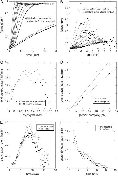
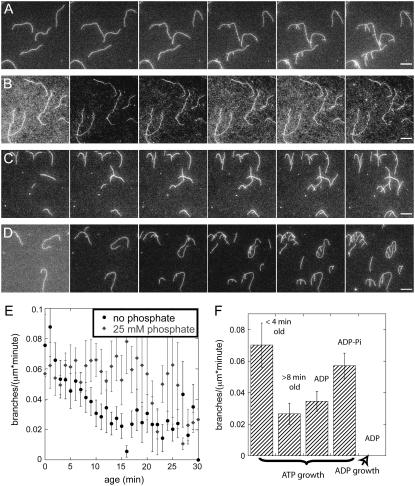
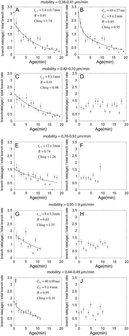
The actin filament network at the leading edge of motile cells relies on localized branching by Arp2/3 complex from "mother" filaments growing near the plasma membrane. The nucleotide bound to the mother filaments (ATP, ADP and phosphate, or ADP) may influence the branch dynamics. To determine the effect of the nucleotide bound to the subunits of the mother filament on the formation and stability of branches, we compared the time courses of actin polymerization in bulk samples measured using the fluorescence of pyrene actin with observations of single filaments by total internal reflection fluorescence microscopy. Although the branch nucleation rate in bulk samples was nearly the same regardless of the nucleotide on the mother filaments, we observed fewer branches by microscopy on ADP-bound filaments than on ADP-P(i)-bound filaments. Observation of branches in the microscope depends on their binding to the slide. Since the probability that a branch binds to the slide is directly related to its lifetime, we used counts of branches to infer their rates of dissociation from mother filaments. We conclude that the nucleotide on the mother filament does not affect the initial branching event but that branches are an order of magnitude more stable on the sides of new ATP- or ADP-P(i) filaments than on ADP-actin filaments.


Figures

FIGURE 1
FIGURE 1
Actin branch formation by Arp2/3 complex observed by fluorescence microscopy on aging mother filaments. Comparison of five separate experiments demonstrates the variability in the observations. (A) Time series of fluorescence micrographs of a branching actin filament. The images in this series were taken 10 s apart. Conditions: 0.6 μM Alexa 488-ATP-actin monomers, 200 nM WASp-VCA, 5 nM Arp2/3 complex in TIRF buffer without phosphate. The mother filament is traced in yellow with a length marker at the barbed end. A branch point is labeled with a green arrow in the frame in which it becomes visible and in the frame immediately before it appears. The length to the branch point from the pointed end is labeled (Lb). The segment of the mother filament that serves as the substrate for the branch labeled in frame t26 (the number refers to the frame number) was first created in the frame labeled t6 (Lb = L6). The difference of these two times yields the age of the mother filament at the time of branching, age = t26 − t6. (B) The observed branch density per minute (branches per micrometer of filament in 60 s) on segments of filaments with ages ranging from 0 to 1100 s. The symbols represent data from five similarly prepared slides. (Inset) These five data sets were normalized by dividing individual branch densities per minute in a given age segment by the total branch density per minute over all ages and times to correct for variations in protein concentrations. (C) Average branch densities per minute observed as a function of the age of the mother filament segment for the five experiments in B. The error bars are ∼30% mean ± 1 SD. (Inset) Normalized mean branch densities per minute have lower standard deviations of 15%.
FIGURE 2
FIGURE 2
Calculation of nucleation rates from the time course of polymerization of bulk samples of actin monomers, Arp2/3 complex, WASp-VCA, and actin filament seeds. The polymer concentration was measured from the fluorescence of pyrene-actin. (AD) Conditions: 2 μM Mg-ATP-actin, 200 nM GST-WASp-VCA, 0–20 nM active Arp2/3 complex, 5 nM actin filament seeds (30 min old, formed from ATP actin monomers in the same buffer used for the nucleation reaction) in polymerization buffer with (open symbols) 25 mM sulfate or (solid symbols) 25 mM phosphate. (A) Time courses of polymerization in phosphate or sulfate buffer with variable concentrations of active Arp2/3 complex: (circles) 0 nM; (squares) 2 nM; (diamonds) 5 nM; (triangles) 10 nM; and (inverted triangles) 20 nM. This stock solution of Arp2/3 complex was 25% active based on the concentration of ends produced with 5 nM of complex. (B) Calculated concentration of barbed ends created by Arp2/3 complex over time in the samples in A. Ends provided by the seeds were subtracted. (C) End creation rate with 2.5 nM active Arp2/3 complex depending on the fraction of the total actin polymerized. (D) Dependence of the end creation rate on the concentration of active Arp2/3 complex with 25 mM sulfate (open circles) or phosphate (solid circles). These rates were calculated at the point where 50% of the actin monomers were polymerized. (E and F) Rates of barbed end formation under the conditions used in TIRF experiments (Figs. 1, 3, and 4). Conditions: 0.6 μM actin monomer, 5 nM Arp2/3 complex (55% active), 200 nM GST-WASp-VCA, seeds grown from ATP-bound monomers for 10 min. Symbols: open diamonds 25 mM sulfate; filled diamonds 25 mM phosphate buffer. (E) End creation rate as a function of time for conditions matching the TIRF experiments formed in 25 mM sulfate (open diamonds) or phosphate (solid diamonds). (F) Rate of ends formed per micrometer of mother filament as a function of time after initiating polymerization in 25 mM phosphate or 25 mM sulfate.
FIGURE 3
FIGURE 3
Effect of nucleotide on the branch density observed by fluorescence microscopy. Conditions: 0.6 μM actin monomer, 5 nM Arp2/3 complex (55% active), 200 nM GST-VCA in TIRF buffer with 25 mM sulfate or phosphate. (AD) Time series of fluorescence micrographs at 60-s intervals in different buffer conditions. (A) ATP-actin monomers in ATP/sulfate buffer with mother filaments polymerized from ATP-actin monomers starting 10 min before the first frame. ATP branches grew from aging mother filaments. (B) ADP-actin monomers in ADP/sulfate buffer with mother filaments polymerized from ADP-actin monomers starting 15 min before the first frame. No ADP branches grew from ADP-actin mother filaments. (C) ATP-actin monomers in ATP/sulfate buffer with mother filaments polymerized from ADP-actin monomers starting 15 min before the first frame. ATP branches grew from ADP mother filaments. (D) ATP-actin monomers in ATP/phosphate buffer with mother filaments polymerized from ATP-actin monomers in phosphate starting 10 min before the first frame. ATP branches grew from ADP-Pi mother filaments. (E) Average observed branch frequency per micron as a function of the age of the mother filament. Mean ± 1 SD. (Solid circles) Branching by ATP-actin monomers in ATP/sulfate buffer from aging mother filaments polymerized from ATP-actin monomers. (Shaded symbols) Branching by ATP-actin monomers in ATP/phosphate buffer from mother filaments polymerized from ATP-actin monomers in phosphate buffer. (F) Summary of the average branch densities per minute observed under various conditions: ATP-actin monomers on aging ATP-actin filaments <4 min old or >8 min old; ATP-actin monomers on ADP-filaments; ATP-actin monomers on ADP-Pi filaments; and ADP-actin on ADP filaments. Averaging was performed over 14–18 experiments containing a total of 2 cm of actin and 1000 events for each condition. Scale bars are 5 μm.
FIGURE 4
FIGURE 4
Normalized branch density per minute observed microscopically as a function of the age of the mother filament with different nucleotide composition and surface adhesiveness. Dividing the individual branch density per minute at each age segment by the total branch density per minute for each experiment provided the normalization. Adhesiveness is inversely related to the mobility of the filaments on the surface, noted in μm/min above each graph. General conditions: 0.6 μM Mg-ATP actin, 5 nM total Arp2/3 complex (50% active), and 200 nM GST-VCA. (AC, E, G, and I) 25 mM sulfate buffer. (A and B) Highly adhesive low mobility (0.38–0.41 μm/min) substrates in sulfate buffer. The decline in the observed branch density per minute with age of the mother filament fits a double exponential (B, t1/2 = 1 min, t1/2 = 12 min, R = 0.95) much better than a single exponential (A, t1/2 = 8.8 min, R = 0.91). (C, E, and G). When the adhesiveness of the substrate was lower, the observed branch density per minute declined with the age of the mother filament as a single exponential noted on each graph. (I) With a higher rate of filament elongation using 1 μM actin monomer, the decline in branch densities per minute fits two exponentials (t1/2 = 1 min, t1/2 = 14 min, R = 0.94) even on a slide of moderate adhesiveness. (D, F, H, and J) 25 mM phosphate buffer ionically balanced with KCl. Observed branch density per minute was independent of the age of the mother filament on all substrates with both concentrations of actin monomers.

Similar articles

Cited by

References

    1. Pollard, T. D., and G. G. Borisy. 2003. Cellular motility driven by assembly and disassembly of actin filaments. Cell. 112:453–465. - PubMed
    1. Machesky, L. M. 1997. Cell motility: complex dynamics at the leading edge. Curr. Biol. 7:R164–R167. - PubMed
    1. Mullins, R. D., and M. D. Welch. 2002. Cellular control of actin nucleation. Annu. Rev. Cell Dev. Biol. 18:247–288. - PubMed
    1. Svitkina, T. M., and G. G. Borisy. 1999. Arp2/3 complex and actin depolymerizing factor/cofilin in dendritic organization and treadmilling of actin filament array in lamellipodia. J. Cell Biol. 145:1009–1026. - PMC - PubMed
    1. Flanagan, L. A., J. Chou, H. Falet, R. Neujahr, J. H. Hartwig, and T. P. Stossel. 2001. Filamin A, the Arp2/3 complex, and the morphology and function of cortical actin filaments in human melanoma cells. J. Cell Biol. 155:511–517. - PMC - PubMed

Substances

LinkOut - more resources