

Reversible model of RNA toxicity and cardiac conduction defects in myotonic dystrophy
- PMID: 16878132
- PMCID: PMC2909745
- DOI: 10.1038/ng1857
Reversible model of RNA toxicity and cardiac conduction defects in myotonic dystrophy
Abstract
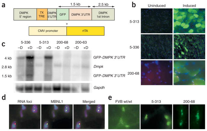
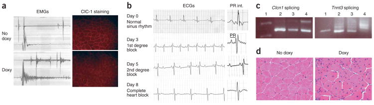
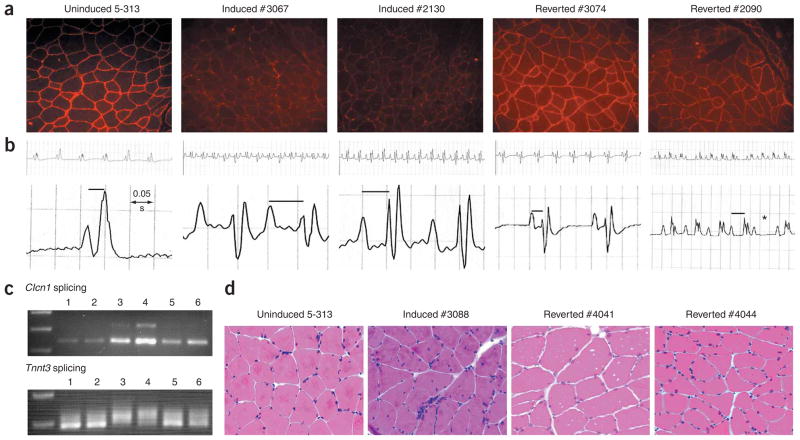
Myotonic dystrophy (DM1), the most common muscular dystrophy in adults, is caused by an expanded (CTG)n tract in the 3' UTR of the gene encoding myotonic dystrophy protein kinase (DMPK), which results in nuclear entrapment of the 'toxic' mutant RNA and interacting RNA-binding proteins (such as MBNL1) in ribonuclear inclusions. It is unclear if therapy aimed at eliminating the toxin would be beneficial. To address this, we generated transgenic mice expressing the DMPK 3' UTR as part of an inducible RNA transcript encoding green fluorescent protein (GFP). We were surprised to find that mice overexpressing a normal DMPK 3' UTR mRNA reproduced cardinal features of myotonic dystrophy, including myotonia, cardiac conduction abnormalities, histopathology and RNA splicing defects in the absence of detectable nuclear inclusions. However, we observed increased levels of CUG-binding protein (CUG-BP1) in skeletal muscle, as seen in individuals with DM1. Notably, these effects were reversible in both mature skeletal and cardiac muscles by silencing transgene expression. These results represent the first in vivo proof of principle for a therapeutic strategy for treatment of myotonic dystrophy by ablating or silencing expression of the toxic RNA molecules.
Conflict of interest statement
The authors declare that they have no competing financial interests.
Figures

Comment in
-
Reversal of fortune.Nat Genet. 2006 Sep;38(9):976-7. doi: 10.1038/ng0906-976. Nat Genet. 2006. PMID: 16941004 No abstract available.
Similar articles
-
Age of onset of RNA toxicity influences phenotypic severity: evidence from an inducible mouse model of myotonic dystrophy (DM1).PLoS One. 2013 Sep 5;8(9):e72907. doi: 10.1371/journal.pone.0072907. eCollection 2013. PLoS One. 2013. PMID: 24039817 Free PMC article.
-
Myotonic dystrophy in transgenic mice expressing an expanded CUG repeat.Science. 2000 Sep 8;289(5485):1769-73. doi: 10.1126/science.289.5485.1769. Science. 2000. PMID: 10976074
-
Systemic delivery of a Peptide-linked morpholino oligonucleotide neutralizes mutant RNA toxicity in a mouse model of myotonic dystrophy.Nucleic Acid Ther. 2013 Apr;23(2):109-17. doi: 10.1089/nat.2012.0404. Epub 2013 Jan 11. Nucleic Acid Ther. 2013. PMID: 23308382
-
Tackling the pathogenesis of RNA nuclear retention in myotonic dystrophy.Biol Cell. 2010 Jul 23;102(9):515-23. doi: 10.1042/BC20100040. Biol Cell. 2010. PMID: 20690904 Review.
-
Myotonic dystrophy: clinical and molecular parallels between myotonic dystrophy type 1 and type 2.Curr Neurol Neurosci Rep. 2002 Sep;2(5):465-70. doi: 10.1007/s11910-002-0074-6. Curr Neurol Neurosci Rep. 2002. PMID: 12169228 Review.
Cited by
-
Disease Phenotypes in a Mouse Model of RNA Toxicity Are Independent of Protein Kinase Cα and Protein Kinase Cβ.PLoS One. 2016 Sep 22;11(9):e0163325. doi: 10.1371/journal.pone.0163325. eCollection 2016. PLoS One. 2016. PMID: 27657532 Free PMC article.
-
MBNL and CELF proteins regulate alternative splicing of the skeletal muscle chloride channel CLCN1.Nucleic Acids Res. 2009 Oct;37(19):6477-90. doi: 10.1093/nar/gkp681. Epub 2009 Aug 31. Nucleic Acids Res. 2009. PMID: 19720736 Free PMC article.
-
A tail-anchored myotonic dystrophy protein kinase isoform induces perinuclear clustering of mitochondria, autophagy, and apoptosis.PLoS One. 2009 Nov 25;4(11):e8024. doi: 10.1371/journal.pone.0008024. PLoS One. 2009. PMID: 19946639 Free PMC article.
-
Transcription-induced DNA toxicity at trinucleotide repeats: double bubble is trouble.Cell Cycle. 2011 Feb 15;10(4):611-8. doi: 10.4161/cc.10.4.14729. Epub 2011 Feb 15. Cell Cycle. 2011. PMID: 21293182 Free PMC article.
-
Mechanisms of RNA-mediated disease.J Biol Chem. 2009 Mar 20;284(12):7419-23. doi: 10.1074/jbc.R800025200. Epub 2008 Oct 28. J Biol Chem. 2009. PMID: 18957432 Free PMC article. Review.
References
-
- Mahadevan M, et al. Myotonic dystrophy mutation: an unstable CTG repeat in the 3′ untranslated region of the gene. Science. 1992;255:1253–1255. - PubMed
-
- Day JW, Ranum LP. RNA pathogenesis of the myotonic dystrophies. Neuromuscul Disord. 2005;15:5–16. - PubMed
-
- Amack JD, Paguio AP, Mahadevan MS. Cis and trans effects of the myotonic dystrophy (DM) mutation in a cell culture model. Hum Mol Genet. 1999;8:1975–1984. - PubMed
Publication types
MeSH terms
Substances
Grants and funding
LinkOut - more resources
Full Text Sources
Other Literature Sources
Molecular Biology Databases
