. 2006 Aug;80(16):8211-24.
doi: 10.1128/JVI.02528-05.

Eclipse phase of herpes simplex virus type 1 infection: Efficient dynein-mediated capsid transport without the small capsid protein VP26

Affiliations

Eclipse phase of herpes simplex virus type 1 infection: Efficient dynein-mediated capsid transport without the small capsid protein VP26

Katinka Döhner et al. J Virol. 2006 Aug.

Abstract

Cytoplasmic dynein,together with its cofactor dynactin, transports incoming herpes simplex virus type 1 (HSV-1) capsids along microtubules (MT) to the MT-organizing center (MTOC). From the MTOC, capsids move further to the nuclear pore, where the viral genome is released into the nucleoplasm. The small capsid protein VP26 can interact with the dynein light chains Tctex1 (DYNLT1) and rp3 (DYNLT3) and may recruit dynein to the capsid. Therefore, we analyzed nuclear targeting of incoming HSV1-DeltaVP26 capsids devoid of VP26 and of HSV1-GFPVP26 capsids expressing a GFPVP26 fusion instead of VP26. To compare the cell entry of different strains, we characterized the inocula with respect to infectivity, viral genome content, protein composition, and particle composition. Preparations with a low particle-to-PFU ratio showed efficient nuclear targeting and were considered to be of higher quality than those containing many defective particles, which were unable to induce plaque formation. When cells were infected with HSV-1 wild type, HSV1-DeltaVP26, or HSV1-GFPVP26, viral capsids were transported along MT to the nucleus. Moreover, when dynein function was inhibited by overexpression of the dynactin subunit dynamitin, fewer capsids of HSV-1 wild type, HSV1-DeltaVP26, and HSV1-GFPVP26 arrived at the nucleus. Thus, even in the absence of the potential viral dynein receptor VP26, HSV-1 used MT and dynein for efficient nuclear targeting. These data suggest that besides VP26, HSV-1 encodes other receptors for dynein or dynactin.


Figures

FIG. 1.
FIG. 1.
Different viral particles. An HSV-1 inoculum may consist of (1) vesicles with viral membrane proteins, (2) vesicles with viral membrane and tegument proteins (L-particles), capsids (3) without or (4) with viral DNA, (5) enveloped particles with tegument and capsid but without DNA, or (6) noninfectious or (7) infectious virions. Antibodies raised against viral membrane proteins detect all particles except for capsids, and antitegument antibodies recognize all particles except vesicles and capsids without tegument. Capsid antibodies detect the capsid-containing particles. DNA-containing particles are identified by PCR, whereas only an infectious virion will result in the formation of a plaque.
FIG. 2.
FIG. 2.
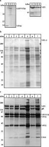
Preparations of the same HSV-1 strain vary in the protein/PFU ratio. (a) Viral particles harvested from the media of cells infected with HSV-1 wt strain F (1), HSV-GFPVP26 (2), or HSV1-ΔVP26 (3) were subjected to linear 8 to 16% SDS-PAGE, blotted, and probed with antibodies generated against recombinant His-tagged VP26 (left) or against VP5 (NC-1; right). HSV-1 wt (1) and HSV1-GFPVP26 (2), but not HSV1-ΔVP26 (3), contained anti-VP26-positive bands at the respective molecular masses of VP26 (12 kDa) and GFPVP26 (39 kDa) (left). Anti-VP5 was used as a loading control (right). (b and c) Gradient-purified virions of HSV-1 wt strain F (lanes 1 and 5), HSV-1 wt strain KOS (lane 2), HSV1-ΔVP26 (lanes 3 and 6), or HSV1-GFPVP26 (lanes 4 and 7) were subjected to linear 7.5 to 18% SDS-PAGE, and the gels were either stained with Coomassie (b) or blotted and probed with antibodies (c) to VP26, VP5, US11, gB, VP16, VP13/14, and Remus V, an antibody that recognizes several capsid, tegument, and glycoproteins of HSV-1. HSV-1 wt strain F, HSV-1 wt strain KOS, HSV1-ΔVP26, or HSV1-GFPVP26 had very similar overall protein compositions (b and c). Preparations with a low genome/PFU ratio (lanes 1 to 4) loaded at 1.3 × 10⁸ PFU per lane contained less protein than preparations with a higher genome/PFU ratio (lanes 5 to 7). Note that in lane 6 only 4 × 10⁷ PFU, about 25% compared to the other lanes, was loaded, resulting in a protein concentration similar to the 100% loaded for HSV-1 wt strain F (lane 1).
FIG. 3.
FIG. 3.
Particle composition of different HSV-1 preparations. (a to e) Virus preparations labeled with anti-gD followed by 10-nm protein A-gold were analyzed by electron microscopy after negative staining. (a) HSV-1 wt (F), good; genome/PFU = 23. (b) HSV-1 wt (F), bad; genome/PFU = 132. (c) HSV-1 wt (KOS), genome/PFU = 20. (d) HSV1-ΔVP26, genome/PFU = 48. (e) HSV1-GFPVP26, genome/PFU = 18. (f to k) Many particles showed a typical herpesvirus morphology with a contrasted capsid structure within an envelope (f and g), other structures looked more like vesicles without internal contrast (h and i), and there were also capsid-like structures without a contrasted envelope (j and k). A large proportion of all particles was labeled by gD (f, h, and j). (l and m) Quantification of different viral particles. The particle concentration increased with the genome/PFU ratio (l), but with the exception of HSV1-GFPVP26 the different particles were present in similar ratios (m). The error bars represent the standard errors of the means. Bars, 500 nm (a to e) and 50 nm (f to k).
FIG. 4.
FIG. 4.
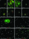
Efficient capsid transport of high-quality preparations to the nucleus (N) (a to h). A larger proportion of capsids (red) derived from preparations with a low genome/PFU ratio (a, e, and g) reached the nucleus within 3 h p.i. than from preparations with a higher genome/PFU ratio (b, c, f, and h; for quantification, see Table 1). HSV-1 wt (a; genome/PFU = 20) and HSV1-GFPVP26 (g; genome/PFU = 18) preparations of high quality showed the most efficient nuclear targeting. Nuclear targeting of good HSV1-ΔVP26 preparations (e; genome/PFU = 48) was somewhat lower. Cells infected with virus preparations of high quality (a and g) showed little labeling for the viral membrane protein gD (green) compared to cells infected with virus preparations with high viral genome/PFU ratios at 3 h p.i. (genome/PFU = 132 [b and c] or 69 [f]). PtK2 cells were infected with 50 PFU/cell in the presence of cycloheximide, fixed, permeabilized, and labeled with anti-HC (red) and anti-gD MAb DL6 (green). (i to k) Early separation of the capsids from the envelope. At 30 min p.i., most capsids (red) of HSV-1 wt (i) and HSV1-GFPVP26 (k) did not colocalize (arrowhead) with the viral glycoprotein gD (green). In contrast, about 50% of HSV1-ΔVP26 capsids (j) colocalized with gD (l; arrow). After 30 min of infection with HSV1-GFPVP26, the capsid and GFP signals partially colocalized (arrow; yellow), several capsids showed no GFP fluorescence (arrowhead; red), and several GFP particles were not labeled by the anticapsid antibodies (green). Vero cells were infected with 50 PFU/cell in the presence of cycloheximide, fixed, permeabilized, and labeled with anticapsid antibody (anti-HC; red) and anti-gD (DL6; pseudocolored in green in panels i to k). GFP was detected by its intrinsic fluorescence (l; green).
FIG. 5.
FIG. 5.
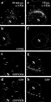
VP26 of HSV-1 wt remained attached to nuclear capsids, while a fraction of GFPVP26 was lost during transport. At 30 min p.i., VP26 (a) and GFPVP26 (c and d) spots were randomly distributed. The digital image processing was adjusted such that no signal was detected with anti-VP26 after infection with HSV1-ΔVP26 (b and f). Most VP26 spots had accumulated at the nuclear envelope 3 h after infection with HSV-1 wt (KOS) (e), while a significant fraction of the GFPVP26 signal was lost (g and h). The remaining GFPVP26 signal was localized at the nuclear envelope or in the cell periphery. The GFP signal at the nucleus (arrows in h) was often weaker than the GFP signal in the cell periphery at 30 min p.i. (arrows in d). Vero cells were infected in the presence of cycloheximide with 25 PFU/cell, fixed, permeabilized, and labeled with anti-VP26 (amino acids 95 to 112).
FIG. 6.
FIG. 6.
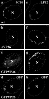
VP5 epitopes on HSV-1 wt, HSV1-ΔVP26, and HSV1-GFPVP26. The anti-VP5 MAb 5C10 detected incoming HSV-1 wt (a) but not HSV1-ΔVP26 (b) capsids. MAb 5C10 also detected some HSV1-GFPVP26 capsids (c; arrow), which mostly (d; arrow) but not always (c; arrowhead) colocalized with the GFP fluorescence. In contrast, the anti-VP5 MAb LP12 labeled HSV1-ΔVP26 (f) and HSV1-GFPVP26 (g) capsids with higher efficiency than HSV-1 wt (e). The MAb LP12 detected more nuclear HSV1-GFPVP26 capsids (g) than the MAb 5C10 (c) and more than were detected by their GFP fluorescence (h). Vero cells were infected for 3 h in the presence of cycloheximide with 25 PFU/cell, fixed, permeabilized, and labeled with MAb 5C10 (a to c) or LP12 (e to g).
FIG. 7.
FIG. 7.
HSV-1 wt, HSV1-ΔVP26, and HSV1-GFPVP26 required MT for efficient immediate-early viral gene expression. For all three KOS viruses, there was less ICP4 and ICP0 expressed when the cells had been infected in the presence of 50 μM nocodazole. Vero cells were infected for 3 h with 5 PFU/cell. Cell lysates were loaded onto linear 4 to 12% SDS gels, blotted, and probed with anti-ICP4 (upper panel) and anti-ICP0 (middle panel), and antiactin was used as a loading control (lower panel).
FIG. 8.
FIG. 8.
HSV-1 wt, HSV1-ΔVP26, and HSV1-GFPVP26 capsids require MT for efficient nuclear targeting. After 3 h, many HSV-1 wt (KOS) (a), HSV1-ΔVP26 (b), and HSV1-GFPVP26 (c) capsids had accumulated at the nucleus (N). In the absence of MT, the capsids remained randomly distributed over the entire cytoplasm (d to f). Vero cells were infected in the presence of cycloheximide for 3 h with 50 PFU/cell in the absence (a to c) or presence of 50 μM nocodazole (d to f). The cells were fixed and labeled with anticapsid antibodies (anti-HC).
FIG. 9.
FIG. 9.
Dynamitin reduced nuclear targeting in the presence and absence of VP26. In Vero cells expressing dynamitin-GFP (marked by N in panels a to c), less HSV-1 wt (a), HSV1-ΔVP26 (b), and HSV1-GFPVP26 (c) capsids reached the nucleus than in untransfected cells. The expression of GFP (marked by N in panels d to f) had no effect on nuclear targeting of HSV-1 wt (d), HSV1-ΔVP26 (e), or HSV1-GFPVP26 (f) capsids. Vero cells were transfected with dynamitin-GFP (a to c) or GFP (d to f) for 30 h, infected with 50 PFU/cell for 3 h in the presence of cycloheximide, fixed, and labeled with an anticapsid antibody (anti-HC). Dynamitin-GFP and GFP were detected by their intrinsic fluorescence (not shown). Arrows point to capsids in the cell periphery, and arrowheads point to capsids distributed in the cytoplasm.

Similar articles

Cited by

References

    1. Antinone, S. E., G. T. Shubeita, K. E. Coller, J. I. Lee, S. Haverlock-Moyns, S. P. Gross, and G. A. Smith. 2006. The herpesvirus capsid surface protein, VP26, and the majority of the tegument proteins are dispensable for capsid transport toward the nucleus. J. Virol. 80:5494-5498. - PMC - PubMed
    1. Bananis, E., S. Nath, K. Gordon, P. Satir, R. J. Stockert, J. W. Murray, and A. W. Wolkoff. 2004. Microtubule-dependent movement of late endocytic vesicles in vitro: requirements for dynein and kinesin. Mol. Biol. Cell 15:3688-3697. - PMC - PubMed
    1. Batterson, W., D. Furlong, and B. Roizman. 1983. Molecular genetics of herpes simplex virus. VIII. Further characterization of a temperature-sensitive mutant defective in release of viral DNA and in other stages of the viral reproductive cycle. J. Virol. 45:397-407. - PMC - PubMed
    1. Booy, F. P., B. L. Trus, W. W. Newcomb, J. C. Brown, J. F. Conway, and A. C. Steven. 1994. Finding a needle in a haystack: detection of a small protein (the 12-kDa VP26) in a large complex (the 200-MDa capsid of herpes simplex virus). Proc. Natl. Acad. Sci. USA 91:5652-5656. - PMC - PubMed
    1. Burkhardt, J. K., C. J. Echeverri, T. Nilsson, and R. B. Vallee. 1997. Overexpression of the dynamitin (p50) subunit of the dynactin complex disrupts dynein-dependent maintenance of membrane organelle distribution. J. Cell Biol. 139:469-484. - PMC - PubMed

Publication types

MeSH terms