. 2006 Aug;80(16):8199-210.
doi: 10.1128/JVI.00457-06.

Betaherpesvirus-conserved cytomegalovirus tegument protein ppUL32 (pp150) controls cytoplasmic events during virion maturation

Affiliations

Betaherpesvirus-conserved cytomegalovirus tegument protein ppUL32 (pp150) controls cytoplasmic events during virion maturation

David P AuCoin et al. J Virol. 2006 Aug.

Abstract

The UL32 gene of human cytomegalovirus (CMV) encodes a prominent betaherpesvirus-conserved virion tegument protein, called pp150 (basic phosphoprotein/ppUL32), that accumulates within a cytoplasmic inclusion adjacent to the nucleus at late times during infection. Using a UL32 deletion mutant (DeltaUL32-BAC) (where BAC is bacterial artificial chromosome), we demonstrate that pp150 is critical for virion maturation in the cytoplasmic compartment. Cotransfection of a pp150 expression plasmid with DeltaUL32-BAC DNA led to complementation of the replication defect with focus formation due to secondary spread. Deletion of the amino terminus of pp150 or disruption of the betaherpesvirus conserved regions, CR1 and CR2, revealed these regions to be critical for replication. In contrast, deletion of the carboxyl terminus only partially compromised maturation while disruption of glycosylation sites had no effect. An African green monkey CMV UL32 homolog complemented DeltaUL32-BAC replication but murine CMV M32 failed to complement, consistent with evolutionary divergence of rodent and primate cytomegaloviruses. Infection with DeltaUL32-BAC showed normal expression of all kinetic classes of viral genes and replication of viral DNA, with accumulation of viral DNA-containing particles in the cytoplasm; however, mutant virus did not spread to adjacent cells. In contrast to this block in virion infectivity, cell-to-cell transfer of pp65-containing particles was observed, suggesting that release of dense bodies continued in the absence of pp150. These observations demonstrate that pp150 is critical for virion egress, possibly at the stage of final envelopment.


Figures

FIG. 1.
FIG. 1.
UL32-BAC structure and complementation. (A) Schematic of the HCMV TownevarATCCshort strain, Towne-BAC-derived ΔUL32-BAC genome, with the locations of the BAC-GFP insertion replacing US1 to US12 and the KanMX4 insertion replacing the entire betaherpesvirus-conserved UL32 ORF (13) indicated by expanded regions. The numbered open boxes indicate the locations of herpesvirus core genes, and the gray shaded area indicates the location of the betaherpesvirus-conserved genes (35, 36). (B) Ethidium bromide-stained images of electrophoretically separated (larger fragments, left; smaller fragments, right) HindIII restriction digests of 5 μg of Towne-BAC (lane 1), rescued-UL32-BAC (lane 2), or ΔUL32-BAC (lane 3). The 11.7-kbp fragment present in Towne-BAC or rescued-UL32-BAC is replaced by 8.3- and 1.85-kbp fragments in ΔUL32-BAC, as indicated by an asterisk on the left of the DNA fragment and by the sizes on the right side of each panel. (C) GFP expression following transfection of HFFs with rescued-UL32-BAC (a focus is shown at ×50 magnification), Towne-BAC (a focus is shown at ×50 magnification), and ΔUL32-BAC (a single cell is shown at ×100 magnification) at day 10 posttransfection. (D) GFP expression following transfection (as described for panel C) with Towne-BAC, rescued-UL32-BAC, and ΔUL32-BAC. Detection of GFP, pp150 antigen, and DNA (Hoechst) was performed on day 8 posttransfection (×300 magnification). A merge is shown to localize the pp150 accumulation. (E) Detection of GFP (top panel) and myc-tagged pp150 (bottom panel) in a secondary spread assay at 8 days following cotransfection of HFFs with ΔUL32-BAC and WT pp150 expression plasmid (pON2780) (×100 magnification). The arrowhead identifies the myc epitope-tagged pp150-positive cell.
FIG. 2.
FIG. 2.
Complementation of ΔUL32-BAC by mutant forms of pp150. (A) IFA of pON2781-encoded amino-terminal truncation of pp150 (top panels) and pON2782-encoded carboxyl-terminal truncation (bottom panels) following cotransfection with ΔUL32-BAC in the secondary spread assay at 10 days posttransfection. A small focus of cells that was generated following cotransfection of the carboxyl-terminal deletion mutant is identified by an arrowhead. Magnification,×80.(B) Immunoblot of 293T cell lysates at 48 h posttransfection with 2 μg of pON2780 (lane 1), pON2781 (lane 2), and pON2782 (lane 3). Proteins were separated by SDS-polyacrylamide gel electrophoresis, transferred to a nylon membrane, and immunoblotted for myc-tagged pp150. β-Actin served as a loading control. (C) IFA of secondary spread, noting the percentage of GFP-positive single cells that developed into foci as described for panel A. Error bars represent the standard deviations of the means of triplicate samples. Photomicrographs above each bar in the graph show representative images of foci or single cells (where foci do not form) for cotransfection combinations indicated. Magnification, ×20.
FIG. 3.
FIG. 3.
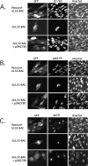
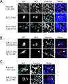
Expression of IE, early, and late viral antigens by ΔUL32-BAC. HFFs were transfected with rescued-UL32-BAC, ΔUL32-BAC, or ΔUL32-BAC plus WT pp150 expression plasmid pON2780. (A) Immunofluorescent analysis of cells fixed 8 days posttransfection and stained with FITC-conjugated MAb 810 to detect IE1/IE2. (B) Immunofluorescent analysis of cells fixed 8 days posttransfection and stained with an HCMV ppUL44 MAb followed by detection with Alexa Fluor 594-conjugated secondary antibody. (C) Immunofluorescent analysis of cells fixed 10 days posttransfection and stained with HCMV pp28 (UL99) MAb, followed by secondary detection with Alexa Fluor 594-conjugated secondary antibody. Hoechst staining was used to localize nuclear DNA. Magnification, ×300.
FIG. 4.
FIG. 4.
Localization of newly synthesized viral DNA. (A) Immunofluorescent images of HFFs transfected with rescued-UL32-BAC (top panels) or ΔUL32-BAC plus WT pp150 (bottom panels), followed by a BrdU (10 μM) pulse for 12 h and a 48-h chase. BrdU was localized with an Alexa Fluor 594-conjugated MAb. A false color merge was used to localize BrdU-stained viral DNA (red) in a juxtanuclear position relative to IE1/IE2-positive nuclei (blue). The merged images contained in boxes are enlarged in column 4. Cells were fixed in methanol:acetic acid (3:1). Magnification, ×400 for columns 1 to 3 and ×1,000 for column 4.
FIG. 5.
FIG. 5.
Localization of MCP within the cytoplasm of ΔUL32-BAC-transfected or -infected cells. (A) Immunofluorescent images of HFFs fixed with 3.7% formaldehyde at day 10 following transfection with rescued-UL32-BAC (top panels), ΔUL32-BAC (middle panels), and ΔUL32-BAC plus WT pp150 (bottom panels). (B) Immunofluorescent images of HFFs infected with supernatant virus from ΔUL32-BAC plus WT pp150-cotransfected cells [ΔUL32 virus (cotransfect)] fixed on day 10 postinfection (top panels) and HFFs infected with supernatant virus recovered from the ΔUL32-BAC-transfected pp150-expressing HFF line [ΔUL32 virus (UL32-HFF)] fixed at day 7 postinfection (bottom panels). (C) Immunofluorescent images of MCP accumulation at day 8 postinfection with rescued-UL32 (top panels) and ΔUL32 (bottom panels) virus fixed with methanol:acetic acid (3:1). GFP-positive cells are shown in the far left panel for all samples fixed with 3.7% formaldehyde (methanol:acetic acid fixation destroys GFP detection). All staining for MCP was done with MAb 28-4 along with Alexa Fluor 594 secondary antibody. Hoechst was used to stain nuclei, and a false color merge with MCP antigen localization is shown. The arrowhead indicates cytoplasmic localization of MCP. All images were collected with equivalent exposure times. Magnification, ×200.
FIG. 6.
FIG. 6.
Localization and accumulation of gB in the cytoplasm of ΔUL32-BAC-transfected and -infected cells. Immunofluorescent images of HFFs from a secondary spread assay at day 10 posttransfection with rescued-UL32-BAC or ΔUL32-BAC plus WT pp150 (pON2780). Detection of gB was accomplished with an anti-HCMV gB MAb along with an Alexa Fluor 594 secondary antibody. Three individual plaques are shown for each construct. All images were collected with equivalent exposure times. Magnification, ×105.
FIG. 7.
FIG. 7.
Localization and distribution of pp65 in ΔUL32-BAC-transfected and ΔUL32 virus-infected cells. Immunofluorescent images of rescued-UL32 (top panels) or ΔUL32 (middle panels) transfection at day 10 posttransfection compared to ΔUL32 virus infection (bottom panels) at day 7 postinfection. Detection of pp65 was accomplished with MAb 28-19 followed by Alexa Fluor 594 secondary antibody. Magnification, ×400.

Similar articles

Cited by

References

    1. Baines, J. D., R. J. Jacob, L. Simmerman, and B. Roizman. 1995. The herpes simplex virus 1 UL11 proteins are associated with cytoplasmic and nuclear membranes and with nuclear bodies of infected cells. J. Virol. 69:825-833. - PMC - PubMed
    1. Baines, J. D., and B. Roizman. 1992. The UL11 gene of herpes simplex virus 1 encodes a function that facilitates nucleocapsid envelopment and egress from cells. J. Virol. 66:5168-5174. - PMC - PubMed
    1. Baldick, C. J., Jr., A. Marchini, C. E. Patterson, and T. Shenk. 1997. Human cytomegalovirus tegument protein pp71 (ppUL82) enhances the infectivity of viral DNA and accelerates the infectious cycle. J. Virol. 71:4400-4408. - PMC - PubMed
    1. Bartz, S. R., and M. A. Vodicka. 1997. Production of high-titer human immunodeficiency virus type 1 pseudotyped with vesicular stomatitis virus glycoprotein. Methods 12:337-342. - PubMed
    1. Baxter, M. K., and W. Gibson. 2001. Cytomegalovirus basic phosphoprotein (pUL32) binds to capsids in vitro through its amino one-third. J. Virol. 75:6865-6873. - PMC - PubMed

Publication types

MeSH terms

LinkOut - more resources