. 2006 Dec 1;400(2):255-65.
doi: 10.1042/BJ20060316.

A critical beta6-beta7 loop in the pleckstrin homology domain of ceramide kinase

Affiliations

A critical beta6-beta7 loop in the pleckstrin homology domain of ceramide kinase

Philipp Rovina et al. Biochem J. .

Abstract

CerK (ceramide kinase) produces ceramide 1-phosphate, a sphingophospholipid with recognized signalling properties. It localizes to the Golgi complex and fractionates essentially between detergent-soluble and -insoluble fractions; however, the determinants are unknown. Here, we made a detailed mutagenesis study of the N-terminal PH domain (pleckstrin homology domain) of CerK, based on modelling, and identified key positively charged amino acid residues within an unusual motif in the loop interconnecting beta-strands 6 and 7. These residues are critical for CerK membrane association and polyphosphoinositide binding and activity. Their mutagenesis results in increased thermolability, sensitivity to proteolysis, reduced apparent molecular mass as well as propensity of the recombinant mutant protein to aggregate, indicating that this loop impacts the overall conformation of the CerK protein. This is in contrast with most PH domains whose function strongly relies on charges located in the beta1-beta2 loop.


Figures
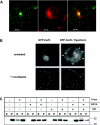
Figure 1
Figure 1. Subcellular localization and solubilization of WT CerK
(A) COS-1 cells overexpressing GFP-tagged WT-CerK were incubated for 30 min with 5 μM BODIPY®-TR-ceramide. After medium exchange, cells were analysed by fluorescence microscopy. Left-hand panel, GFP fluorescence; middle panel, BODIPY® fluorescence; right-hand panel, merge. (B) COS-1 cells overexpressing GFP-tagged WT-CerK were treated or not with 33 μM nocodazole for 3 h. Subsequently, cells were osmotically challenged by diluting the medium 1:4 in water and incubated for another 30 min before fluorescence microscopy. White arrows indicate plasma membrane localization. (C) COS-1 cells overexpressing C-terminally FLAG-tagged CerK were fractionated as described in the Experimental section, in the absence or presence of 2% Triton X-114, 2 mM EGTA or 500 mM KCl, as indicated. At the end of the harvesting period, cells were high-speed centrifuged and the resulting supernatants (S) and pellets (P) were analysed with SDS/PAGE and anti-FLAG Western blotting.
Figure 2
Figure 2. Model and ESP calculations for human CerK PH domain
(A) Model of CerK PH domain based on TAPP2 (1V5P.A). (B) ESP calculations. The representation is given in colour-coded isosurfaces at a counter level of ±144 mV. The red surface encompasses all co-ordinates in the vicinity of the protein that exhibit a negatively signed ESP of −144 mV. The blue surface is similar but represents a layer of positive ESPs of +144 mV. The protein structure was colour-coded from red (N-terminus) to blue (C-terminus).
Figure 3
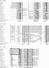
Figure 3. Structure-based multiple sequence alignment of CerK PH domains
CerK PH domain sequences of multiple species are aligned according to a multiple structure alignment of known PH domains. Highly conserved residues are white on black background, conserved negative residues (DE, conserved marker ‘–’), positive (HKR, ‘+’), tiny (AGS, ‘t’), polar (CDEHKNQRST, ‘p’) are boldface on white background, conserved aliphatic (ILV, ‘I’), hydrophobic (ACFGHILMTVWY, ‘h’), aromatic (FHWY, ‘a’) and big (EFHIKLMQRWY, ‘b’) residues are set on light grey background (italic font for big residues), conserved charged (DEHKR, ‘c’) residues on dark grey background, and conserved small (DEHKR, ‘s’) residues are shown in grey font [51]. The asterisks (*) denote mutagenized residues of the present study. The secondary structure elements of the final model are shown in the last row (E=strand and H=helix), following the default Chroma consensus string (80%). The unique and conserved β6–β7 loop of CerK PH domains from various species is boxed. The structure identifiers denote the four-letter PDB code, the chain (A or B) and the NMR model number (1), if applicable.
Figure 4
Figure 4. Subcellular localization, fractionation and PPIn binding of CerK PH domain-mutant proteins
(A) COS-1 cells were transiently transfected with plasmids encoding CerK WT and mutant alleles N-terminally fused to GFP. Cells were analysed using fluorescent microscopy 24 h after transfection. Pictures shown are representative of most of the cellular population, observed in several experiments. Golgi/cytoplasm fluorescence ratios were determined as described in the Experimental section, and are given as the means for measurements obtained with at least eight cells. (B) Cell fractionation of WT CerK compared with a CerK allele lacking the PH domain, to the 90/91/96/98 mutant of this study as well as to the kinase-dead CerK G198D mutant (Kin). All constructs were 6×His-FLAG-tagged at the C-terminus. Cell fractionation was performed as described in the Experimental section and obtained fractions were resolved by SDS/PAGE followed by Western blotting using an anti-FLAG antibody. (C) Lipid-protein overlays. Upper panel, left: in vitro translated and 35S-labelled CerK, or purified GST–CerK, binding to PPIn arrays, were assayed as described in the Experimental section, followed by autoradiography or anti-GST Western blotting respectively. Upper panel, right: control GST protein domains (see the Experimental section for full description), probed with anti-GST Western blotting [34]. Lower panel: phospholipid strip overlay analysis using C-terminally FLAG-tagged immunoprecipitated proteins: full-length CerK, or its PH domain only (CerK-PH), or CerK lacking its PH domain (ΔPH-CerK), or the K90V,R91A CerK mutant protein, followed by anti-FLAG Western blotting. On the right, the PH domain of PLCδ1 was used as a control.
Figure 5
Figure 5. Activity of CerK PH domain mutant proteins
(A) Different mutant alleles of CerK were expressed in COS cells and assayed in vitro as crude cell lysates, in comparison with WT CerK. Results shown represent the means for at least three experiments (−S.D.), performed in triplicates. *** indicates statistical significance with P<0.001 (t test). Expression of mutant proteins with a heavily compromised PH domain reached on average approx. 50% of WT CerK levels (see Supplementary Figure 1 at http://www.BiochemJ.org/bj/400/bj4000255add.htm). (B) In-cell kinase assays were performed for N-terminally GFP-tagged WT CerK and key mutants, using 32P labelling followed by lipid extraction (densitometric measurements as percentage relative to WT-CerK are indicated underneath).
Figure 6
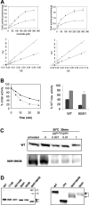
Figure 6. Biochemical characterization of CerK PH domain mutant proteins
(A) Substrate kinetic studies were performed using C8-ceramide and ATP as substrates and a CerK construct harbouring a 6×His–FLAG tag at the C-terminus. WT and the 90/91 CerK mutant were overexpressed in COS-1 cells and lysates were directly used for the in vitro kinase assays. For each kinetic a Lineweaver–Burk plot is shown, which was used for evaluation of Km values. This is one of two similar experiments, displayed as means±S.D. for triplicate evaluations. (●) CerK WT. (○) CerK 90/91. (B) Left panel: whole cell lysates of COS-1 cells overexpressing WT CerK (solid line) or the CerK 90/91 mutant (broken line), C-terminally FLAG–6×His-tagged proteins, were pre-incubated at 30 °C for the indicated times, before assaying for the remaining activity. Right panel: The presence of 20% glycerol (grey bars) prevents the inactivation of the CerK 90/91 mutant protein. This is one of two experiments with similar results. (C) WT and mutant CerK, both C-terminally 6×His–FLAG-tagged, were overexpressed in COS-1 cells and harvested in lysis buffer. Lysates were incubated with different amounts of trypsin (0, 0.001, 0.01 and 1 μg/ml). After a 30 min incubation, samples were processed for PAGE analysis. Western blotting was performed using an anti-FLAG antibody. This is one of two experiments with similar results. (D) Left panel: N-terminally GFP-tagged CerK and mutant alleles were overexpressed in COS-1 cells. SDS/PAGE was performed on lysates followed by Western-blot analysis using an antibody against the GFP tag. The presence of protein multimers is indicated by arrows heads. Right panel: in vitro translated [35S]methionine-labelled CerK WT and mutants were analysed by SDS/PAGE followed by autoradiography.

Similar articles

Cited by

References

    1. Sugiura M., Kono K., Liu H., Shimizugawa T., Minekura H., Spiegel S., Kohama T. Ceramide kinase, a novel lipid kinase. J. Biol. Chem. 2002;277:23294–23300. - PubMed
    1. Tuson M., Marfany G., Gonzalez-Duarte R. Mutation of CERKL, a novel human ceramide kinase gene, causes autosomal recessive retinitis pigmentosa (RP26) Am. J. Hum. Genet. 2004;74:128–138. - PMC - PubMed
    1. Bornancin F., Mechtcheriakova D., Stora S., Graf C., Wlachos A., Devay P., Urtz N., Baumruker T., Billich A. Characterization of a ceramide kinase-like protein. Biochim. Biophys. Acta. 2005;1687:31–43. - PubMed
    1. Waggoner D. W, Johnson L. B., Mann P. C., Morris V., Guastella J., Bajjalieh S. M. MuLK, a eukaryotic multi-substrate lipid kinase. J. Biol. Chem. 2004;279:38228–38235. - PubMed
    1. Van Overloop H., Gijsbers S., Van Veldhoven P. P. Further characterization of human ceramide kinase. J. Lipid Res. 2006;47:268–283. - PubMed

MeSH terms