Different mitochondrial intermembrane space proteins are released during apoptosis in a manner that is coordinately initiated but can vary in duration
- PMID: 16864784
- PMCID: PMC1518810
- DOI: 10.1073/pnas.0603007103
Different mitochondrial intermembrane space proteins are released during apoptosis in a manner that is coordinately initiated but can vary in duration
Abstract
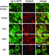
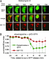
The release of mitochondrial intermembrane space proteins to the cytosol is a key event during apoptosis. We used in situ fluorescent labeling of proteins tagged with a short tetracysteine-containing sequence to follow the release of Smac, Omi, adenylate kinase-2, cytochrome c, and apoptosis-inducing factor (AIF) during apoptosis and compared the release with that of cytochrome c tagged with GFP in individual cells observed over time. We observed a caspase-independent, simultaneous release of cytochrome c, Smac, Omi, and adenylate kinase-2. Although AIF release also was caspase-independent and commenced with that of the other proteins, it proceeded much more slowly and incompletely from mitochondria, perhaps because of a requirement for a secondary event. These results suggest that these proteins are released through the same mitochondrial pore and that apoptosis may not be regulated through a selective release of individual mitochondrial proteins. The timing and extent of AIF release makes it unlikely that it is involved in the induction of apoptosis, either upstream or downstream of mitochondrial outer membrane permeabilization.
Conflict of interest statement
Conflict of interest statement: No conflicts declared.
Figures


Similar articles
-
Apoptosis is regulated by the VDAC1 N-terminal region and by VDAC oligomerization: release of cytochrome c, AIF and Smac/Diablo.Biochim Biophys Acta. 2010 Jun-Jul;1797(6-7):1281-91. doi: 10.1016/j.bbabio.2010.03.003. Epub 2010 Mar 6. Biochim Biophys Acta. 2010. PMID: 20214874
-
Toxic proteins released from mitochondria in cell death.Oncogene. 2004 Apr 12;23(16):2861-74. doi: 10.1038/sj.onc.1207523. Oncogene. 2004. PMID: 15077149 Review.
-
Endogenously released Smac is insufficient to mediate cell death of human lung carcinoma in response to etoposide.Exp Cell Res. 2004 Aug 1;298(1):83-95. doi: 10.1016/j.yexcr.2004.04.007. Exp Cell Res. 2004. PMID: 15242764
-
Mitochondrial release of pro-apoptotic proteins: electrostatic interactions can hold cytochrome c but not Smac/DIABLO to mitochondrial membranes.J Biol Chem. 2005 Jan 21;280(3):2266-74. doi: 10.1074/jbc.M411106200. Epub 2004 Nov 9. J Biol Chem. 2005. PMID: 15537572
-
Central roles of apoptotic proteins in mitochondrial function.Oncogene. 2013 May 30;32(22):2703-11. doi: 10.1038/onc.2012.348. Epub 2012 Aug 6. Oncogene. 2013. PMID: 22869150 Review.
Cited by
-
Orphan proteins of unknown function in the mitochondrial intermembrane space proteome: New pathways and metabolic cross-talk.Biochim Biophys Acta. 2016 Nov;1863(11):2613-2623. doi: 10.1016/j.bbamcr.2016.07.004. Epub 2016 Jul 15. Biochim Biophys Acta. 2016. PMID: 27425144 Free PMC article. Review.
-
Singlet Oxygen, Photodynamic Therapy, and Mechanisms of Cancer Cell Death.J Oncol. 2022 Jun 25;2022:7211485. doi: 10.1155/2022/7211485. eCollection 2022. J Oncol. 2022. PMID: 35794980 Free PMC article. Review.
-
The boundary of life and death: changes in mitochondrial and cytosolic proteomes associated with programmed cell death of Arabidopsis thaliana suspension culture cells.Front Plant Sci. 2023 Aug 1;14:1194866. doi: 10.3389/fpls.2023.1194866. eCollection 2023. Front Plant Sci. 2023. PMID: 37593044 Free PMC article.
-
Inhibition of Akt sensitises neuroblastoma cells to gold(III) porphyrin 1a, a novel antitumour drug induced apoptosis and growth inhibition.Br J Cancer. 2009 Jul 21;101(2):342-9. doi: 10.1038/sj.bjc.6605147. Epub 2009 Jun 23. Br J Cancer. 2009. PMID: 19550420 Free PMC article.
-
Optical control of MAP kinase kinase 6 (MKK6) reveals that it has divergent roles in pro-apoptotic and anti-proliferative signaling.J Biol Chem. 2020 Jun 19;295(25):8494-8504. doi: 10.1074/jbc.RA119.012079. Epub 2020 May 5. J Biol Chem. 2020. PMID: 32371393 Free PMC article.
References
-
- Danial N. N., Korsmeyer S. J. Cell. 2004;116:205–219. - PubMed
-
- Green D. R. Cell. 2005;121:671–674. - PubMed
-
- Verhagen A. M., Ekert P. G., Pakusch M., Silke J., Connolly L. M., Reid G. E., Moritz R. L., Simpson R. J., Vaux D. L. Cell. 2000;102:43–53. - PubMed
-
- Du C., Fang M., Li Y., Li L., Wang X. Cell. 2000;102:33–42. - PubMed
-
- Suzuki Y., Imai Y., Nakayama H., Takahashi K., Takio K., Takahashi R. Mol. Cell. 2001;8:613–621. - PubMed
Publication types
MeSH terms
Substances
Grants and funding
LinkOut - more resources
Full Text Sources
Other Literature Sources

