. 2006 Aug 9;25(15):3640-51.
doi: 10.1038/sj.emboj.7601216. Epub 2006 Jul 20.

Complex interaction of Drosophila GRIP PDZ domains and Echinoid during muscle morphogenesis

Affiliations

Complex interaction of Drosophila GRIP PDZ domains and Echinoid during muscle morphogenesis

Laura E Swan et al. EMBO J. .

Abstract

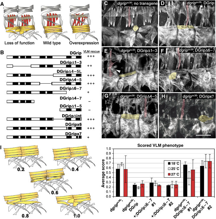
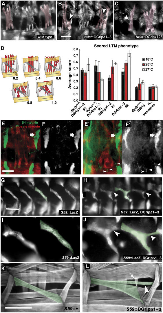
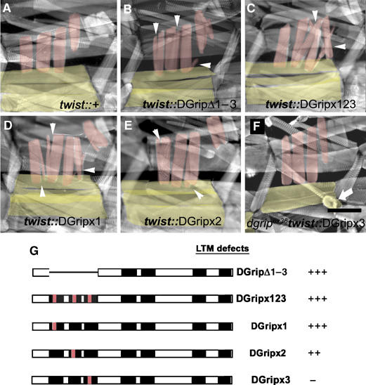
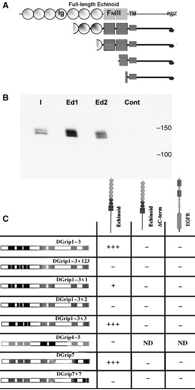
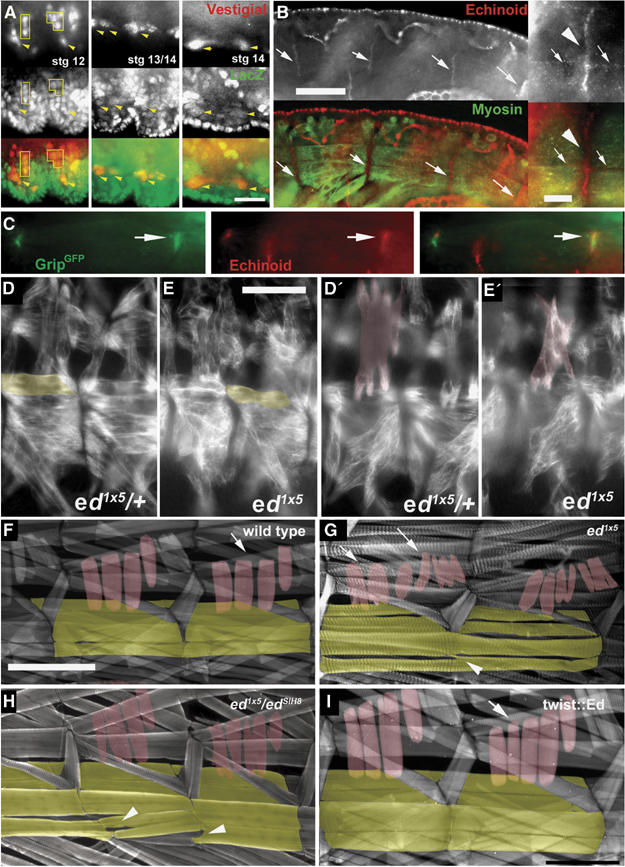
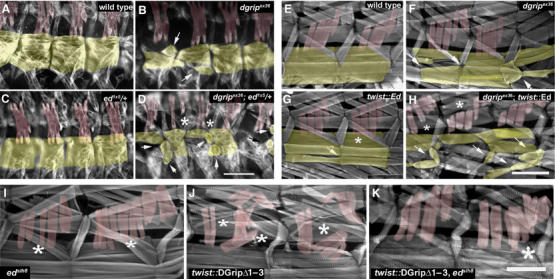
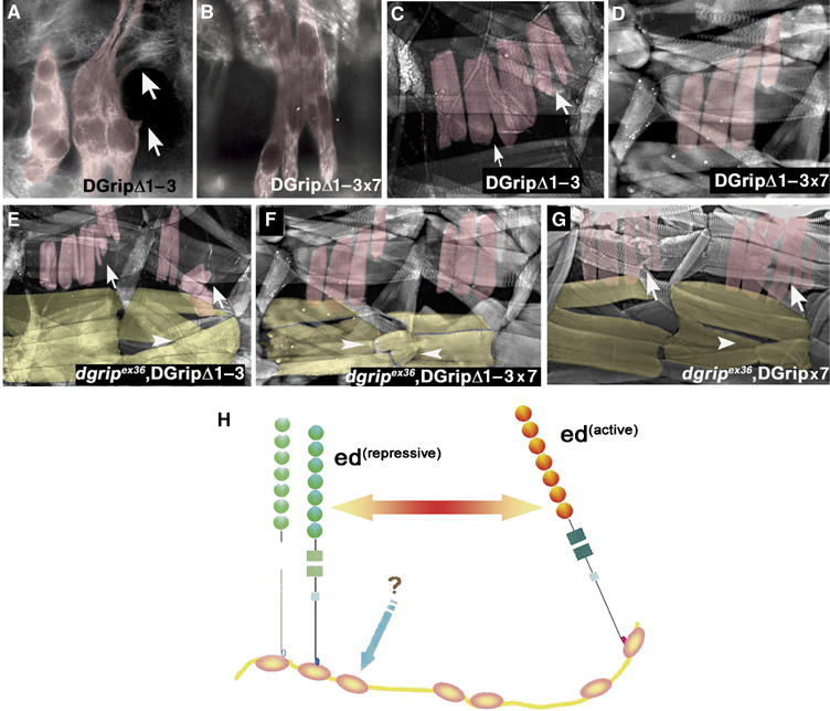
Glutamate receptor interacting protein (GRIP) homologues, initially characterized in synaptic glutamate receptor trafficking, consist of seven PDZ domains (PDZDs), whose conserved arrangement is of unknown significance. The Drosophila GRIP homologue (DGrip) is needed for proper guidance of embryonic somatic muscles towards epidermal attachment sites, with both excessive and reduced DGrip activity producing specific phenotypes in separate muscle groups. These phenotypes were utilized to analyze the molecular architecture underlying DGrip signaling function in vivo. Surprisingly, removing PDZDs 1-3 (DGripDelta1-3) or deleting ligand binding in PDZDs 1 or 2 convert DGrip to excessive in vivo activity mediated by ligand binding to PDZD 7. Yeast two-hybrid screening identifies the cell adhesion protein Echinoid's (Ed) type II PDZD-interaction motif as binding PDZDs 1, 2 and 7 of DGrip. ed loss-of-function alleles exhibit muscle defects, enhance defects caused by reduced DGrip activity and suppress the dominant DGripDelta1-3 effect during embryonic muscle formation. We propose that Ed and DGrip form a signaling complex, where competition between N-terminal and the C-terminal PDZDs of DGrip for Ed binding controls signaling function.


Figures

Figure 1
Figure 1
A structure–function map for DGrip in morphogenesis of VLM. (A) Scheme of muscle phenotypes evoked by DGrip loss of function and overexpression. Loss of DGrip function primarily affects VLM (yellow, see (C)) whereas LTM (red) remain unaffected. Strong overexpression of DGrip using 24B-gal4 disturbs LTM morphology (Swan et al, 2004). (B) Scheme of rescue activities of indicated DGrip constructs. Rescue of VLM morphology in dgripex36 mutant background is shown. Pan-muscular expression of wild-type DGrip using twist-gal4 fully rescues the dgripex36 VLM phenotype (+++), while other constructs have a reduced rescue ability (++, +) or exert no effect (−) on muscle rescue. All constructs were characterized in at least two independent lines. (C–H) Muscle myosin stainings in stage 16 embryos. (C) dgripex36, twist-gal4 muscles show typical defects in VLM morphology. One VLM (yellow) and one LTM (red) are labeled for ease of identification. (D) Re-expression of wild-type DGrip using twist-gal4 fully rescues these defects, whereas expression of DGripΔ1–3 in the dgripex36, twist-gal4 background (E) provokes strong dominant LTM (arrowhead) and slight VLM morphology defects. (F, H) Expression of DGrip missing the C-terminal PDZDs (DGripΔ6–7 (F) and DGripx7 (H)) results in only partial rescue of dgripex36 VLMs, with many VLMs appearing atypically round (asterisk in (F) compare to asterisk in (D)). (G) Constructs missing PDZDs 4 and 5 (DGripΔ4–5) behave like wild-type DGrip, fully rescuing the dgripex36 defect, without provoking LTM defects. Scale bar in (H): 30 μm. (I) Quantification of rescue activity for DGripΔ6–7. Left: scheme of VLM defects used as ‘clinical score' (between 0.2 and 1) for quantification. Right: Average scores from over 30 larval hemisegments per condition are plotted, raising temperatures used indicated in colors. While DGripΔ6–7 hardly rescues dgripex36 VLM defects, no dominant effects of DGripΔ6–7 expression are observed in dgripex36/+ heterozygous background.
Figure 2
Figure 2
DGripΔ1–3 provokes defects during embryonic muscle guidance. (A–C) Muscle myosin stainings in stage 16 embryos, some LTMs colored in red. (A) wild type; (B) DGripΔ1–3- and (C) DGripx123-expression in wild-type background with the pan-muscular driver twist-gal4 causes LTM defects (arrowheads). LTM defects include splitting into multiple projections and bending away from their target tendon cells. (D) Quantification of LTM morphology defects after DGripΔ1–3 expression scored by average clinical score (n>30, larval hemisegments per condition). At different expression levels (controlled by raising temperature) and with two independent transgenes, DGripΔ1–3 provokes LTM defects neither present in animals comparably expressing wild-type DGrip nor in dgrip mutants. (E, F′) Wild-type embryo (E, F) and twist∷dgripΔ1–3 (E′, F′) embryos costained for integrin (green) and muscle myosin (red) (E, E′), (F and F′) show integrin channel only. In twist-gal4∷dgripΔ1–3 embryos, LTMs form aberrant, integrin positive attachments in ectopic positions (arrowheads). The large integrin-positive attachment sites of segment border attaching muscles are labeled by arrows. (G–I) Genetically labeling embryonic muscle 5 (individual muscle 5 labeled in green) with the S59-gal4 driver reveals guidance defects in DGripΔ1–3 expressing muscles. (H, J) S59-gal4∷GripΔ1–3, lacZ muscles shortly after guidance process showing extra filopodia-like projections (arrowheads) not present in wild-type muscle 5 (G, I). (K, L) Ectopic projections (arrowheads) of muscle 5 (green) established in embryogenesis are still present in larval stages of S59-gal4∷DGripΔ1–3 larvae (L, labeled in green) but not in S59-gal4/+ controls (K). In addition, other muscles can ectopically adhere to DGripΔ1–3 expressing muscles (L, arrow). Scale bar in (B): 20 μm; scale bar in (E): 15 μm; scale bar in (G): 30 μm in (K): 150 μm.
Figure 3
Figure 3
Removal of PDZDs 1–3, or of ligand binding surfaces of PDZD 1 or 2, results in dominantly active Dgrip. The dominant DGripΔ1–3 phenotype is recapitulated by specifically point mutating the ligand binding sites of individual PDZDs. (A–F) Phalloidin labeling of 3rd instar larvae, VLMs in yellow, LTMs in red. (A) Typical bar-shaped LTMs and VLMs in control larvae. (B) Ectopic LTM projections (arrowheads) and mild morphological defects of VLMs in DGripΔ1–3 expressing animal. This phenotype is fully recapitulated (arrowheads) in DGripx123-expressing larvae, where the ligand binding capability of the PDZDs 1–3 is disturbed by point mutations. Mutation of PDZD 1 only (D) results in a similar phenotype, expression of DGrip point-mutated at PDZD 2 only (E) produces slightly weaker dominant defects. (F) Mutation of PDZD 3 does not cause dominant defects, but does not allow proper rescue of dgripex36 VLMs (arrowhead). (G) Summary of LTM defects caused by twist-gal4 driven expression of the indicated dgrip constructs. Scale bar in (F): 150 μm.
Figure 4
Figure 4
The cell-adhesion molecule Echinoid physically interacts with Dgrip. (A) Y2H screen performed using the first three PDZDs of DGrip as bait returned four independent isolates of the Immunoglobulin (Ig) and Fibronectin type III (FNIII)-domain containing cell adhesion molecule Echinoid (Ed). All isolates contained the transmembrane (TM) region and the entire cytosolic tail, including the EIIV PDZ-ligand motif. (B) Full-length, C-terminally myc-tagged DGrip expressed in Sf.9 cells specifically binds to a 10aa peptide representing the C-terminus of Ed. Shown is the input (I), binding of DGrip-myc to the Ed peptide (Ed1/2, two independent experiments), and binding to a 10aa scrambled control peptide (cont). (C) Y2H experiments reveal a specific pattern of Ed binding to DGrip PDZDs. The Ed cytosolic tail strongly interacts with a construct containing the first three PDZDs of DGrip, or containing PDZD 7 only (+++). This interaction was abolished by point mutation of PDZDs 2 or 7 (−), strongly reduced by point mutation of PDZD 1 (+) and unaffected by point mutation of PDZD 3 (+++). This interaction depended on the EIIV motif at the C-terminal of Ed, as Ed-ΔC-term did not interact with DGrip constructs. DGrip did not interact with the EGFR C-terminus, used here as a control, which has a C-terminal type I PDZ-ligand motif (ETRV).
Figure 5
Figure 5
Echinoid in muscle morphogenesis of the Drosophila embryo. (A) Expression of lacZ (green) from the ed locus in P(lacZ)edk01120 combined with labeling of muscle precursors with Vestigial (red). Scale bar in (A): 10 μm. (B) Anti-Ed antibodies (red) stain the ends of morphologically mature VLMs (arrows) visualized by antibodies against muscle myosin (green). Scale bar in (B): 30 μm; Inset: magnified view of Ed accumulation at muscle ends (arrowhead). Some Ed protein is also found at other parts of the muscle membrane (arrows). Scale bar in inset: 5 μm. (C) Ed protein colocalizes with muscle expressed twist-gal4∷DGrip-GFP at muscle ends (arrows). (D–H) ed mutants displaying morphological defects of both VLMs (yellow) and LTMs (red) in embryos (D–E′) and larvae (F–H). (D, D′) ed1x5/+ control embryo showing normal VLMs and LTMs, respectively. (E, E′) ed1x5 embryo shows defects in both VLM and LTM morphology. The same muscle field is shown in two focal planes. (F) Control larva. (G) The strong ed allele ed1x5 produces few homozygous larvae, which survive to 2nd instar. In these larvae, defects in VLMs (arrowhead) and LTMs (arrows) are evident, with both muscle groups forming aberrant projections and ectopic adhesion points. (H) Similar muscle phenotypes are also observed in 3rd instar larvae of the genotype ed1x5 over the strong hypomorphic allele edSlH8 (arrows and arrowheads indicate VLMs and LTMs respectively). (I) Only minor defects of LTMs are evoked by pan-muscular expression of Ed. Scale bar in (E): 35 μm; Scale bar in (F): 200 μm.
Figure 6
Figure 6
Functional interactions between Echinoid and DGrip in muscle morphogenesis. (A–D) Heterozygosity for ed mutant allele enhances defects in VLM and provokes LTM defects as shown by muscle myosin stainings of stage 16 embryos (A) wild type; (B) dgripex36 mutants with characteristic defects in VLM morphology (arrow); (C) ed1x5/+ embryos show no defects in LTMs or VLMs. (D) dgripex36; ed1x5/+ embryos exhibit more severe VLM defects (arrows) than dgripex36 embryos, where sometimes LTMs might be missing (asterisks). More severe examples of dgripex36; ed1x5/+ embryos (not shown) exhibit completely deranged somatic musculature, where muscle identification is no longer possible. Scale bar in (D): 50 μm. (E–H) Muscle-specific overexpression of Ed enhances defects in dgripex36 mutants. (E) Control larvae, showing bar-like morphology in VLM (yellow) and LTM (red). (F) dgripex36 larvae; (G) twist-gal4∷UAS-ed larvae exhibit few, mild defects in LTM and VLM morphology. Arrow indicates VLM with weakly distorted morphology, asterisks mark slightly split LTMs. (H) twist-mediated expression of Echinoid in the dgripex36 background greatly enhances defects; VLMs are more severely deranged than in dgripex36, and often appear to adhere to other muscles (arrows), whereas LTMs split (asterisks). Scale bar in (H): 300 μm. (I–K) Homozgosity for edSlH8 suppresses DGripΔ1–3 activity. (I) edSlH8 larvae show defects typical for ed zygotic alleles with slight LTM splitting (asterisks) and some malformation of VLMs. (J) twist-gal4∷UAS-dgripΔ1–3 controls with severe malformation of LTMs (asteriks). (K) edSlH8; twist-gal4∷UAS-dgripΔ1–3 larvae consistently showed far milder LTM (asteriks) defects than twist-gal4∷UAS-dgripΔ1–3 processed in parallel (n>30 hemisegments per genotype). Scale bar in (K) 150 μm.
Figure 7
Figure 7
Mutation of the Echinoid-binding DGrip PDZD 7 represses DGripΔ1–3 activity. This figure depicts muscle myosin stainings in embryos (A, B) and phalloidin labeling in larvae (C, D). Point mutation of PDZD 7 reduces DGripΔ1–3 activity. (A–D) The dominant activity of the DGripΔ1–3 protein ((A) embryo; (C) larva), which causes abnormal muscle projections (arrows) is reduced by point mutation of the PDZD 7 ligand binding surface, producing DGripΔ1–3x7, which shows only slight defects in LTM morphology in embryo (B) or larva (D). (E, F) DGripΔ1–3x7 shows only limited rescue ability in dgripex36 VLMs (F, arrowheads) when compared to DGripΔ1–3 (E, arrowheads). (G) DGripx7 produces mild dominant defects of LTMs (arrows) and impaired rescue of VLMs (arrowheads). (H) Model of DGrip–Echinoid functional interaction during muscle morphogenesis. DGrip may act by maintaining the equilibrium between active and repressive Echinoid signaling. Ed binds DGrip at PDZD 2 (and possibly 1), where it is repressed. Interaction of an unknown protein with PDZD 3 relieves this repression, allowing Ed to bind PDZD 7 and activating the complex.

Similar articles

Cited by

References

    1. Ahmed A, Chandra S, Magarinos M, Vaessin H (2003) Echinoid mutants exhibit neurogenic phenotypes and show synergistic interactions with the Notch signaling pathway. Development 130: 6295–6304 - PubMed
    1. Artero R, Furlong EE, Beckett K, Scott MP, Baylies M (2003) Notch and Ras signaling pathway effector genes expressed in fusion competent and founder cells during Drosophila myogenesis. Development 130: 6257–6272 - PubMed
    1. Bai J, Chiu W, Wang J, Tzeng T, Perrimon N, Hsu J (2001) The cell adhesion molecule Echinoid defines a new pathway that antagonizes the Drosophila EGF receptor signaling pathway. Development 128: 591–601 - PubMed
    1. Baran R, Jin Y (2002) Getting a GRIP on liprins. Neuron 34: 1–2 - PubMed
    1. Brand AH, Perrimon N (1993) Targeted gene expression as a means of altering cell fates and generating dominant phenotypes. Development 118: 401–415 - PubMed

MeSH terms