

doi: 10.1128/JB.00250-06.
A parA homolog selectively influences positioning of the large chromosome origin in Vibrio cholerae
Affiliations
- PMID: 16855253
- PMCID: PMC1540020
- DOI: 10.1128/JB.00250-06
Item in Clipboard
A parA homolog selectively influences positioning of the large chromosome origin in Vibrio cholerae
J Bacteriol.
2006 Aug.
Abstract
A Vibrio cholerae deletion mutant lacking VS2773, a parA partitioning gene homolog located in a parAB operon on the large chromosome, displays altered positioning of the large chromosome origin. Deletion of a second parA homolog on the large chromosome (VC2061) does not affect its origin positioning. The origin position of the small chromosome is unchanged by either or both of these deletions, suggesting that VC2773 function is specific to the replicon on which it is carried. VC2773 and VC2772 form a parABS system with inverted repeats found near the large chromosome origin.
Figures
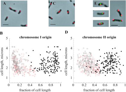
Positions of V. cholerae chromosome origins in wild-type strain N16961. (A and B) Positioning of the large chromosome origin in cells. (C and D) Positioning of the small chromosome origin. Graphs show positions of signals in cells containing one (red) or two (black) fluorescent foci; they depict the lengths of the cells on the y axis and the positions of the fluorescent foci along the length of the cell on the x axis. Photomicrographs depict the Cy3 probe signals superimposed on phase-contrast images of cell bodies. Bars, 2 μm. (E) Examples of dually labeled cells, where the oriCI probe signal is red (Cy3) and the oriCII probe signal is green (fluorescein).
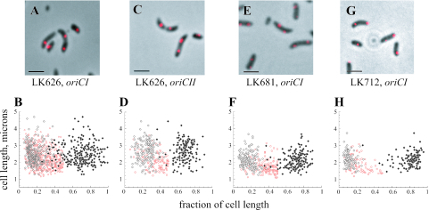
Positions of V. cholerae chromosome origins in strains LK626, LK681, and LK712. Graphs show positions of signals in cells containing one (red) or two (black) fluorescent foci. The labeling of axes and scale are the same as those in Fig. 1. (A and B) Positioning of the large chromosome origin in LK626. (C and D) Positioning of the small chromosome origin in LK626. (E and F) LK681 (N16961 with pJK4) labeled with the oriCI probe. (G and H) LK712 (LK626 with “knocked-in” VC2773) labeled with the oriCI probe.

Isolation of the promoter for the V. cholerae chromosome 1 parAB locus. (A) Northern blot showing transcripts detected using a parB probe in three separate samples of each strain. The positions of RNA markers and their sizes in kb are depicted to the side of the blot. Arrowheads, transcripts; asterisk, background band from rRNA. (B) Beta-galactosidase activities (in Miller units) assayed in bacteria grown in LB at 37°C to an OD600 of 0.4 to 0.6, using the following strains: wild-type parent strain KFV10, KFV10 with a plasmid containing a promoterless lacZ gene (pRKlac290), and KFV10 with pSD19 (pRKlac290 containing the upstream region of parA) (averages of three experiments are shown).
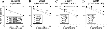
Stabilization of mini-F plasmids containing putative parS sequences by coresident plasmids carrying the VC2773-VC2772 parAB locus. Experiments were performed multiply, and the graphs are from a representative experiment. Bacteria were grown in M9-CSA with chloramphenicol to maintain the mini-F derivative; at an OD of ∼0.3, they were back diluted into medium lacking chloramphenicol (time zero) and plated at the specified generation times. Colonies were patched onto chloramphenicol to assay for the presence of the mini-F plasmid. The y axes in all panels show the percentages of colonies still retaining the mini-F derivative. (A) Stability of the sop+ mini-F plasmid pDAG114, as well as its unstable derivative pDAG203, in the presence of a coresident pBluescript vector or pSD25 (carrying both parA and parB). (B to D) Stabilities of pDS38, pSD39, and pSD44 (pDAG203 carrying IR1, IR2, and IR3, respectively). Each plasmid was assayed in the presence of empty pBluescript vector, pSD9 (with an in-frame deletion of VC2773, thus carrying parB), pSD21 (with an in-frame deletion of VC2772, thus carrying parA), or pSD25 (carrying both parA and parB). The legend for each graph indicates the par genes supplied in trans.
Similar articles
-
The chromosome partitioning proteins Soj (ParA) and Spo0J (ParB) contribute to accurate chromosome partitioning, separation of replicated sister origins, and regulation of replication initiation in Bacillus subtilis.Mol Microbiol. 2006 May;60(4):853-69. doi: 10.1111/j.1365-2958.2006.05140.x. Mol Microbiol. 2006. PMID: 16677298
-
Distinct segregation dynamics of the two Vibrio cholerae chromosomes.Mol Microbiol. 2005 Jan;55(1):125-36. doi: 10.1111/j.1365-2958.2004.04379.x. Mol Microbiol. 2005. PMID: 15612922
-
ParABS systems of the four replicons of Burkholderia cenocepacia: new chromosome centromeres confer partition specificity.J Bacteriol. 2006 Feb;188(4):1489-96. doi: 10.1128/JB.188.4.1489-1496.2006. J Bacteriol. 2006. PMID: 16452432 Free PMC article.
-
Management of multipartite genomes: the Vibrio cholerae model.Curr Opin Microbiol. 2014 Dec;22:120-6. doi: 10.1016/j.mib.2014.10.003. Curr Opin Microbiol. 2014. PMID: 25460805 Review.
-
Bacterial chromosome segregation by the ParABS system.Open Biol. 2020 Jun;10(6):200097. doi: 10.1098/rsob.200097. Epub 2020 Jun 17. Open Biol. 2020. PMID: 32543349 Free PMC article. Review.
Cited by
-
The three vibrio cholerae chromosome II-encoded ParE toxins degrade chromosome I following loss of chromosome II.J Bacteriol. 2011 Feb;193(3):611-9. doi: 10.1128/JB.01185-10. Epub 2010 Nov 29. J Bacteriol. 2011. PMID: 21115657 Free PMC article.
-
Centromere binding and evolution of chromosomal partition systems in the Burkholderiales.J Bacteriol. 2012 Jul;194(13):3426-36. doi: 10.1128/JB.00041-12. Epub 2012 Apr 20. J Bacteriol. 2012. PMID: 22522899 Free PMC article.
-
DNA motifs that sculpt the bacterial chromosome.Nat Rev Microbiol. 2011 Jan;9(1):15-26. doi: 10.1038/nrmicro2477. Nat Rev Microbiol. 2011. PMID: 21164534 Review.
-
Evidence for a DNA-relay mechanism in ParABS-mediated chromosome segregation.Elife. 2014 May 23;3:e02758. doi: 10.7554/eLife.02758. Elife. 2014. PMID: 24859756 Free PMC article.
-
Evidence for two different regulatory mechanisms linking replication and segregation of vibrio cholerae chromosome II.PLoS Genet. 2013 Jun;9(6):e1003579. doi: 10.1371/journal.pgen.1003579. Epub 2013 Jun 20. PLoS Genet. 2013. PMID: 23818869 Free PMC article.
References
-
- Clark, D., and O. Maaloe. 1967. DNA replication and the cell cycle in Escherichia coli. J. Mol. Biol. 23:99-112.
Publication types
MeSH terms
Substances
LinkOut - more resources
Full Text Sources
