. 2006 Jul 19;26(29):7756-66.
doi: 10.1523/JNEUROSCI.1560-06.2006.

Interaction of survival and death signaling in basal forebrain neurons: roles of neurotrophins and proneurotrophins

Affiliations

Interaction of survival and death signaling in basal forebrain neurons: roles of neurotrophins and proneurotrophins

Marta Volosin et al. J Neurosci. .

Abstract

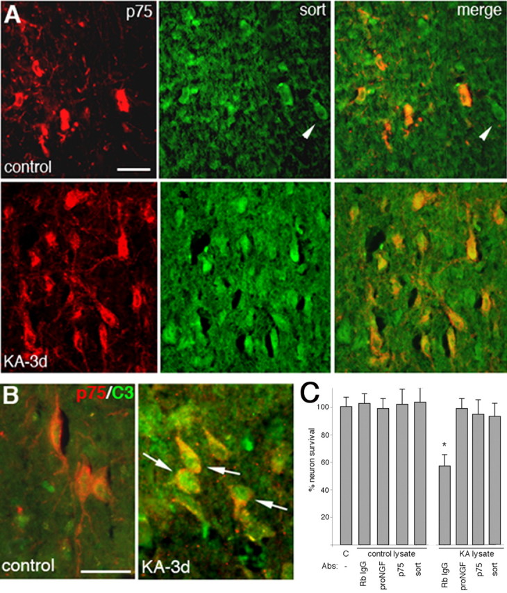
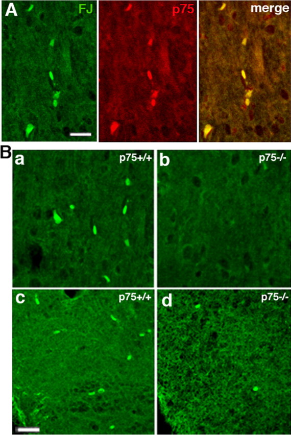
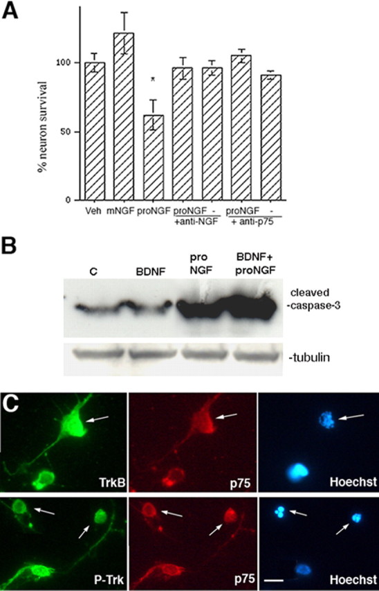
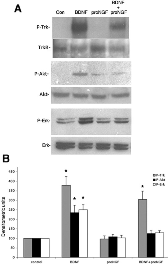
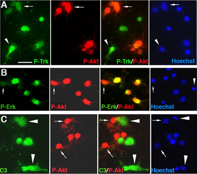
Proneurotrophins bind with high affinity to p75 neurotrophin receptor (p75NTR) and lack the capacity to bind Trk receptors, suggesting that proneurotrophins can elicit apoptosis via p75NTR even in cells expressing survival-promoting Trk receptors. In the CNS, basal forebrain (BF) neurons are particularly vulnerable to degeneration in Alzheimer's disease, and are among the few populations of brain neurons that express p75NTR throughout life. These neurons also express Trk receptors and may be concomitantly exposed to both proneurotrophins and mature neurotrophins during development, disease, or after injury. We investigated the interaction of mature and proneurotrophin signaling in these CNS neurons. Kainic acid-induced seizures elicited production of pro-NGF by BF astrocytes before caspase activation in p75NTR-positive BF neurons, demonstrating local production of proneurotrophins under pathological conditions and suggesting apoptotic signaling in vivo. Mechanisms of proneurotrophin-induced death were analyzed in cultured BF neurons, and required both p75NTR and its coreceptor sortilin. Surprisingly, exposure to both mature neurotrophins and proneurotrophins demonstrated that Trk phosphorylation did not prevent pro-NGF-induced apoptosis via p75NTR. However, activation of PI3K (phosphatidylinositol 3-kinase)/Akt and MEK (mitogen-activated protein kinase kinase)/Erk pathways prevented pro-NGF-induced apoptosis, revealing a novel critical checkpoint in survival versus apoptotic signaling downstream of Trk activation, and suggesting that pro-NGF blocks survival signaling by preventing Akt and Erk activation. This study shows that proneurotrophins are produced in the brain under pathological conditions, and can elicit apoptosis of BF neurons even when Trk receptors are activated.


Figures

Figure 1.
Figure 1.
Pro-NGF elicits loss of BF neurons in culture. BF neurons were grown for 5 d and treated overnight with the following: vehicle (imidazole), mature NGF (100 ng/ml), pro-NGF (1 ng/ml), pro-NGF (1 ng/ml) in the presence of blocking antibodies to NGF (Sigma; which also block pro-NGF; diluted in the medium 1:500) or p75 (9651 (Huber and Chao, 1995; Friedman, 2000); diluted 1:500 in the medium), or the antibodies alone. Data are expressed as mean percentage of control ± SEM in triplicate cultures from five independent experiments (n = 15). The asterisk indicates a value different from control at p < 0.05. Veh, Vehicle; mNGF, mature NGF.
Figure 2.
Figure 2.
Sortilin is required for proneurotrophin-induced death of BF neurons. A, Pro-NGF and pro-BDNF elicit a loss of BF neurons that can be prevented by either anti-p75NTR or anti-sortilin. BF neurons were cultured for 5 d and treated as indicated overnight. Pro-NGF and pro-BDNF were used at 1 ng/ml, and the antibodies were diluted 1:500 in the media. The antibodies alone had no effect on neuronal survival. B, Neurotensin blocks pro-NGF and pro-BDNF-induced neuronal loss in a dose-dependent manner. BF neurons were cultured for 5 d and treated overnight with pro-NGF (1 ng/ml) or pro-BDNF (1 ng/ml) in the presence or absence of 4 or 40 μm neurotensin. For A and B, data are expressed as mean percentage of control ± SEM in triplicate cultures from three independent experiments (n = 9). The asterisks indicate values different from control at p < 0.05. C, Immunostaining demonstrates colocalization of sortilin with p75 on cultured BF neurons. The arrows indicate p75/sortilin-positive apoptotic neurons, and the arrowheads show healthy p75/sortilin-positive neurons. Scale bar, 10 μm.
Figure 3.
Figure 3.
Pro-NGF is induced in the BF in vivo by seizures. a–c show sections from a control rat injected with saline, and d–f show sections from a rat injected with kainic acid 1 d after seizures. Sections were labeled for pro-NGF (a, d) and GFAP (b, e). The merged images demonstrate the presence of pro-NGF in astrocytes (f) of KA-treated rats compared with control rats (c). Scale bar, 100 μm.
Figure 4.
Figure 4.
In vivo induction of p75NTR and sortilin in BF neurons after seizures, and activation of caspases in p75NTR-positive neurons. A, Expression of p75NTR (red) and sortilin (green) after kainic acid-induced seizures (bottom row) compared with saline-injected controls (top row). Note that sortilin is expressed in all the p75NTR-positive neurons (orange/yellow) as well as in some neurons that lack p75NTR (arrowheads). Scale bar, 100 μm. B, Rats injected with saline or kainic acid were analyzed after 3 d by immunostaining for p75NTR (red) and cleaved caspase-3 (green). Arrows indicate several p75NTR-positive neurons also labeled for cleaved caspase-3. Scale bar, 25 μm. C, Lysates were prepared from saline (C) or KA-treated rats, incubated with anti-pro-NGF or rabbit (Rb) IgG and added overnight to cultured BF neurons. Lysates from KA-treated, but not control, rats induced a 40% loss of BF neurons that was prevented by preincubation with anti-pro-NGF in three separate experiments with three different sets of lysates. Neuronal death induced by lysates from KA-treated rats was also prevented by the presence of anti-p75NTR (9651) or anti-sortilin. The asterisk indicates a value different from other treatment groups at p < 0.05.
Figure 5.
Figure 5.
Kainic acid induced fewer dying neurons in the basal forebrain of p75−/− mice than wild-type mice. A, In wild-type mice, kainic acid induced Fluoro-Jade B (FJ) labeling, indicative of dying neurons, in p75+ cells labeled with the 9651 antibody. B, Fluoro-Jade B labeling demonstrated that kainic acid induced death of fewer neurons after 1 d in the p75−/− mice compared with wild-type mice in both the medial septal area (a, b) and diagonal band (c, d). Scale bar, 100 μm.
Figure 6.
Figure 6.
Pro-NGF-activated signaling in BF neurons. A, BF neurons were cultured for 5 d and treated with pro-NGF for the indicated times. Cells were harvested for Western blot and probed for P-JNK, stripped, and reprobed for total JNK. One representative experiment of three is shown. Numbers indicate the average fold increase from the three experiments in band density compared with control treatment and normalized for total JNK. B, BF neurons were treated with the indicated factors for 4 h and analyzed by Western blot for cleaved caspase-6 (top) and cleaved caspase-3 (bottom). Blots were stripped and reprobed for tubulin. One representative experiment of three is shown. Veh, Vehicle; matNGF, mature NGF.
Figure 7.
Figure 7.
BDNF pretreatment of BF neurons does not protect against pro-NGF-induced apoptosis. A, BF neurons were grown in the presence of BDNF (10 ng/ml) for 5 d and treated overnight with pro-NGF alone, or in the presence of anti-p75NTR or anti-NGF as in Figure 1. Data are expressed as mean percentage of control ± SEM in triplicate cultures from four independent experiments (n = 12). The asterisks indicate values different from control at p < 0.05. Veh, Vehicle; mNGF, mature NGF. B, Western blot demonstrating that cleavage of caspase-3 was induced only by proneurotrophins and was not blocked by pretreatment with mature BDNF. The blot was stripped and reprobed for tubulin. C, Control. C, BF neurons were cultured on slide wells and treated with BDNF and pro-NGF. Neurons were double immunostained for full-length TrkB (anti-TrkBin) (top) or P-Trk (bottom) and p75NTR, and labeled with Hoechst to identify apoptotic nuclei. Scale bar, 10 μm.
Figure 8.
Figure 8.
Western blot analysis of distinct signaling pathways activated by mature neurotrophins or proneurotrophins. Cultured BF neurons were treated for 15 min with mature BDNF or pro-NGF or both. A, Cells were analyzed by Western blot for P-Trk, P-Akt, and P-Erk. Blots were then stripped and reprobed for TrkB, total Akt, and total Erk, respectively. Con, Control. B, Quantitation of P-Trk (gray bars), P-Akt (black bars), and P-Erk (open bars) relative to total TrkB, Akt, and Erk, respectively, from three independent experiments. The asterisks indicate values significantly different from control at p < 0.05. Error bars indicate SEM.
Figure 9.
Figure 9.
Pro-NGF induces apoptosis of BDNF-treated BF neurons with activated Trk receptors but not with phosphorylated Akt or Erk. A, Neurons were double immunostained for P-Trk and P-Akt and labeled with Hoechst. Anti-P-Akt labeled a subset of the P-Trk-positive neurons that were not apoptotic (arrows). The arrowheads indicate apoptotic neurons that are P-Trk positive and P-Akt negative. B, Neurons were double labeled for P-Akt and P-Erk. P-Erk was present in most but not all of the P-Akt-positive population, which were all healthy. An arrow indicates a P-Akt-positive neuron that lacks P-Erk. An arrowhead indicates an apoptotic cell that had neither P-Akt nor P-Erk. C, Neurons were double labeled for P-Akt and cleaved caspase-3 (C3) and labeled with Hoechst. The arrows indicate healthy neurons, and the arrowheads indicate apoptotic neurons. All of the cleaved caspase-3-positive neurons were apoptotic and lacked P-Akt, whereas all of the P-Akt-positive neurons were healthy and lacked cleaved caspase-3. Scale bar: (in A) A–C, 20 μm.
Figure 10.
Figure 10.
Akt is a critical mediator of BF neuron survival. A, BF neurons were grown in culture for 5 d and infected during the last 36 h with 50 MOI of adenovirus expressing either GFP or constitutively activated ras (ras V12) that was myc tagged. Neurons infected with control virus were identified by GFP labeling, and those infected with the rasV12 virus were identified by immunostaining for the myc tag. Uninfected and infected cultures were treated with pro-NGF for 5 h, fixed, and labeled with Hoechst to identify apoptotic nuclei. Data are expressed as percentage of apoptotic neurons. Triplicate cultures were analyzed in two independent experiments (n = 6). The asterisk indicates a value significantly different from all other groups at p < 0.05. Error bars indicate SEM. C, Control. B, BF neurons were grown for 5 d and treated overnight with the PI3K inhibitor LY294002 (LY) (50 μm) or the Mek1 inhibitor PD98059 (PD) (10 μm) alone or in combination with pro-NGF. Triplicate cultures were analyzed in three independent experiments (n = 9) and expressed as percentage of apoptotic neurons. The asterisk indicates a value significantly different from all other groups at p < 0.05. The double asterisk indicates a value significantly different from pro-NGF alone at p < 0.05. Error bars indicate SEM.

Similar articles

Cited by

References

    1. Alderson RF, Alterman AL, Barde Y-A, Lindsay RM (1990). Brain-derived neurotrophic factor increases survival and differentiated functions of rat septal cholinergic neurons in culture. Neuron 5:297–306. - PubMed
    1. Allendoerfer KL, Cabelli RJ, Escandon E, Kaplan DR, Nikolics K, Shatz CJ (1994). Regulation of neurotrophin receptors during the maturation of the mammalian visual system. J Neurosci 14:1795–1811. - PMC - PubMed
    1. Barker PA (2004). p75NTR is positively promiscuous: novel partners and new insights. Neuron 42:529–533. - PubMed
    1. Beattie MS, Harrington AW, Lee R, Kim JY, Boyce SL, Longo FM, Bresnahan JC, Hempstead BL, Yoon SO (2002). ProNGF induces p75-mediated death of oligodendrocytes following spinal cord injury. Neuron 36:375–386. - PMC - PubMed
    1. Bhakar AL, Howell JL, Paul CE, Salehi AH, Becker EB, Said F, Bonni A, Barker PA (2003). Apoptosis induced by p75NTR overexpression requires Jun kinase-dependent phosphorylation of Bad. J Neurosci 23:11373–11381. - PMC - PubMed

Publication types

MeSH terms