. 2006 Sep 15;281(37):27029-38.
doi: 10.1074/jbc.M601128200. Epub 2006 Jul 18.

HIV-1 Nef selectively activates Src family kinases Hck, Lyn, and c-Src through direct SH3 domain interaction

Affiliations

HIV-1 Nef selectively activates Src family kinases Hck, Lyn, and c-Src through direct SH3 domain interaction

Ronald P Trible et al. J Biol Chem. .

Abstract

Nef is an HIV-1 virulence factor that promotes viral pathogenicity by altering host cell signaling pathways. Nef binds several members of the Src kinase family, and these interactions have been implicated in the pathogenesis of HIV/AIDS. However, the direct effect of Nef interaction on Src family kinase (SFK) regulation and activity has not been systematically addressed. We explored this issue using Saccharomyces cerevisiae, a well defined model system for the study of SFK regulation. Previous studies have shown that ectopic expression of c-Src arrests yeast cell growth in a kinase-dependent manner. We expressed Fgr, Fyn, Hck, Lck, Lyn, and Yes as well as c-Src in yeast and found that each kinase was active and induced growth suppression. Co-expression of the negative regulatory kinase Csk suppressed SFK activity and reversed the growth-inhibitory effect. We then co-expressed each SFK with HIV-1 Nef in the presence of Csk. Nef strongly activated Hck, Lyn, and c-Src but did not detectably affect Fgr, Fyn, Lck, or Yes. Mutagenesis of the Nef PXXP motif essential for SH3 domain binding greatly reduced the effect of Nef on Hck, Lyn, and c-Src, suggesting that Nef activates these Src family members through allosteric displacement of intramolecular SH3-linker interactions. These data show that Nef selectively activates Hck, Lyn, and c-Src among SFKs, identifying these kinases as proximal effectors of Nef signaling and potential targets for anti-HIV drug discovery.


Figures
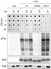
FIGURE 1
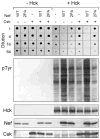
FIGURE 1. Hck induces yeast growth suppression in a kinase-dependent manner
Yeast cultures were transformed with galactose-inducible expression plasmids for wild-type Hck (WT), a kinase-dead mutant (K269D), a mutant lacking the C-terminal Csk phosphorylation site (Y501F), or the empty expression plasmid (−Hck). Cells were co-transformed with galactose-inducible vectors for wild-type (WT) or kinase-dead (KD) Csk as indicated or with the empty vector as a negative control (−). Top: liquid cultures were grown with glucose as the sole carbon source to repress protein expression and normalized to equal densities. Cells were then spotted onto agar selection plates containing galactose as the sole carbon source and incubated for 3 days at 30 °C. Cultures were spotted in 4-fold dilutions to enhance visualization of the growth-suppressive phenotype. Plates were scanned, and yeast patches appear as dark circles. Lower panels: immunoblots from cultures shown at the top. Transformed cells were grown in liquid culture in the presence of galactose at 30 °C for 18 h. Protein extracts were separated via SDS-PAGE and immunoblotted for tyrosine-phosphorylated proteins (pTyr) as well as for Hck and Csk.
FIGURE 2
FIGURE 2. HIV-1 Nef activates Hck in a PXXP-dependent manner
Yeast cultures were transformed with galactose-inducible expression plasmids for wild-type Hck (+Hck) in the absence (−) or presence of wild-type (WT) or PXXP mutant (2PA) forms of HIV-1 Nef. Cells were co-transformed with galac-tose-inducible expression vectors for Csk or the corresponding empty vector as indicated. Control cultures without Hck are shown on the left (−Hck). Top: liquid cultures were grown with glucose as the sole carbon source to repress protein expression and normalized to equal densities. Cells were then spotted onto agar selection plates containing galactose as the sole carbon source and incubated for 3 days at 30 °C. Cultures were spotted in 4-fold dilutions to enhance visualization of the growth-suppressive phenotype. Plates were scanned and yeast patches appear as dark circles. Lower panels: Immunoblots from cultures shown at the top. Transformed cells were grown in liquid culture in the presence of galactose at 30 °C for 18 h. Protein extracts were separated via SDS-PAGE and immunoblotted for tyrosine-phosphorylated proteins (pTyr) as well as for Hck, Nef, and Csk.
FIGURE 3
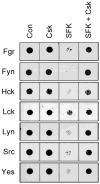
FIGURE 3. Csk reverses growth suppression of yeast by SFKs
Yeast cultures were transformed with the SFKs indicated on the left either alone (SFK) or in the presence of Csk (SFK + Csk). Cells transformed with empty vectors (Con) or with Csk alone (Csk) were included as controls in each experiment. Liquid cultures were grown with glucose as the sole carbon source to repress protein expression and normalized to equal densities. Cells were then spotted onto agar selection plates containing galactose as the sole carbon source and incubated for 3 days at 30 °C. Plates were scanned, and yeast patches appear as dark circles. Cultures were spotted in 4-fold dilutions to enhance visualization of the growth-suppressive phenotype. The dilutions showing the clearest differences in growth in the presence and absence of Csk are presented.
FIGURE 4
FIGURE 4. Csk suppresses SFK activity in yeast
Yeast cultures were transformed with expression vectors for each of the SFKs shown at the top in the presence (+) or absence (−) of Csk. Cells were grown in liquid culture in the presence of galactose at 30 °C for 18 h. Protein extracts were separated via SDS-PAGE, and immunoblotted for tyrosine-phosphorylated proteins (pTyr) as well as for each Src family member (SFK) and Csk.
FIGURE 5
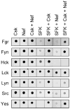
FIGURE 5. HIV-1 Nef selectively induces growth suppression in yeast co-expressing down-regulated forms of Hck, Lyn, and c-Src
Yeast cultures were transformed with the SFKs indicated on the left either alone (SFK) or in the presence of Csk (SFK + Csk), HIV-1 Nef (SFK + Nef), or both (SFK + Csk + Nef). Cells transformed with Csk alone (Csk), Nef alone (Nef) or both (Csk + Nef) were included as controls in each experiment. Liquid cultures were grown with glucose as the sole carbon source to repress protein expression and normalized to equal densities. Cells were then spotted onto agar selection plates containing galactose as the sole carbon source and incubated for 3 days at 30 °C. Plates were scanned, and yeast patches appear as dark circles. Cultures were spotted in 4-fold dilutions to enhance visualization of the growth-suppressive phenotype. The dilutions showing the greatest differences in growth are presented.
FIGURE 6
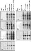
FIGURE 6. HIV-1 Nef selectively activates Hck, Lyn, and c-Src in yeast
Yeast cultures were transformed with expression vectors for each of the SFKs indicated either alone (SFK) or in the presence of Csk (SFK + Csk), HIV-1 Nef (SFK + Nef), or both (SFK + Csk + Nef). Cells transformed with Csk alone (Csk), Nef alone (Nef), or both (Csk + Nef) were included as negative controls. Cells were grown in liquid culture in the presence of galactose at 30 °C for 18 h. Protein extracts were separated via SDS-PAGE and immu-noblotted for tyrosine-phosphorylated proteins (pTyr). Control immuno-blots confirmed expression of each SFK, Csk, and Nef (data not shown).
FIGURE 7
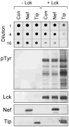
FIGURE 7. Lck is activated by herpesvirus saimiri Tip but not HIV-1 Nef in yeast
Yeast cultures were transformed with galactose-inducible expression plasmids for Lck (+Lck) or the empty expression plasmid as negative control (−Lck). Cells were co-transformed with galactose-inducible vectors for HIV-1 Nef or FLAG-tagged herpervirus saimiri Tip as indicated or with the empty vector (Con). Top: liquid cultures were grown with glucose as the sole carbon source to repress protein expression and normalized to equal densities. Cells were then spotted onto agar selection plates containing galactose as the sole carbon source and incubated for 3 days at 30 °C. Cultures were spotted in 4-fold dilutions to enhance visualization of the growth-suppressive phenotype. Plates were scanned, and yeast patches appear as dark circles. Lower panels: immunoblots from cultures shown at the top. Transformed cells were grown in liquid culture in the presence of galactose at 30 °C for 18 h. Protein extracts were separated via SDS-PAGE and immunoblotted for tyrosine-phosphorylated proteins (pTyr) as well as for Lck, Nef, and Tip. Tip was visualized using an anti-FLAG antibody and runs as multiple bands in the presence of Lck due to tyrosine phosphorylation.
FIGURE 8
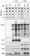
FIGURE 8. HIV-1 Nef-mediated activation of Lyn and c-Src is PXXP-dependent
Yeast cultures were transformed with galactose-inducible expression plasmids for Lyn (+Lyn) and c-Src (+Src) in the absence (−) or presence of wild-type (WT) or PXXP mutant (2PA) forms of HIV-1 Nef. Cells expressing the Nef proteins alone were included as a negative control. Top: liquid cultures were grown with glucose as the sole carbon source to repress protein expression and normalized to equal densities. Cells were then spotted onto agar selection plates containing galactose as the sole carbon source and incubated for 3 days at 30 °C. Cultures were spotted in 4-fold dilutions to enhance visualization of the growth-suppressive phenotype. Plates were scanned, and yeast patches appear as dark circles. Lower panels: immunoblots from cultures shown at the top. Transformed cells were grown in liquid culture in the presence of galactose at 30 °C for 18 h. Protein extracts were separated via SDS-PAGE and immunoblotted for tyrosine-phosphorylated proteins (pTyr) as well as for Lyn, Src, Csk, and Nef.
FIGURE 9
FIGURE 9. Activation of SFKs by HIV-1 Nef in vitro
Recombinant Hck, Lyn, and c-Src were purified from Sf9 insect cells in their down-regulated forms and assayed for kinase activity with a peptide substrate in vitro either alone or in the presence of a 5- or 10-fold molar excess of purified recombinant Nef. Details of the FRET-based tyrosine kinase assay used for this experiment can be found under “Materials and Methods.” Each condition was repeated in quadruplicate, and the extent of phosphorylation is expressed as mean percent phosphorylation relative to a control phosphopeptide ± S.D. The overall experiment was repeated twice with comparable results.

Similar articles

Cited by

References

    1. Fackler OT, Baur AS. Immunity. 2002;16:493–497. - PubMed
    1. Peter F. Immunity. 1998;9:433–437. - PubMed
    1. Joseph AM, Kumar M, Mitra D. Curr HIV Res. 2005;3:87–94. - PubMed
    1. Daniel MD, Kirchhoff F, Czajak SC, Sehgal PK, Desrosiers RC. Science. 1992;258:1938–1941. - PubMed
    1. Kestler HW, III, Ringler DJ, Mori K, Panicali DL, Sehgal PK, Daniel MD, Desrosiers RC. Cell. 1991;65:651–662. - PubMed

Publication types

LinkOut - more resources