. 2006 Aug;80(15):7729-39.
doi: 10.1128/JVI.00425-06.

Ribosomal protein S6 associates with alphavirus nonstructural protein 2 and mediates expression from alphavirus messages

Affiliations

Ribosomal protein S6 associates with alphavirus nonstructural protein 2 and mediates expression from alphavirus messages

Stephanie A Montgomery et al. J Virol. 2006 Aug.

Abstract

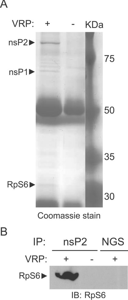
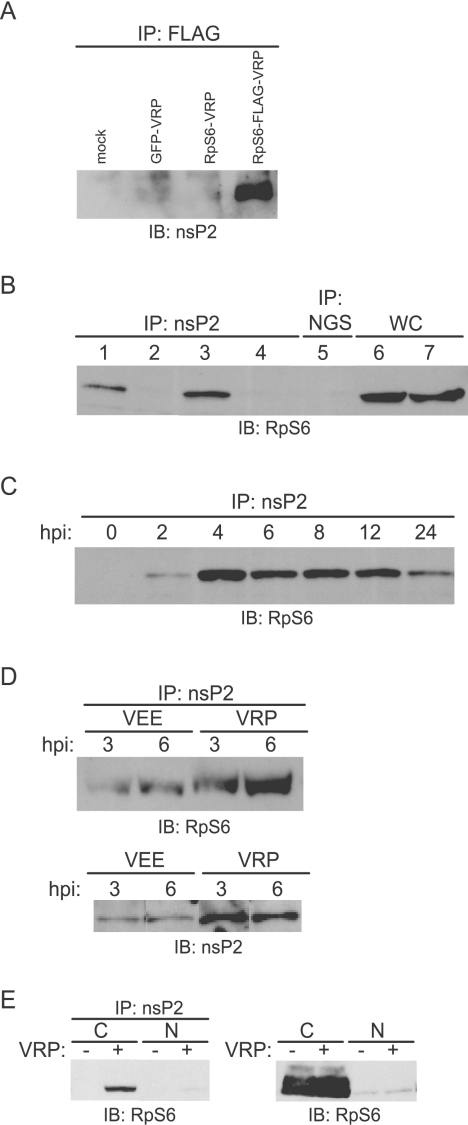
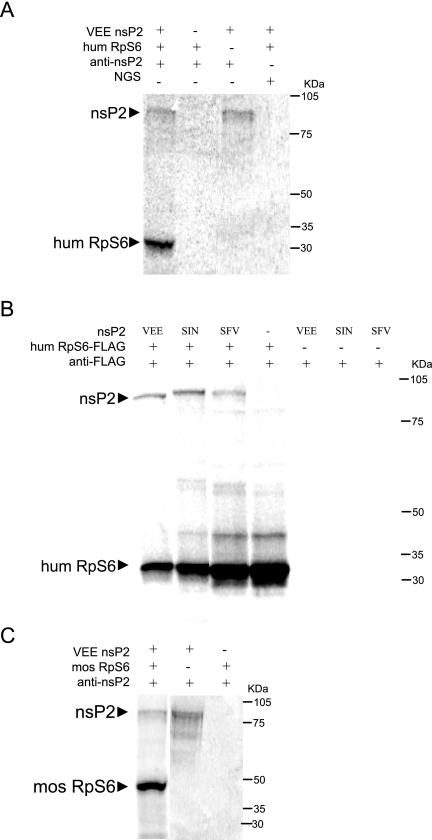
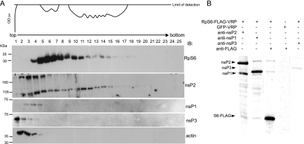
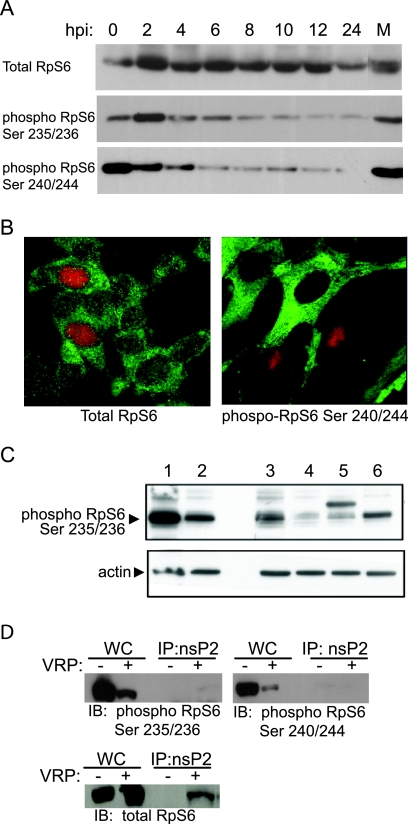
Although alphaviruses dramatically alter cellular function within hours of infection, interactions between alphaviruses and specific host cellular proteins are poorly understood. Although the alphavirus nonstructural protein 2 (nsP2) is an essential component of the viral replication complex, it also has critical auxiliary functions that determine the outcome of infection in the host. To gain a better understanding of nsP2 function, we sought to identify cellular proteins with which Venezuelan equine encephalitis virus nsP2 interacted. We demonstrate here that nsP2 associates with ribosomal protein S6 (RpS6) and that nsP2 is present in the ribosome-containing fractions of a polysome gradient, suggesting that nsP2 associates with RpS6 in the context of the whole ribosome. This result was noteworthy, since viral replicase proteins have seldom been described in direct association with components of the ribosome. The association of RpS6 with nsP2 was detected throughout the course of infection, and neither the synthesis of the viral structural proteins nor the presence of the other nonstructural proteins was required for RpS6 interaction with nsP2. nsP1 also was associated with RpS6, but other nonstructural proteins were not. RpS6 phosphorylation was dramatically diminished within hours after infection with alphaviruses. Furthermore, a reduction in the level of RpS6 protein expression led to diminished expression from alphavirus subgenomic messages, whereas no dramatic diminution in cellular translation was observed. Taken together, these data suggest that alphaviruses alter the ribosome during infection and that this alteration may contribute to differential translation of host and viral messages.


Figures

FIG. 1.
FIG. 1.
RpS6 coimmunoprecipitates with nsP2 in vivo. (A) Coomassie blue-stained gel of VRP-infected cell lysates immunoprecipitated with anti-nsP2 antibody. GFP-VRP (5005-3000) was used to infect BHK-21 cells at an MOI of 10 IU/cell. At 24 hpi, cells were lysed in NP-40 lysis buffer. Approximately 8 × 10⁵ cell equivalents were immunoprecipitated anti-nsP2 antibody, separated on an 8% SDS-PAGE gel, and stained with Coomassie blue to visualize proteins that coimmunoprecipitate with nsP2. As labeled, nsP2, nsP1, and RpS6 were identified by MALDI-TOF/MS. (B) Immunoblot for human RpS6 on GFP-VRP- or mock-infected lysates that were immunoprecipitated by using anti-nsP2 antibody. Normal goat serum (NGS) serves as an isotype-matched control for immunoprecipitation. RpS6 is specifically detected in VRP-infected lysates immunoprecipitated for nsP2.
FIG. 2.
FIG. 2.
Characterization of the RpS6-nsP2 interaction in vivo. Unless otherwise stated, cells were infected at an MOI of 10 IU/cell with GFP-VRP (5005-3000) or mock infected and lysed in NP-40 lysis buffer at 8 hpi. Lysates were then immunoprecipitated using anti-nsP2 antibody and subsequently subjected to immunoblot analysis using anti-RpS6 antibody. (A) Demonstration of the interaction in the reciprocal manner. HEK-293 cells were mock infected or infected with either GFP-VRP, RpS6-VRP, or RpS6-FLAG-VRP. At 8 hpi, the cells were lysed in NP-40 lysis buffer, and lysates were immunoprecipitated with anti-FLAG antibody and subsequently examined by immunoblot for nsP2. nsP2 coimmunoprecipitates specifically in VRP-infected lysates immunoprecipitated for the FLAG tag present on the RpS6 fusion protein. (B) The interaction between RpS6 and nsP2 occurs in multiple cell types. Lanes 1 and 2 contain samples using HEK-293 (human) cell lines and lanes 3 to 7 are from BHK-21 (hamster) cell lines. Samples in lanes 1, 3, 5, and 6 are infected with VRP; lanes 2, 4, and 7 contain mock-infected samples. As shown, the samples in lanes 1 to 4 were immunoprecipitated with anti-nsP2 antibody, the sample in lane 5 was immunoprecipitated with isotype-matched normal goat sera (NGS), and lanes 6 and 7 contain a direct load of whole-cell lysate (WC). (C) RpS6 coimmunoprecipitates with nsP2 throughout the course of infection. HEK-293 cells were infected and samples were harvested over a time course at the time points indicated. The nsP2-RpS6 interaction is continuously detected. (D) In the upper panel, RpS6 coimmunoprecipitates with nsP2 in VEE-infected cells. HEK-293 cells were infected at an MOI of 10 PFU/cell with VEE virus (V3000) or at 10 IU/cell with GFP-VRP (5005-3000). The presence of the viral structural proteins in VEE virus does not affect the ability of RpS6 to specifically coimmunoprecipitate with nsP2. In the lower panel, total cell lysates from VEE- or VRP-infected cells at 3 and 6 hpi were immunoblotted for nsP2. Note that more nsP2 has accumulated at each time point in the VRP-infected lysates, which corresponds to more RpS6 coimmunoprecipitating with nsP2 from VRP-infected lysates. (E) For the left panel, nuclear and cytoplasmic fractions were prepared from GFP-VRP- or mock-infected cells before immunoprecipitation for nsP2. The RpS6 that coimmunoprecipitates with nsP2 is predominantly cytoplasmic. For the right panel, equivalent amounts of crude cytoplasmic and nuclear lysates are subjected to immunoblot analysis using anti-RpS6 antibody, revealing that in both VRP-infected and mock-infected lysates there is much less RpS6 present in the nucleus compared to that in the cytoplasm.
FIG. 3.
FIG. 3.
In vitro interactions of nsP2 and RpS6. (A) nsP2 and human RpS6 interact when the two proteins are synthesized together in vitro. nsP2 and RpS6 were translated alone or together from individual pCI-neo-based plasmids in the presence of 35S label and subsequently immunoprecipitated using anti-nsP2 antibody or isotype-matched antibody (NGS). The specific coimmunoprecipitation RPS6 with nsP2 demonstrates that the two proteins interact in the absence of other viral proteins. (B) Similar to VEE, nsP2 from SIN and SFV interact with human RpS6. nsP2 from either VEE, SIN, or SFV was cotranslated with RpS6-FLAG. Immunoprecipitation using anti-FLAG antibody reveals that all of the alphavirus nsP2s coimmunoprecipitate with RpS6-FLAG. (C) Mosquito RpS6 associates with nsP2. RpS6 from either human (hum RpS6) or Aedes albopictus (mos RpS6) was cotranslated with VEE nsP2. Subsequent immunoprecipitation using anti-VEE nsP2 antibody showed that mosquito RpS6 coimmunoprecipitates with nsP2.
FIG. 4.
FIG. 4.
Other proteins associated with the complex containing nsP2 and RpS6. (A) RpS6 and nsP2 are present in the same fractions of a polysome gradient. HEK-293 cells were infected with GFP-VRP at an MOI of 5. At 8 hpi, cells were lysed and lysates were run over a 10 to 40% continuous sucrose gradient. An aliquot of each fraction of the polysome gradient was examined by immunoblotting using anti-RpS6, anti-nsP2, anti-nsP1, anti-nsP3, or anti-actin antibody. The polysome profile of this gradient, as monitored by measuring the optical density at 254 nm, is shown. The presence of RpS6 marks fractions that contain ribosomes, whereas the presence of actin demonstrates fractions containing most other cellular proteins. Note that nsP2, but not nsP1 and nsP3, comigrates with RpS6. The upper band in the nsP2 blot is the P123 precursor. (B) nsP1 coimmunoprecipitates with RpS6 and nsP2. HEK-293 cells were infected with RpS6-FLAG-VRP or GFP-VRP (as indicated) at an MOI of 10, and protein synthesis was monitored by using 35S label. At 8 hpi, cell lysates were made and subsequently used for immunoprecipitation. Immunoprecipitation with anti-nsP2 antibody reveals that nsP1 and RpS6-FLAG coimmunoprecipitate with nsP2, immunoprecipitation with anti-nsP1 antibody reveals that nsP2 and RpS6-FLAG coimmunoprecipitate with nsP1, and immunoprecipitation with anti-FLAG antibody reveals that nsP1 and nsP2 coimmunoprecipitate with RpS6-FLAG. Cells infected with GFP-VRP and immunoprecipitated with anti-FLAG antibody show that nsP1 and nsP2 coimmunoprecipitation is specific. There was no coimmunoprecipitation of the other radiolabeled proteins when lysates were immunoprecipitated with anti-nsP3 antibody.
FIG. 5.
FIG. 5.
VRP infection and RpS6 phosphorylation. (A) RpS6 phosphorylation is diminished within hours of VRP infection. Western blots for total RpS6 (upper panel) and phosphorylated RpS6 at Ser235/236 (middle panel) or Ser 240/244 (lower panel) are shown. Lysates are from GFP-VRP-infected (MOI of 10) HEK-293 cells at the time points indicated or from mock-infected (M) cells. (B) Similar to VRP infection, SFV4 reduces the amount of phosphorylated RpS6. Cells infected with SFV-NP, an SFV replicon that expresses influenza virus NP, at 8 hpi. NP, a nuclear protein, is visualized by Cy3 staining (red). In the left panel, total RpS6 is FITC stained (green); in the right panel, phospho-RpS6 (Ser240/Ser 244) is FITC stained. (C) Lysates of SFV-NP-infected cells at 14 hpi were immunoblotted for phospho-RpS6 (Ser240/Ser 244), demonstrating diminished RpS6 phosphorylation after SFV infection. Lane 1, cell lysates with phosphorylated RpS6 after 6 h of incubation in medium containing 20% fetal calf serum; lane 2, cell lysates with diminished amount of phosphorylated RpS6 after incubation in medium containing 0% fetal calf serum; lane 3, mock infection; lane 4, lysates from SFV-NP-infected cells; lane 5, cells infected with SFV-capsid enhancer-NP (note the nonspecific capsid band above RpS6 band); lane 6, cells infected with influenza virus. The lower gel shows actin. SFV replicon infection reduces phosphorylated RpS6 (lanes 4 and 5), whereas influenza virus infection does not diminish phosphorylated RpS6 as greatly (lane 6). (D) The population of RpS6 that associates with nsP2 is predominantly unphosphorylated. An immunoblot for total RpS6 and phosphorylated RpS6 on HEK-293 cell lysates immunoprecipitated with anti-nsP2 antibody is shown. Cells were infected at an MOI of 10 IU/cell with GFP-VRP (5005-3000) or were mock infected. At 8 hpi the cells were lysed in NP-40 lysis buffer, and a tripled amount of VRP- or mock-infected lysate was immunoprecipitated with anti-nsP2 antibody. The VRP- and mock-infected immunoprecipitates were then equally divided among three gels and analyzed by immunoblotting with one of two phospho-specific RpS6 antibodies, anti-RPS6 Ser235/236 or anti-RpS6 Ser 240/244, as well as an antibody that recognizes the total amount of RpS6. The RpS6 that coimmunoprecipitates with nsP2 is predominantly unphosphorylated at Ser235/Ser 236 or Ser 240/Ser 244. Note that the amount of phosphorylated RpS6 in VRP-infected cells is diminished compared to the amount of phosphorylated RpS6 in mock-infected cells (compare the whole-cell lysates labeled WC).
FIG. 6.
FIG. 6.
Diminished RpS6 protein level reduces expression of virally encoded protein. (A) HeLa cells were mock transfected or transfected with either a nontargeting siRNA pool (NTsiRNA) or an RpS6 siRNA pool (S6siRNA). At 48 h posttransfection, cell lysates were collected and immunoblotted for RpS6 or tubulin. (B) At 48 h after HeLa cells were transfected with siRNA, they were infected at an MOI of 5 with GFP-VRP. At 18 hpi the cells were harvested and subjected to FACS analysis. The numbers on the profiles (4.30, 4.35, and 4.04) indicate the percentage of cells infected as determined by GFP fluorescence. Although similar percentages of cells were infected in the mock-transfected (upper panel), nontargeting siRNA-transfected (middle panel), and RpS6 siRNA-transfected (lower panel) groups, RpS6 knockdown led to diminished fluorescence. The FACS profiles shown are representative of 10 samples. (C) Median GFP intensity of cells infected with GFP-VRP after transfection with NTsiRNA (□) or S6siRNA (▪). The intensity of virally encoded GFP is decreased 10-fold with the diminished RpS6 level. The data are representative of triplicate experiments. (D) At 48 h after transfection with siRNA, cellular translation was measured by using a 1-h pulse with [35S]methionine-[35S]cysteine. Extracts were analyzed by trichloroacetic acid precipitation and quantitation by a scintillation counter. The results are averages of quadruplicate samples and representative of three experiments. Host protein synthesis is minimally reduced after transfection with RpS6 siRNA, whereas cycloheximide treatment (CHX) dramatically reduces 35S-label incorporation.

Similar articles

Cited by

References

    1. Ahola, T., A. Lampio, P. Auvinen, and L. Kääriäinen. 1999. Semliki Forest virus mRNA capping enzyme requires association with anionic membrane phospholipids for activity. EMBO J. 18:3164-3172. - PMC - PubMed
    1. Baric, R. S., L. J. Carlin, and R. E. Johnston. 1983. Requirement for host transcription in the replication of Sindbis virus. J. Virol. 45:200-205. - PMC - PubMed
    1. Barton, D. J., S. G. Sawicki, and D. L. Sawicki. 1991. Solubilization and immunoprecipitation of alphavirus replication complexes. J. Virol. 65:1496-1506. - PMC - PubMed
    1. Borchers, C., J. F. Peter, M. C. Hall, T. A. Kunkel, and K. B. Tomer. 2000. Identification of in-gel digested proteins by complementary peptide-mass fingerprinting and tandem mass spectrometry data obtained on an electrospray ionization quadrupole time-of-flight mass spectrometer. Anal. Chem. 72:1163-1168. - PubMed
    1. Cardinali, B., M. Di Cristiana, and P. Pierandrei-Amaldi. 1999. Resistance of ribosomal protein mRNA translation to protein synthesis shutoff induced by poliovirus. J. Virol. 73:7070-7076. - PMC - PubMed

Publication types

MeSH terms

LinkOut - more resources