. 2006 Aug;80(15):7382-93.
doi: 10.1128/JVI.02052-05.

Autoactivation of the Epstein-Barr virus oncogenic protein LMP1 during type II latency through opposite roles of the NF-kappaB and JNK signaling pathways

Affiliations

Autoactivation of the Epstein-Barr virus oncogenic protein LMP1 during type II latency through opposite roles of the NF-kappaB and JNK signaling pathways

Gautier Goormachtigh et al. J Virol. 2006 Aug.

Abstract

Epstein-Barr virus (EBV) is associated with several human malignancies where it expresses limited subsets of latent proteins. Of the latent proteins, latent membrane protein 1 (LMP1) is a potent transforming protein that constitutively induces multiple cell signaling pathways and contributes to EBV-associated oncogenesis. Regulation of LMP1 expression has been extensively described during the type III latency of EBV. Nevertheless, in the majority of EBV-associated tumors, the virus is commonly found to display a type II latency program in which it is still unknown which viral or cellular protein is really involved in maintaining LMP1 expression. Here, we demonstrate that LMP1 activates its own promoter pLMP1 through the JNK signaling pathway emerging from the TES2 domain. Our results also reveal that this activation is tightly controlled by LMP1, since pLMP1 is inhibited by LMP1-activated NF-kappaB signaling pathway. By using our physiological models of EBV-infected cells displaying type II latency as well as lymphoblastoid cell lines expressing a type III latency, we also demonstrate that this balanced autoregulation of LMP1 is shared by both latency programs. Finally, we show that this autoactivation is the most important mechanism to maintain LMP1 expression during the type II latency program of EBV.


Figures

FIG. 1.
FIG. 1.
LMP1 upregulates its own promoter in a dose-dependent manner. (A) Dose-dependent transactivation of the LMP1 promoter by LMP1. HEK 293 cells were cotransfected with 80 ng of the firefly luciferase-based pLMP-luc reporter plasmid in conjunction with increasing doses (0 to 0.8 μg) of LMP1 expression vector. For each point, the amounts of DNA were completed with the corresponding empty vector (pSVHA). At 48 h posttransfection, we measured firefly luciferase activities, which were then normalized for transfection efficiency (on the basis of Renilla luciferase activity measured from cotransfected pRLnull reporter, which was included in all transfections). The promoter activity was expressed as activation (n-fold) over the corresponding control empty vector. Representative results are shown as means ± SD values (error bars) of an experiment with three replicate samples. (B) Dose-dependent expression of LMP1. Equal amounts (1 μg) of protein extracts from the cells described above were analyzed by Western blotting with the S12 antibody.
FIG. 2.
FIG. 2.
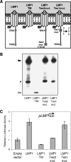
Mutation in TES2 abrogates the upregulating effect of LMP1 on its own promoter. (A) Schematic representation of the mutants used. Wild-type LMP1 is composed of a short N-terminal cytoplasmic region fused to the HA peptide, six transmembrane domains, and a long C-terminal cytoplasmic region responsible of signaling. Domains involved in signal transduction (transforming effector sites TES1 and TES2) are depicted by light gray boxes. LMP1-TM is deleted of the C-terminal cytoplasmic region of LMP1. For LMP1-Tes1mut and LMP1-Tes2mut, point mutations generated in either TES1 or TES2 are indicated. (B) Expressed levels of wild-type LMP1 and its mutants. HEK 293 cells were transiently transfected with 0.4 μg of plasmid DNA for each construct. Equal amounts (1 μg) of protein extracts from the cells indicated were analyzed by Western blotting with anti-HA antibody. The filled arrowhead indicates the wild-type and point-mutated versions of LMP1, while the open arrowhead points to LMP1-TM. The filled star designates a LMP1 cleavage product. (C) Comparison of the effects of LMP1 mutants on the LMP1 promoter. HEK 293 cells were cotransfected with 80 ng of pLMP-luc and 0.4 μg of expression plasmids encoding LMP1 or its mutant versions as indicated. Cells were harvested and analyzed for luciferase activity as described above. Representative results are shown as means ± SD values (error bars) of an experiment with three replicate samples.
FIG. 3.
FIG. 3.
The JNK signaling pathway induced by LMP1 is essential for pLMP1 upregulation. (A) Inhibition of LMP1 autoactivation by overexpression of JNKAPF. HEK 293 cells were cotransfected with 80 ng of pLMP-luc and 0.24 μg of expression plasmid encoding JNKAPF or its corresponding empty vector together with (black bars) or without (gray bars) 0.24 μg of pSVHA-LMP1. (B) Overexpression of JNKAPF effectively inhibits the LMP1-induced JNK signaling. c-Jun-dependent transcription activity was monitored by using a Gal4-Jun fusion protein and a gal4-luc reporter. HEK 293 cells were cotransfected with 80 ng of a construct encoding Gal4-Jun, 80 ng of a reporter plasmid, gal4-luc, containing Gal4-responsive elements, and 0.24 μg of pSVHA-LMP1 or the corresponding empty vector with or without 0.24 μg of expression plasmid encoding JNKAPF. Data shown are the ratios of luciferase activity measured in the presence of LMP1 to luciferase activity observed in the absence of LMP1. (C) Artificial activation of JNK induces pLMP1 activity and enhances LMP1 autoactivation. HEK 293 cells were cotransfected with 80 ng of pLMP-luc and 0.24 μg of expression plasmid encoding the fusion protein MKK7-JNKfus or its corresponding empty vector together with (black bars) or without (gray bars) 0.24 μg of pSVHA-LMP1. (D) Expression of MKK7-JNKfus activates the JNK signaling pathway. HEK 293 cells were cotransfected with 80 ng of a reporter plasmid AP1-luc, containing AP1-responsive elements, with or without 0.4 μg of expression plasmid encoding MKK7-JNKfus. In each case, cells were harvested and analyzed for luciferase activity and expression of LMP1 and JNKAPF or MKK7-JNKfus (under panels A and C) as described above. Representative results are shown as means ± SD values (error bars) of an experiment with three replicate samples.
FIG. 4.
FIG. 4.
The NF-κB signaling pathway inhibits the LMP1 promoter. (A) Increased pLMP1 activity and enhanced LMP1 autoactivation by IκBm overexpression. HEK 293 cells were cotransfected with 80 ng of pLMP-luc and 0.24 μg of expression plasmid encoding IκBm or its corresponding empty vector together with (black bars) or without (gray bars) 0.24 μg of pSVHA-LMP1. (B) Overexpression of IκBm abolishes the LMP1-induced NF-κB signaling pathway. HEK 293 cells were cotransfected with 80 ng of a reporter construct κB-luc, containing NF-κB-responsive elements, and 0.24 μg of pSVHA-LMP1 or its corresponding empty vector with or without 0.24 μg of expression plasmid encoding IκBm. Data shown are the ratios of luciferase activity measured in the presence of LMP1 to luciferase activity observed in the absence of LMP1. (C) NF-κB p65 overexpression inhibits pLMP1 and impairs LMP1 autoactivation. HEK 293 cells were cotransfected with 80 ng of pLMP-luc and 0.24 μg of expression plasmid encoding p65 or its corresponding empty vector together with (black bars) or without (gray bars) 0.24 μg of pSVHA-LMP1. (D) Activation of NF-κB signaling by overexpression of p65. HEK 293 cells were cotransfected with 80 ng of a reporter plasmid κB-luc with or without 0.24 μg of expression plasmid encoding p65. In each case, cells were harvested and analyzed for luciferase activity and expression of LMP1 and IκBm or p65 (blots at the bottom of panels A and C) as described above. Representative results are shown as means ± SD values (error bars) of an experiment with three replicate samples.
FIG. 5.
FIG. 5.
Opposite roles of the JNK and NF-κB pathways on endogenous LMP1 expression at the mRNA and protein levels. Inhibition of (A) NF-κB activity upregulates, whereas inhibition of (B) JNK activity downregulates the endogenous LMP1 protein levels in PRI and TE1 cell lines. The cells were treated with vehicle (DMSO), sulfasalazine (1 mM and 5 mM for TE1 and PRI cells, respectively), or SP600125 (20 μM) for 16 h. Equal amounts of cells were harvested and directly lysed in 3× Laemmli buffer. Protein lysates were analyzed by Western blotting for the expression of LMP1, IκBα, and EBNA2. Equal loading of proteins in each lane was confirmed by probing the membrane with anti-β-actin antibody. (C) Sulfasalazine increases, whereas SP600125 decreases the LMP1 mRNA levels in PRI cells. PRI cells were treated with either vehicle (DMSO) or sulfasalazine (5 mM) or SP600125 (20 μM) for 16 h. Cells were then harvested, and mRNAs were extracted and subjected to real-time RT-PCR as described in Materials and Methods. LMP1 mRNA levels were normalized to the mRNA levels of three housekeeping genes (β-actin, glyceraldehyde-3-phosphate dehydrogenase, and hypoxanthine phosphoribosyltransferase), and the normalized levels in the untreated condition were arbitrarily assigned a value of 100. (D) The constitutively active form of IκBα inhibits NF-κB target genes and increases the levels of LMP1 transcripts in PRI cells. Where indicated, the PRI-pRT1-IκBm stable cell line were treated with doxycycline (2 μg/ml) (+ Dox) to induce the expression of IκBm. After induction for 24 h, the relative LMP1 mRNA levels were quantified by real-time RT-PCR as described above. After induction for 48 h, protein lysates were collected as described above and analyzed by Western blotting for the expression of EBNA2, ICAM1, and TRAF1.
FIG. 6.
FIG. 6.
The dominant-negative form of LMP1, LMP1-CT, downregulates the mRNA and protein levels of LMP1 in EBV-infected cells. (A) Transient overexpression of LMP1-CT results in a decrease in LMP1 protein levels in TE1 cells. TE1 cells were transiently transfected by nucleofection (see Materials and Methods) with 2 μg of the LMP1-CT expression vector or the corresponding empty vector. Forty hours after nucleofection, protein lysates were collected as described above and analyzed by Western blotting with the S12 antibody. The open arrowhead points to full-length LMP1-CT, while the filled arrowhead indicates the endogenous wild-type LMP1. (B) Conditional LMP1-CT overexpression results in a decrease in LMP1 protein levels in PRI cells. Where indicated, the PRI-pRT1-LMP1-CT stable cell line was treated with doxycycline (2 μg/ml) (+ Dox) to induce the expression of LMP1-CT. After induction for 48 h, protein lysates were collected as described above and analyzed by Western blotting with the S12 antibody. The open arrowhead points to full-length LMP1-CT, while the filled arrowhead indicates endogenous wild-type LMP1. The open star designates a LMP1-CT cleavage product. In the rightmost lane, 1 μg of protein lysate from HEK 293 cells transfected only with the LMP1-CT expression vector was loaded to clearly localize the full-length and cleaved products of LMP1-CT. (C) Induction of LMP1-CT expression results in a disappearance of LMP1 mRNA in TE1 cells. The TE1-pRT1-LMP1-CT stable cell line was treated as described above. After induction for 48 h, LMP1-CT expression was measured by green fluorescent protein (GFP) fluorescence detection with a flow cytometer (inset), and the relative LMP1 mRNA levels were quantified by real-time RT-PCR as described above. The asterisk indicates undetectable levels of amplification. Total RNAs were also subjected to semiquantitative RT-PCR followed by electrophoresis on ethidium bromide-stained 1.5% agarose gel (insets). (D) Induction of LMP1-CT expression results in a decrease in LMP1 mRNA levels in PRI cells. The PRI-pRT1-LMP1-CT stable cell line was treated as described above. After induction for 48 h, the relative LMP1 mRNA levels were quantified by real-time RT-PCR as described above.

Similar articles

Cited by

References

    1. Adamson, A. L., D. Darr, E. Holley-Guthrie, R. A. Johnson, A. Mauser, J. Swenson, and S. Kenney. 2000. Epstein-Barr virus immediate-early proteins BZLF1 and BRLF1 activate the ATF2 transcription factor by increasing the levels of phosphorylated p38 and c-Jun N-terminal kinases. J. Virol. 74:1224-1233. - PMC - PubMed
    1. Adriaenssens, E., A. Mougel, G. Goormachtigh, E. Loing, V. Fafeur, C. Auriault, and J. Coll. 2004. A novel dominant-negative mutant form of Epstein-Barr virus latent membrane protein-1 (LMP1) selectively and differentially impairs LMP1 and TNF signaling pathways. Oncogene 23:2681-2693. - PubMed
    1. Aguilera, C., R. Hoya-Arias, G. Haegeman, L. Espinosa, and A. Bigas. 2004. Recruitment of IκBα to the hes1 promoter is associated with transcriptional repression. Proc. Natl. Acad. Sci. USA 101:16537-16542. - PMC - PubMed
    1. Bennett, B. L., D. T. Sasaki, B. W. Murray, E. C. O'Leary, S. T. Sakata, W. Xu, J. C. Leisten, A. Motiwala, S. Pierce, Y. Satoh, S. S. Bhagwat, A. M. Manning, and D. W. Anderson. 2001. SP600125, an anthrapyrazolone inhibitor of Jun N-terminal kinase. Proc. Natl. Acad. Sci. USA 98:13681-13686. - PMC - PubMed
    1. Bornkamm, G. W., C. Berens, C. Kuklik-Roos, J. M. Bechet, G. Laux, J. Bachl, M. Korndoerfer, M. Schlee, M. Hölzel, A. Malamoussi, R. D. Chapman, F. Nimmerjahn, J. Mautner, W. Hillen, H. Bujard, and J. Feuillard. 7 September 2005, posting date. Stringent doxycycline-dependent control of gene activities using an episomal one-vector system. Nucleic Acids Res. 33:e137. [Online.] doi:10.1093/nar/gni137. - DOI - PMC - PubMed

Publication types

Substances