

Estrogen receptor-alpha methylation predicts melanoma progression
- PMID: 16818643
- PMCID: PMC2856460
- DOI: 10.1158/0008-5472.CAN-06-0801
Estrogen receptor-alpha methylation predicts melanoma progression
Abstract
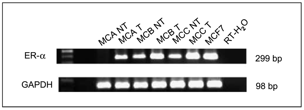
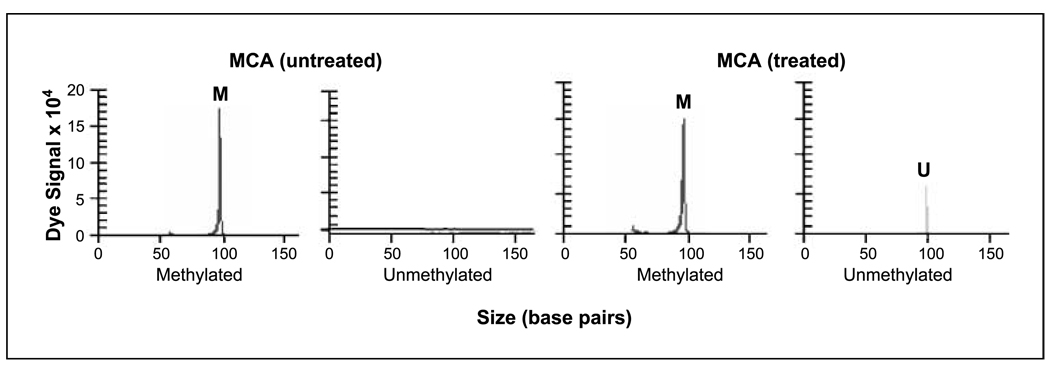
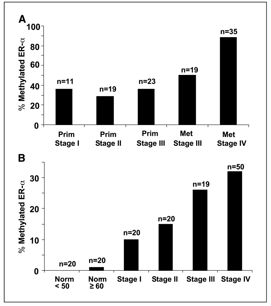
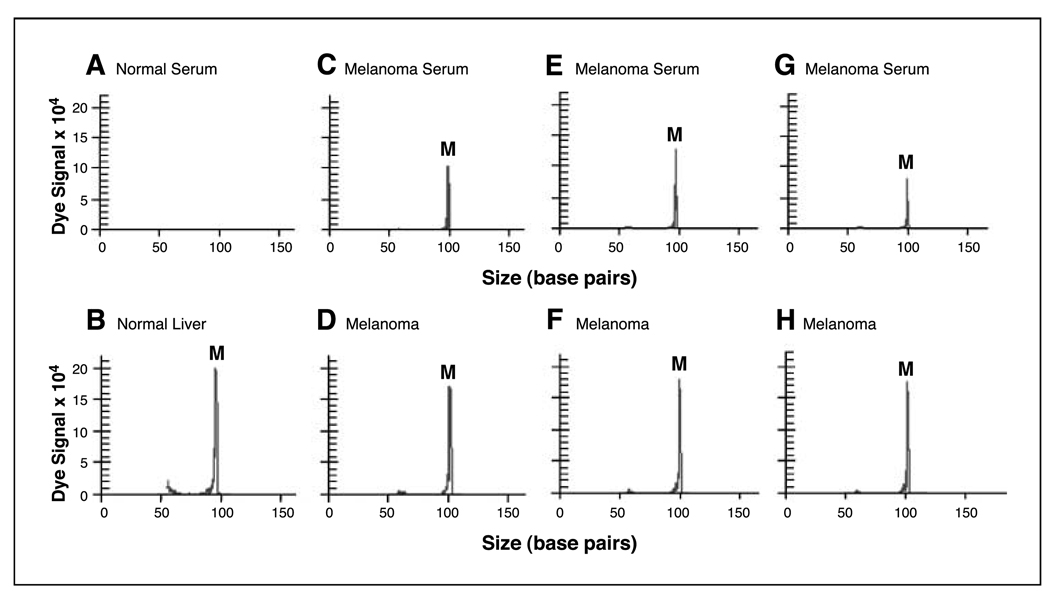
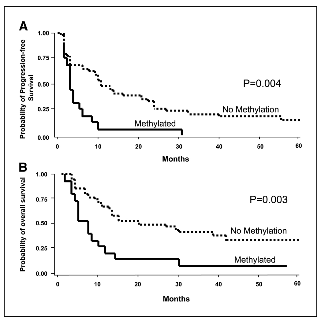
The role of estrogen receptor alpha (ER-alpha) in melanoma is unknown. ER-alpha expression may be regulated in melanoma via hypermethylation of promoter CpG islands. We assessed ER-alpha hypermethylation in primary and metastatic melanomas and sera as a potential tumor progression marker. ER-alpha methylation status in tumor (n = 107) and sera (n = 109) from American Joint Committee on Cancer (AJCC) stage I to IV melanoma patients was examined by methylation-specific PCR. The clinical significance of serum methylated ER-alpha was assessed among AJCC stage IV melanoma patients receiving biochemotherapy with tamoxifen. Rates of ER-alpha methylation in AJCC stage I, II, and III primary melanomas were 36% (4 of 11), 26% (5 of 19), and 35% (8 of 23), respectively. Methylated ER-alpha was detected in 42% (8 of 19) of stage III and 86% (30 of 35) of stage IV metastatic melanomas. ER-alpha was methylated more frequently in metastatic than primary melanomas (P = 0.0003). Of 109 melanoma patients' sera in AJCC stage I, II, III, and IV, methylated ER-alpha was detected in 10% (2 of 20), 15% (3 of 20), 26% (5 of 19), and 32% (16 of 50), respectively. Serum methylated ER-alpha was detected more frequently in advanced than localized melanomas (P = 0.03) and was the only factor predicting progression-free [risk ratio (RR), 2.64; 95% confidence interval (95% CI), 1.36-5.13; P = 0.004] and overall survival (RR, 2.31; 95% CI, 1.41-5.58; P = 0.003) in biochemotherapy patients. Hypermethylated ER-alpha is a significant factor in melanoma progression. Serum methylated ER-alpha is an unfavorable prognostic factor.
Figures
Similar articles
-
Predictive utility of circulating methylated DNA in serum of melanoma patients receiving biochemotherapy.J Clin Oncol. 2005 Dec 20;23(36):9351-8. doi: 10.1200/JCO.2005.02.9876. J Clin Oncol. 2005. PMID: 16361635 Free PMC article. Clinical Trial.
-
Association of circulating tumor cells with serum tumor-related methylated DNA in peripheral blood of melanoma patients.Cancer Res. 2006 Jun 15;66(12):6111-7. doi: 10.1158/0008-5472.CAN-05-4198. Cancer Res. 2006. PMID: 16778184 Free PMC article.
-
AIM1 and LINE-1 epigenetic aberrations in tumor and serum relate to melanoma progression and disease outcome.J Invest Dermatol. 2012 Jun;132(6):1689-97. doi: 10.1038/jid.2012.36. Epub 2012 Mar 8. J Invest Dermatol. 2012. PMID: 22402438 Free PMC article.
-
Promoter hypermethylation: a common cause of reduced p16(INK4a) expression in uveal melanoma.Cancer Res. 2001 Jul 1;61(13):5303-6. Cancer Res. 2001. PMID: 11431374
-
Dipeptidyl peptidase IV (DPPIV), a candidate tumor suppressor gene in melanomas is silenced by promoter methylation.Front Biosci. 2008 Jan 1;13:2435-43. doi: 10.2741/2856. Front Biosci. 2008. PMID: 17981724 Review.
Cited by
-
Insights from Mendelian randomization and genetic correlation analyses into the relationship between endometriosis and its comorbidities.Hum Reprod Update. 2023 Sep 5;29(5):655-674. doi: 10.1093/humupd/dmad009. Hum Reprod Update. 2023. PMID: 37159502 Free PMC article. Review.
-
Emerging technologies for studying DNA methylation for the molecular diagnosis of cancer.Expert Rev Mol Diagn. 2015 May;15(5):647-64. doi: 10.1586/14737159.2015.1027194. Epub 2015 Mar 22. Expert Rev Mol Diagn. 2015. PMID: 25797072 Free PMC article. Review.
-
A four-DNA methylation biomarker is a superior predictor of survival of patients with cutaneous melanoma.Elife. 2019 Jun 6;8:e44310. doi: 10.7554/eLife.44310. Elife. 2019. PMID: 31169496 Free PMC article.
-
Methylation levels of the "long interspersed nucleotide element-1" repetitive sequences predict survival of melanoma patients.J Transl Med. 2011 May 26;9:78. doi: 10.1186/1479-5876-9-78. J Transl Med. 2011. PMID: 21615918 Free PMC article.
-
A Narrative Review of the Role of Estrogen (Receptors) in Melanoma.Int J Mol Sci. 2024 Jun 6;25(11):6251. doi: 10.3390/ijms25116251. Int J Mol Sci. 2024. PMID: 38892441 Free PMC article. Review.
References
-
- Balch CM, Soong SJ, Atkins MB, et al. An evidence-based staging system for cutaneous melanoma. CA Cancer J Clin. 2004;54:131–149. - PubMed
-
- Dulaimi E, Hillinck J, Ibanez de Caceres I, Al-Saleem T, Cairns P. Tumor suppressor gene promoter hyper-methylation in serum of breast cancer patients. Clin Cancer Res. 2004;10:6189–6193. - PubMed
-
- Jeronimo C, Henrique R, Hoque MO, et al. A quantitative promoter methylation profile of prostate cancer. Clin Cancer Res. 2004;10:8472–8478. - PubMed
-
- Lapidus RG, Ferguson AT, Ottaviano YL, et al. Methylation of estrogen and progesterone receptor gene 5′ CpG islands correlates with lack of estrogen and progesterone receptor gene expression in breast tumors. Clin Cancer Res. 1996;2:805–810. - PubMed
-
- Shinozaki M, Hoon DS, Giuliano AE, et al. Distinct hypermethylation profile of primary breast cancer is associated with sentinel lymph node metastasis. Clin Cancer Res. 2005;11:2156–2162. - PubMed
Publication types
MeSH terms
Substances
Grants and funding
LinkOut - more resources
Full Text Sources
Other Literature Sources
Medical
